EOSINOPHIL-ASSOCIATED DERMATOSES
“Eosinophil-associated dermatoses” encompass a wide range of diseases characterized by the presence of a few to many eosinophils and/or evidence of eosinophil degranulation in skin and/or mucous membranes. Disorders characteristically associated with eosinophil infiltration include arthropod bite reactions and ectoparasite infestations (see Chs. 84 & 85), drug eruptions (see Ch. 21), atopic dermatitis (see Ch. 12), helminth infections (see Ch. 83), and Wells syndrome (Fig. 26.2). However, there are a number of other skin diseases where tissue eosinophilia may be present, including bullous pemphigoid, urticaria, and eosinophilic granulomatosis with polyangiitis (EGPA; Churg–Strauss syndrome) (Table 26.1). In addition, tissue eosinophilia can be observed in a wide range of skin disorders, from inflammatory to infectious to neoplastic.
More recently, it has been noted that mild to moderate peripheral eosinophilia (up to 35%–40%), along with tissue infiltration, is a key feature of IgG4-related disease (IgG4-RD; Table 26.2). Initially described as a pancreatic disorder with increased serum IgG4 levels and numerous IgG4- positive plasma cells in affected tissues, the spectrum of IgG4-RD has been expanded to include any organ, including skin. Proposed cutaneous manifestations are granuloma faciale and lesions that resemble angiolymphoid hyperplasia with eosinophilia (see Table 26.2 & Ch. 24).
Wells Syndrome
Synonym: Eosinophilic cellulitis
Key features
The characteristic presentation is recurrent, painful and/or pruritic, edematous to indurated plaques
Histologically, a diffuse dermal infiltrate composed of eosinophils and histiocytes is seen as are foci of amorphous/granular material, termed “flame figures”, which reflect localized degranulation of eosinophils
Flame figures are a characteristic but are not diagnostic feature, as they can be observed in a wide range of eosinophil-rich dermatoses
Distinction between Wells syndrome as a specific entity and a reaction pattern occurring in the setting of another dermatosis may be difficult
Dramatic improvement after administration of systemic corticosteroids
Introduction
Wells syndrome is a cutaneous disorder of unknown etiology characterized by one or more edematous (early) to indurated (late) plaques that resemble well-demarcated cellulitis. The classic histopathologic findings consist of a diffuse dermal infiltrate of eosinophils plus characteristic “flame figures”.
History
Wells described the first patient in 1971 as “recurrent granulomatous dermatitis with eosinophilia”. He later renamed the disease
“eosinophilic cellulitis”. Spigel and Winkelmann proposed the eponym, Wells syndrome, in 1979.
Epidemiology
Over 100 cases of Wells syndrome have been reported to date, ranging from newborns to patients over 70 years of age.
Pathogenesis
The pathogenesis of Wells syndrome is unknown. Local hypersensitivity due to “triggers” has been proposed, including insect bites or stings, drugs, allergic contact dermatitis, an underlying malignancy, and infections (e.g. dermatophytes, viruses, Toxocara canis). Of note, peripheral lymphocytes isolated from patients with Wells syndrome were reported to have exaggerated responses to mosquito salivary gland extracts. Activated eosinophils clearly play a major role in the pathophysiology of Wells syndrome and it is thought that Th2 cells, and IL-5 in particular, are responsible for eosinophil recruitment and activation in this disease.
Clinical features
In Wells syndrome, there are recurrent episodes of prodromal itching or burning, accompanied by markedly edematous plaques. The latter may have an annular or arcuate configuration, and vesicles and bullae can also be seen. Initially, the lesions are bright red, and then they fade to a pink–brown or slate-gray color (Fig. 26.3). The plaques may become indurated, and lesions usually resolve over 4 to 8 weeks. Less common clinical presentations include papules and hemorrhagic bullae. The extremities are most frequently affected, but involvement of the trunk can also be seen. The most common systemic complaint is malaise, with fever in less than a quarter of patients. Peripheral blood eosinophilia is common. Patients are often misdiagnosed as having erysipelas or acute cellulitis. Precipitating events, including arthropod bites and stings or vaccinations, have been described in a minority of patients.
Pathology
An interstitial dermal infiltrate of eosinophils, admixed with lymphocytes and histiocytes is seen (Fig. 26.4). The infiltrate is most prominent in the mid to deep dermis, with occasional involvement of the subcutaneous fat, fascia, and skeletal muscle. The superficial dermis may have massive papillary dermal edema to the point of subepidermal bulla formation. Intraepidermal vesiculation, with eosinophilic spongiosis, may also be present.
In addition to intact eosinophils, extracellular eosinophil granules are present in the dermis. The characteristic brightly eosinophilic flame figures consist of collagen fibers coated with eosinophil granule proteins (Fig. 26.4, inset). Utilizing indirect immunofluorescence assays, extracellular eMBP1 from eosinophil granules has been localized to flame
figures. In addition, a palisade of histiocytes and a few multinucleated giant cells partially surround these flame figures. While flame figures are considered a hallmark of Wells syndrome, they are not a specific finding. Flame figures can also be seen in other disorders in which degranulation of eosinophils occurs including arthropod bite and sting reactions, scabies, and less often, parasitic infections (e.g. onchocerciasis), bullous pemphigoid, pemphigus vegetans, and EGPA.
Differential diagnosis
Wells syndrome often has a striking clinicopathologic presentation. Early on, bacterial cellulitis and erysipelas are the most common clinical mimics. The histopathologic findings of both erysipelas and bacterial cellulitis can also include significant edema, but neutrophils are the predominant inflammatory cell in these soft tissue infections. Other causes of pseudocellulitis, including exaggerated reactions to arthropod bites, should be considered (see Table 74.10).
Toxocara canis and other parasitic infections can present with clinical and pathologic findings resembling Wells syndrome. When a parasitic infection is suspected, laboratory evaluation including stool examinations, specific parasite serologies, and even PCR of stool may be necessary. Other disorders that may present with urticarial plaques due to infiltrates of eosinophils are listed in Table 26.1. Late (mature) lesions of Wells syndrome may clinically, but not histologically, resemble morphea.
Eosinophilic annular erythema (EAE) is also in the differential diagnosis, but this entity is considered by many to be a subset of Wells syndrome. EAE is a figurate erythema that presents with annular and polycyclic skin lesions due to dermal infiltrates of eosinophils. However, flame figures are uncommon. EAE has been observed in patients with a number of conditions including malignancy, autoimmune thyroid disease, borreliosis, chronic gastritis caused by Helicobacter pylori infection, diabetes mellitus, hepatitis C virus, chronic kidney disease, thymoma, and autoimmune pancreatitis.
Treatment
Initial therapy with prednisone 10–80 mg daily results in dramatic improvement within a few days in most patients. Tapering the dose over one month is generally well tolerated. Flares or recurrence may be treated with additional courses of prednisone. Steroid-sparing medications, including minocycline, colchicine, antimalarials, dapsone,
IL-5Rα monoclonal antibodies. Anti-IL-5 antibodies inhibit its biological effects on eosinophils. Benralizumab interacts, via its Fab region, with the α subunit of the IL-5 receptor, thereby neutralizing IL-5 bioactivity. In addition, its Fc region binds to the FcγIIIRa on NK cells which induces apoptosis of eosinophils. See Ch. 138 for discussion of monoclonal antibodies that target the IL-4 receptor and IL-13. From Pelaia C, Paolett G, Puggioni F, et al. Interlukin-5 in the pathophysiology of severe asthma. Front Physiol 2019;10:1514.
griseofulvin, interferon-α and antihistamines, have limited evidence of efficacy. Recent reports suggest benefit with dupilumab, omalizumab (anti-IgE), mepolizumab (anti-IL-5), and benralizumab (anti-α chain of IL-5R) (Fig. 26.5). For milder cases, potent topical corticosteroids may be sufficient.
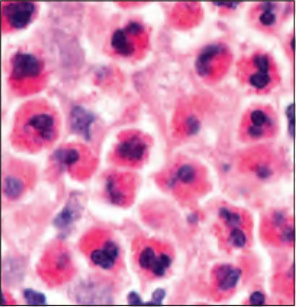
Fig. 26.2 Evaluation of adults with cutaneous infiltrates rich in eosinophils. These disorders are characterized histologically by eosinophil infiltration and/or eosinophil granule protein deposition. Peripheral blood eosinophilia occurs in some patients. Of note, systemic corticosteroids can significantly reduce the peripheral blood eosinophil count. Photomicrograph, courtesy Lorenzo Cerroni, MD.
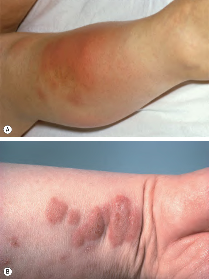
Fig. 26.3 Wells syndrome – clinical features.A Large edematous, erythematous plaque on the leg surrounded by several smaller plaques and nodules. This presentation may be misdiagnosed clinically as infectious cellulitis. B More mature dull pink nodules and plaques. A, Courtesy Luis Requena, MD.
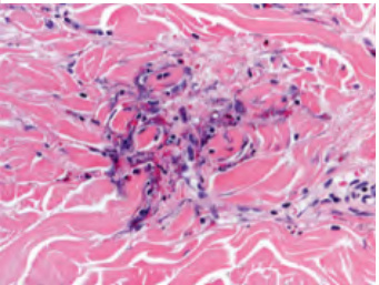
Fig. 26.4 Wells syndrome – histopathologic features. Perivascular and interstitial inflammatory infiltrates that contain eosinophils. There are several flame figures at sites of degranulated eosinophils (arrowheads; inset). Courtesy Lorenzo Cerroni, MD.
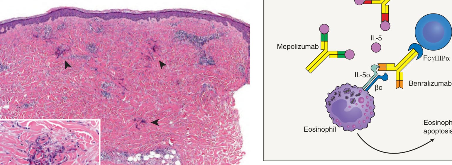
Fig. 26.5 Mechanisms of action of monoclonal antibodies that target IL-5 and
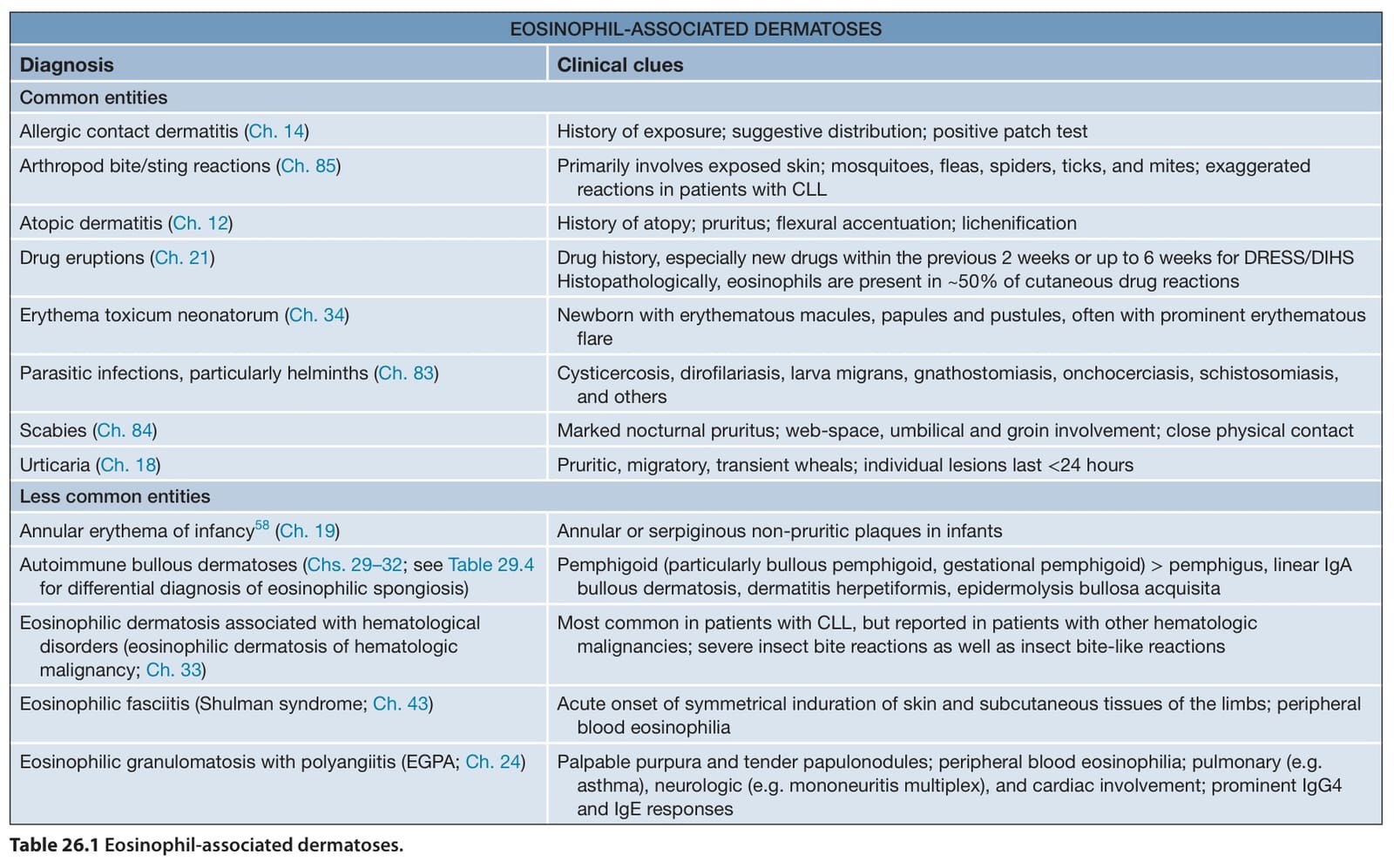
Table 26.1 Eosinophil-associated dermatoses.
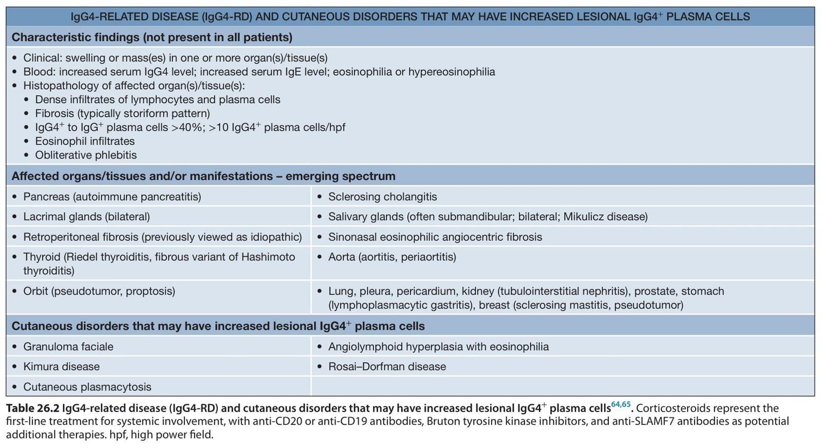
Table 26.2 IgG4-related disease (IgG4-RD) and cutaneous disorders that may have increased lesional IgG4+ plasma cells. Corticosteroids represent the first-line treatment for systemic involvement, with anti-CD20 or anti-CD19 antibodies, Bruton tyrosine kinase inhibitors, and anti-SLAMF7 antibodies as potential additional therapies. hpf, high power field.