HYPEREOSINOPHILIC SYNDROMES
Key features
Mucocutaneous lesions occur in over 50% of patients with hypereosinophilic syndromes (HESs), most commonly presenting as pruritic erythematous papules or nodules, urticaria, angioedema, and/or mucosal ulcers
Classification and treatment of HESs became more precise with discovery of the FIP1L1-PDGFRA fusion gene, whose protein product is a constitutively activated tyrosine kinase; additional chromosomal rearrangements have been identified that involve PDGFRA, PDGFRB, FGFR1, and PCM1-JAK2 which encode plateletderived growth factor receptor (PDGFR)-α, PDGFR-β, fibroblast growth factor receptor 1, and pericentriolar material 1/Janus kinase 2, respectively
Based upon recently adopted terminology and classification criteria, primary (neoplastic) and secondary (reactive) forms of HES have been more clearly defined
The FIP1L1-PDGFRA fusion gene and other genetic abnormalities
(see above) are present in patients with primary (neoplastic) HES, which includes eosinophilic leukemia
The lymphocytic (lymphoid) subtype of secondary (reactive) HES is characterized by a clonal proliferation of T cells with increased production of Th2 cytokines, particularly IL-5
End-organ damage may occur from sustained tissue eosinophilia, including life-threatening endomyocardial fibrosis and/or thrombosis
Introduction
The term “hypereosinophilic syndrome” or “HES” was coined in the late 1960s to refer to a spectrum of eosinophil-associated diseases with common clinical features, including peripheral eosinophilia (>1500 cells/mcl) and internal organ involvement. In the 1990s, a subset of patients with HES was found to have a clonal proliferation of T cells, with increased production of IL-5. In 2003, it was discovered that some patients with the HES variant that responded to imatinib had a FIP1L1-PDGFRA fusion gene due to a chromosomal translocation. Its protein product is a constitutively activated tyrosine kinase that is 100 times more sensitive to inhibition by imatinib than the BCR-ABL kinase present in chronic myelogenous leukemia (CML) patients. Since then, additional genetic abnormalities have been identified and a number of HES variants have been described (Table 26.3), leading to more tailored treatment recommendations (Fig. 26.6).
History
The term “hypereosinophilic syndromes (HESs)” was first coined in 1968 with the diagnostic criteria initially proposed in 1975. This was followed by the description of two major variants of HES – lymphocytic (LHES) and myeloproliferative (MHES). In 2011, experts from multiple disciplines came to a consensus regarding terminology and classification criteria during the Working Conference on Eosinophil Disorders and Syndromes. The currently recognized clinical variants are outlined in Table 26.3. Of note, the World Health Organization’s definition of HES excludes myeloid variants associated with molecular abnormalities.
Epidemiology
Up to 25% of patients have associated HES (see Table 26.3), which has an equal sex distribution, while MHES (FIP1L1-PDGFRA- or other mutation-positive HES) has a striking male predominance (>90%). HES can affect all age groups, but the majority are adults. While individuals with the FIP1L1-PDGFRA fusion gene may be regarded as having chronic eosinophilic leukemia, there are rare patients who develop more aggressive disease with transformation to acute leukemia. Patients with LHES are at risk for developing lymphoma.
Clinical Features
Patients present with signs and symptoms related to the organ systems affected by eosinophilic infiltrates. The most common mucocutaneous findings are pruritic erythematous macules, papules, plaques or nodules on the trunk and extremities or urticaria and angioedema. Other cutaneous manifestations include erythema annulare centrifugum-like lesions, livedo reticularis, retiform purpura, and superficial thrombophlebitis, with the thrombotic entities occurring almost exclusively in primary HES.
Since eosinophils have been implicated as the primary cause of endorgan damage in all forms of HES, clinical improvement usually parallels a decrease in both blood and tissue eosinophilia. However, the underlying pathogenesis of variants other than MHES and LHES still remains unclear. Occasionally, patients with episodic angioedema with eosinophilia (Gleich syndrome) or the nodules, eosinophilia, rheumatism, dermatitis and swelling (NERDS) syndrome may develop T cell clones and fulfill the diagnostic criteria for LHES (see Table 26.3). Therefore, as outlined in Figure 26.6, patients who cannot be classified as either MHES or LHES should continue to be monitored for cytogenetic abnormalities or T cell clonality.
Pathology
The histopathology of cutaneous lesions of HES is nonspecific and varies depending upon the type of skin lesion biopsied. Urticarial lesions have findings similar to ordinary urticaria, i.e. perivascular > interstitial infiltrates of lymphocytes, eosinophils, and occasional neutrophils. Biopsies from papules or plaques occasionally exhibit spongiosis in addition to the dermal infiltrate that usually contains at least a few eosinophils. Flame figures are occasionally present. When studied with immunohistochemical or immunofluorescence stains for specific eosinophil granule proteins, HES mucosal ulcers show extensive deposition of granule proteins in the absence of morphologically identifiable intact eosinophils. Thrombosis of dermal blood vessels has been observed in biopsy specimens from retiform purpura and necrotic skin lesions in HES patients.
Differential Diagnosis
The clinical and histopathologic differential diagnosis of HES includes other eosinophil-associated dermatoses (see Table 26.1), along with other dermatoses in which eosinophil degranulation occurs but infiltrates of intact eosinophils are not observed. The footprint of eosinophil participation is deposition of granular contents (e.g. eMBP1), which remain in the tissue while the cell itself is not morphologically identifiable, although parts of the eosinophil may be recognizable by electron microscopy. However, most other eosinophil-associated dermatoses will not be associated with peripheral blood hypereosinophilia of >1.5 cells × 10/L.
In contrast to Wells syndrome, patients with primary HES tend to be more systemically ill, with signs and symptoms of multi-organ involvement. Occasionally, dermal flame figures are seen in skin lesions of HES. EGPA can resemble HES clinically and is considered by some authors to be an HES-associated disease. Patients with EGPA have asthma as well as prominent IgG4 and IgE responses (see Ch. 24). While patients with EGPA have vasculitis of small and medium-sized vessels, vasculitis is an uncommon feature of HES. However, thromboembolic phenomena may produce lesions that mimic vasculitis (see Ch. 22).
Parasitic infections and infestations may closely resemble HES. A history of travel to endemic areas or a suggestive dietary history implicates helminthiasis. In such patients, serologic testing for anti-Strongy-loides antibodies and three stool samples for ova and parasites should be obtained. A total serum IgE level >500 IU/ml is often seen in helminth infections. Inappropriate treatment of Strongyloides stercoralis with systemic corticosteroids or other immunosuppressives may result in severe sequelae, including death.
In patients with HES and isolated urticarial plaques with or without angioedema, the differential diagnosis includes ordinary urticaria. In such patients, demonstration of internal organ involvement favors HES. In addition, HES with episodic angioedema may resemble hereditary or acquired angioedema clinically, but complement studies help to distinguish these entities (see Fig. 18.19). Furthermore, patients with hereditary angioedema often have a family history of the disease and rarely are their peripheral blood eosinophil counts as high as in HES.
Treatment
If patients do not present with life-threatening manifestations, high-dose corticosteroids (e.g. prednisone 1 mg/kg/day), if indicated, can be initiated after the evaluation is complete (see Fig. 26.6). Patients should be administered ivermectin if there has been exposure to Strongyloides.
If FIP1L1-PDGFRA or other genetic abnormalities involving PDGFRA or PDGFRB are present, imatinib therapy is warranted (see Fig. 26.6). As a tyrosine kinase inhibitor, imatinib prevents autophosphorylation of the fusion protein and phosphorylation of other substrates, resulting in termination of the signaling cascade that induces genes related to eosinophil proliferation and activation. Patients respond to varying doses of imatinib, depending upon an individual patient’s sensitivity and ability of the given dosage to suppress the mutant clone. Imatinib doses ranging from 400 mg daily to 100 mg weekly or less have proven effective. However, maintenance therapy is required to avoid relapse. Because endomyocardial disease may worsen during the first several days of imatinib administration, serum levels of troponin and
N-terminal pro-brain natriuretic peptide (NT-proBNP) should be monitored. Preemptive and concurrent treatment with corticosteroids is also recommended to maintain cardiac function. Other kinase inhibitors (nilotinib, dasatinib, and sorafenib) may also be useful in patients who do not respond to imatinib or become resistant as a consequence of mutations such as T674I in FIP1L1-PDGFRA.
Secondary HES is best managed by treating the underlying disease, once determined. When the underlying disease fails to respond to first-line therapies and the patient lacks PDGFRA or PDGFRB fusion genes, then corticosteroids are begun, usually prednisone at 1 mg/kg/ day; ~70% of patients will respond, with the peripheral eosinophilia returning to normal levels. For patients who fail corticosteroid monotherapy or develop significant long-term side effects, there are several other therapeutic options (see Fig. 26.6). For example, patients with no
Interferon (IFN)-α (12–50 × 10 U/week) has been beneficial in patients with both primary and secondary HES. Its mode of action is likely via myelosuppression but may include effects on Th2 helper cells by changing the cytokine milieu (including a decrease in IL-5 levels). For some patients, pegylated interferon (peginterferon-α 2a), administered weekly, is better tolerated. One concern regarding the use of IFN- α, based upon the findings of an in vitro study in which IFN-α acted as a growth factor for CD3−CD4+ cells, is probably minimized by concomitant corticosteroid therapy.
Two monoclonal antibodies against human IL-5 approved for the treatment of asthma, mepolizumab and reslizumab, and a monoclonal antibody directed against the alpha chain of the IL-5R, benralizumab, have been used to treat HES in clinical trials (see Fig. 26.5). Analyses of cytokine production by lymphocytes before and after treatment with mepolizumab showed a striking reduction in Th2 cytokine production (IL-13, IL-10) in addition to the expected IL-5 reduction. In a randomized, double-blind, placebo-controlled trial involving FIP1L1-PDGFRA-negative patients who were already receiving prednisone (20–60 mg/ day), administration of mepolizumab (750 mg IV every 4 weeks for 9 cycles) led to twice as many individuals (84% vs 43%) being successfully tapered to a stable prednisone dose of ≤10 mg/day.
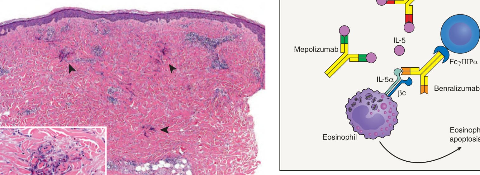
Fig. 26.5 Mechanisms of action of monoclonal antibodies that target IL-5 and
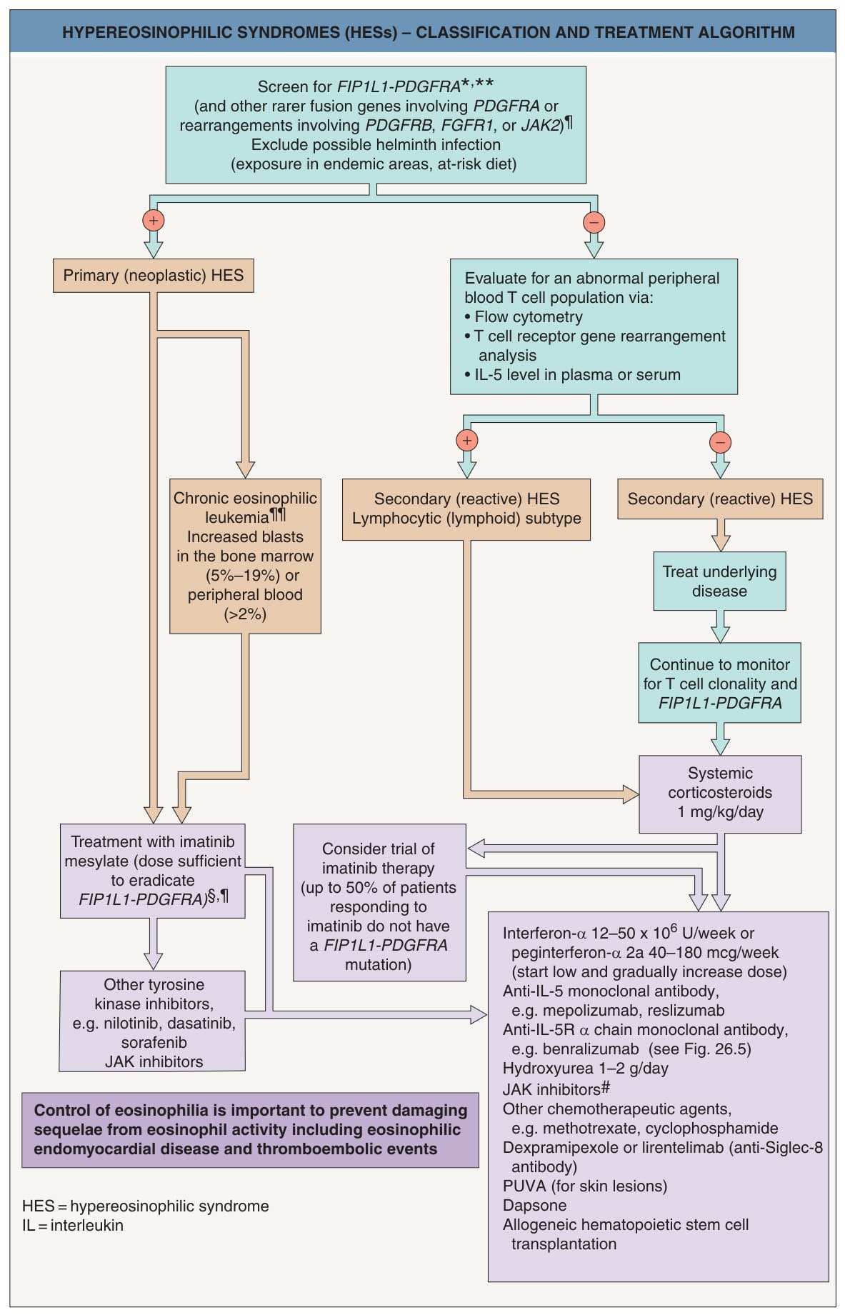
**Fig. 26.6 Hypereosinophilic syndromes (HESs) – classification and treatment algorithm. *An 800 kb deletion on chromosome 4 produces a fusion gene composed of a portion of the PDGFRA (platelet-derived growth factor receptor α) gene that encodes its kinase domain linked to a previously uncharacterized gene that resembles Fip1, which encodes an essential component of the Saccharomyces cerevisiae polyadenylation machinery. The resultant product is a constitutively activated tyrosine kinase that is a target of the tyrosine kinase inhibitor, imatinib. Screening can be performed on a peripheral blood sample via reverse transcriptase- PCR or fluorescence in situ hybridization (FISH). Bone marrow biopsy with cytogenetic studies should be done to exclude hematologic disorders that are accompanied by eosinophilia. Increased bone marrow mast cells and increased serum tryptase may be observed. ¶PDGFRB, FGFR1, and JAK2 encode platelet-derived growth factor receptor-β, fibroblast growth factor receptor-1, and Janus kinase 2, respectively; patients with genetic abnormalities involving PDGFRA and PDGFRB typically respond to imatinib but not those involving FGFR1. Patients may become resistant due to new mutations (during blast crisis) or tandem mutations (during chronic phase). ¶¶May have other clonal cytogenetic and molecular genetic abnormalities; in the presence of eosinophilia, considered sufficient for diagnosis even in the absence of excess blasts. §Plus systemic corticosteroids if cardiac involvement. including those with JAK2 mutations. Data from refs detectable FIP1L1-PDGFRA fusion gene have achieved partial remission from imatinib, although the mechanism of response in these patients is unclear. Perhaps some patients had diagnostically occult PDGFRA or PDGFRB rearrangements. Thus, imatinib represents a reasonable therapeutic option, given its relative safety.
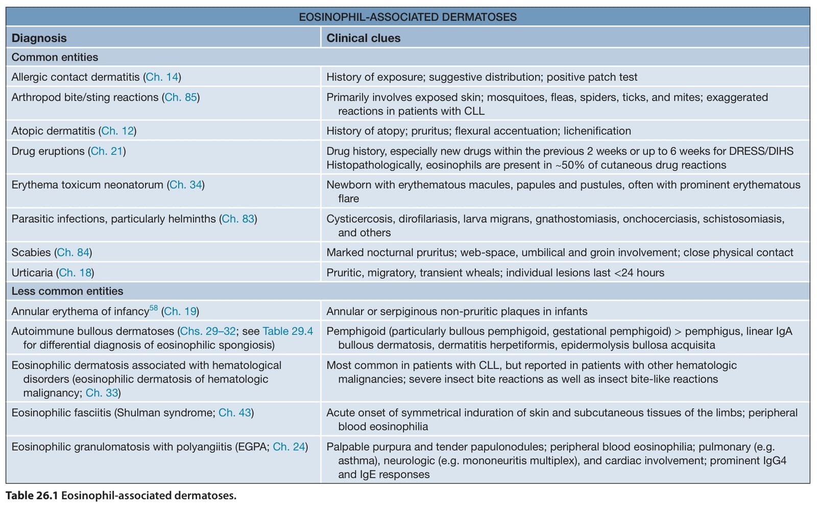
Table 26.1 Eosinophil-associated dermatoses.
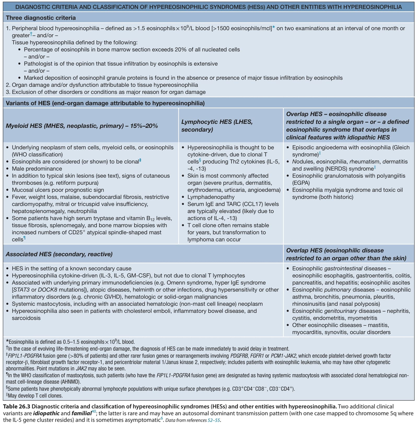
Table 26.3 Diagnostic criteria and classification of hypereosinophilic syndromes (HESs) and other entities with hypereosinophilia. Two additional clinical variants are idiopathic and familial; the latter is rare and may have an autosomal dominant transmission pattern (with one case mapped to chromosome 5q where the IL-5 gene cluster resides) and it is sometimes asymptomatic.Data from references
The main treatment goals via single- or multiple-agent regimens are relief of symptoms and maintaining the peripheral eosinophil count in the range of 1–2 × 10/L blood or less. HES patients also need to be routinely monitored for organ involvement, particularly cardiac disease, in addition to progression to lymphoma or leukemia. Patients who fail corticosteroid monotherapy have a worse prognosis.
Additional table on entities in which flame figures may be seen, available in our eBook (see inside front cover for access code).