GESTATIONAL PEMPHIGOID
Synonyms: Pemphigoid gestationis Herpes gestationis
Key features
Rare, pruritic, vesiculobullous eruption that develops during late pregnancy or the immediate postpartum period
Linear C3 deposition along the basement membrane zone (BMZ) by direct IF
IgG1 autoantibodies are directed against a transmembrane hemidesmosomal protein (BP180; BPAG2; collagen XVII)
Increased risk of prematurity and small-for-gestational age neonates; the risk correlates with disease severity
Commonly recurs in subsequent pregnancies
Introduction
Gestational pemphigoid is a rare, self-limited, autoimmune bullous disease. It is the most clearly characterized dermatosis of pregnancy and the only one that may also affect the skin of the newborn.
History
Milton first coined the term “herpes gestationis” in 1872 and Bulkley (1874) canonized the term “as embodying the clinical characters of the eruption and signifying at the same time the sex and state of the body in which it appears”. Little additional insight was gained until IF techniques revealed complement deposition along the BMZ in 1973, a feature now accepted as essential for the diagnosis of gestational pemphigoid.
Epidemiology
The incidence of gestational pemphigoid has been estimated at 1 : 1700–1 : 50 000 pregnancies, correlating with the prevalence of
HLA-DR3 and -DR4 in different populations. While occurring primarily during pregnancy and the immediate postpartum period, gestational pemphigoid has rarely developed in association with trophoblastic tumors (hydatidiform mole, choriocarcinoma). However, no case of a pemphigoid-like disease has been reported in men with choriocarcinoma, who have a biochemically similar yet entirely syngenic tumor; of note, the nuclear genome in placental tissue (and hence choriocarcinoma in women) is primarily paternal in origin. Patients with a history of gestational pemphigoid appear to be at increased risk for the development of Graves disease.
Pathogenesis
Historically, gestational pemphigoid was thought to be caused by an anti-BMZ “serum factor” (the “herpes gestationis [HG] factor”) that induces C3 deposition along the dermal–epidermal junction. This factor is now known to be complement-fixing autoantibodies of the IgG1 subclass directed against a 180 kDa transmembrane hemidesmosomal protein (BP180; BPAG2; collagen XVII). As in patients with bullous pemphigoid (BP), it is the non-collagenous (NC) segment closest to the plasma membrane of the basal keratinocyte, NC16A, that constitutes the immunodominant region of BP180 (see Fig. 31.9). Circulating antibodies are almost exclusively directed against this domain, as demonstrated by ELISA and immunoblot studies of maternal or neonatal sera.
What initiates the production of autoantibody remains enigmatic. Because the antibodies also bind to amniotic basement membrane (a structure derived from fetal ectoderm and antigenically similar to skin), attention has focused on immunogenetics and potential cross-reactivity between placental tissue and skin. Immunogenetic studies have revealed a marked increase in HLA antigens DR3 or DR4, and, curiously, nearly 50% of patients have the simultaneous presence of both. There is essentially a 100% incidence of anti-HLA antibodies in patients with a history of gestational pemphigoid. Since the only source of disparate HLA antigens is typically the placenta (which is primarily of paternal origin), the universal finding of anti-HLA antibodies implies a high frequency of immunologic insult during gestation. Women with gestational pemphigoid also have increased expression of MHC class II antigens (DR, DP, DQ) within the villous stroma of their chorionic villi. It has therefore been proposed that gestational pemphigoid is a disease, initiated by the aberrant expression of MHC class II antigens (of paternal haplotype), that serves to initiate an allogeneic response to placental BMZ, which then cross-reacts with skin.
Clinical Features
Gestational pemphigoid may develop during any trimester as well as immediately postpartum, but classically it presents during late pregnancy. There is an abrupt onset of cutaneous lesions on the trunk, in particular the abdomen and often within or immediately adjacent to the umbilicus (Fig. 27.1). Rapid progression to a generalized pemphigoid-like eruption then occurs, with pruritic urticarial papules and plaques, followed by clustered (herpetiform) vesicles or tense bullae on an erythematous base. The eruption may involve the entire body, sparing only the mucous membranes. While the clinical presentation and course may vary considerably, spontaneous improvement during late gestation is common. This is followed, however, by a flare at the time of delivery in 75% of patients. Such flares may be dramatic, with an explosive onset of blistering within hours.
Most disease activity spontaneously remits during the weeks to months following delivery, but there are isolated reports of a protracted course postpartum. Flares and/or recurrences in association with menstruation are common, and in 25%–50% of patients, they may also be induced by oral contraceptives (Table 27.2). Gestational pemphigoid may not develop during the patient’s first pregnancy, but once established, it is quite likely to recur in subsequent pregnancies, usually with
an earlier onset and more severe course. “Skipped” pregnancies have been observed in 5%–8% of women.
Approximately 10% of newborns develop mild skin involvement due to passive transfer of maternal antibodies and this resolves spontaneously within days to weeks (see Ch. 34). There seems to be an increased risk of prematurity and small-for-gestational age neonates, presumably due to chronic placental insufficiency. More recently it was shown that this risk correlates with disease severity, i.e. occurrence of blistering and early onset, and not with the use of systemic corticosteroids.
Pathology
The classic histologic finding of a subepidermal vesicle is seen in the minority of patients. Instead, a nonspecific mixed cellular infiltrate containing a variable number of eosinophils is more common. The presence of eosinophils is the most constant histologic feature of gestational pemphigoid (Fig. 27.2).
The essential component for the diagnosis of gestational pemphigoid is a linear deposition of C3 along the BMZ of perilesional skin by direct IF microscopy (Fig. 27.3). This is observed in 100% of patients, and linear IgG deposition is seen in 30% of patients. When salt-split skin specimens are employed for conventional indirect IF, deposition of IgG along the bottom of the epidermal fragment is observed in 30% of patients. However, complement-added indirect IF reveals the circulating anti-BMZ IgG1 autoantibodies in essentially all patients. Determination of antibody titers via BP180-NC16A ELISA may be helpful in following disease activity and monitoring therapy.
microscopy. Linear deposits of C3 are seen along the basement membrane zone. Courtesy Immunofluorescence Department, St John’s Institute of Dermatology, St Thomas’ Hospital, London, UK.
An increased incidence of anti-thyroid antibodies has been documented, but clinically apparent thyroid dysfunction is uncommon. Routine laboratory investigations are normal.
Differential Diagnosis
The most frequent considerations in the differential diagnosis are PEP and drug eruptions. PEP is a particularly challenging exclusion, given the difficulty of distinguishing PEP and urticarial lesions of gestational pemphigoid. Direct and indirect IF and, more recently, the BP180- NC16A ELISA are key to the differentiation and are especially relevant in helping the patient plan for future pregnancies.
Treatment
The primary goal in treating this self-limited disease is to relieve pruritus and suppress blister formation. In mild cases, the use of potent topical corticosteroids combined with emollients and systemic antihistamines may be adequate. However, systemic corticosteroids remain the cornerstone of therapy (Table 27.3). Most patients respond to 0.5 mg/kg of prednisolone daily; the dose is tapered as soon as blister formation is suppressed. The common flare associated with delivery usually requires a temporary increase in dosage. Those rare patients with refractory disease may benefit from plasmapheresis during pregnancy. Persistent disease after delivery is uncommon and is treated like BP.
Anecdotal alternatives to corticosteroids (dapsone, doxycycline, or minocycline ± nicotinamide, pyridoxine, cyclosporine) or adjuvants (methotrexate, cyclophosphamide, gold, IVIg) have been tried.
None of these medications, with the possible exception of cyclosporine and IVIg, are safe prior to term and thus should be avoided.
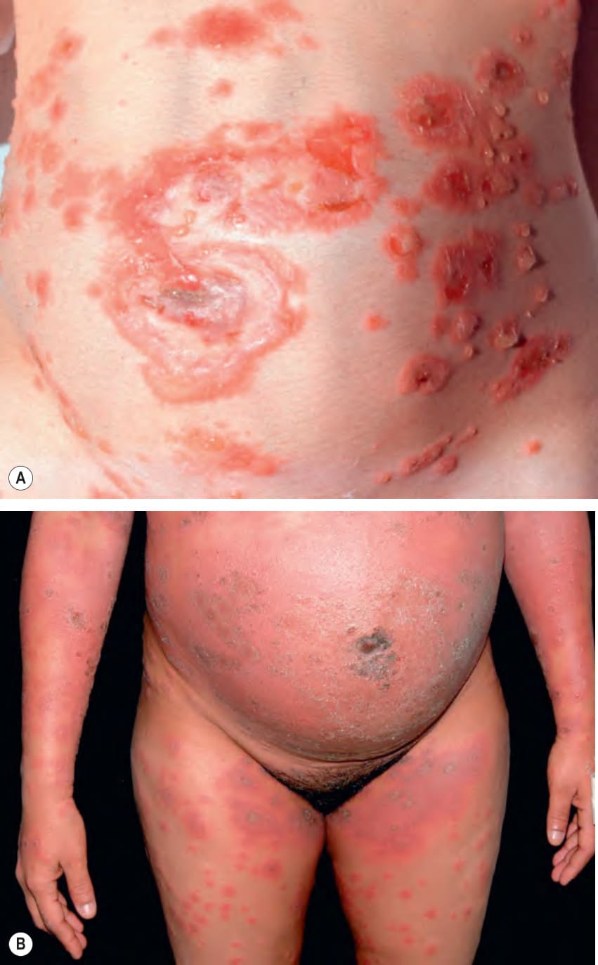
Fig. 27.1 Gestational pemphigoid.A Intact tense bullae arising within areas of edematous erythema as well as erosions due to ruptured bullae; lesions typically involve the umbilical region. B Confluent crusted erythematous plaques studded with small vesicles; umbilical involvement is again noted. Dusky urticarial lesions are also present on the thighs. B, Courtesy Luis Requena, MD.
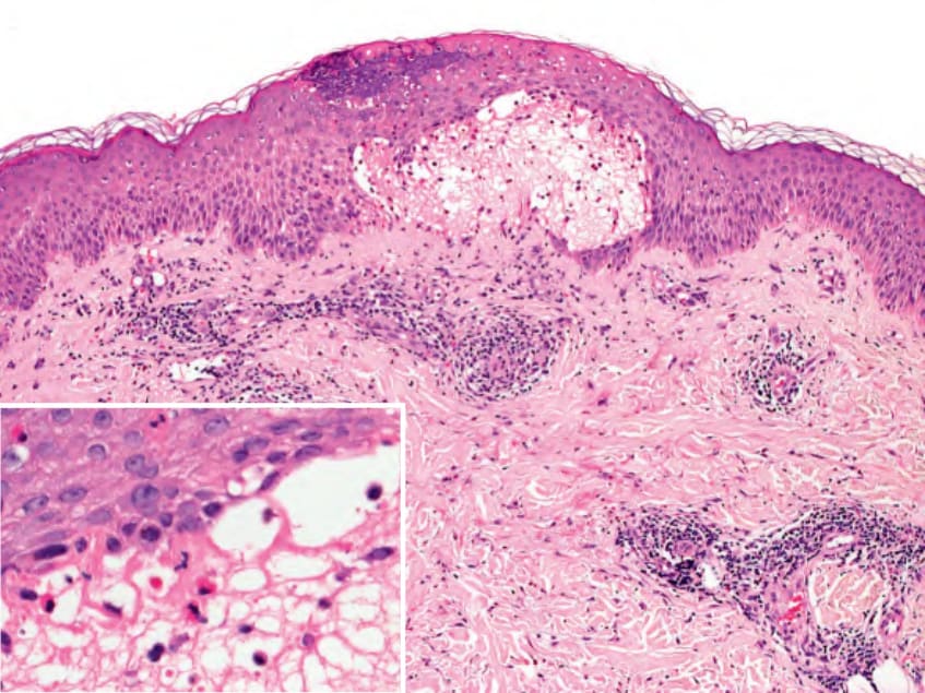
Fig. 27.2 Gestational pemphigoid – histopathologic features of an early lesion. Focal subepidermal vesicle accompanied by a superficial and mid dermal perivascular and interstitial mixed inflammatory infiltrate. Eosinophils are seen within the blister cavity and the epidermis (inset). Courtesy Lorenzo Cerroni, MD.
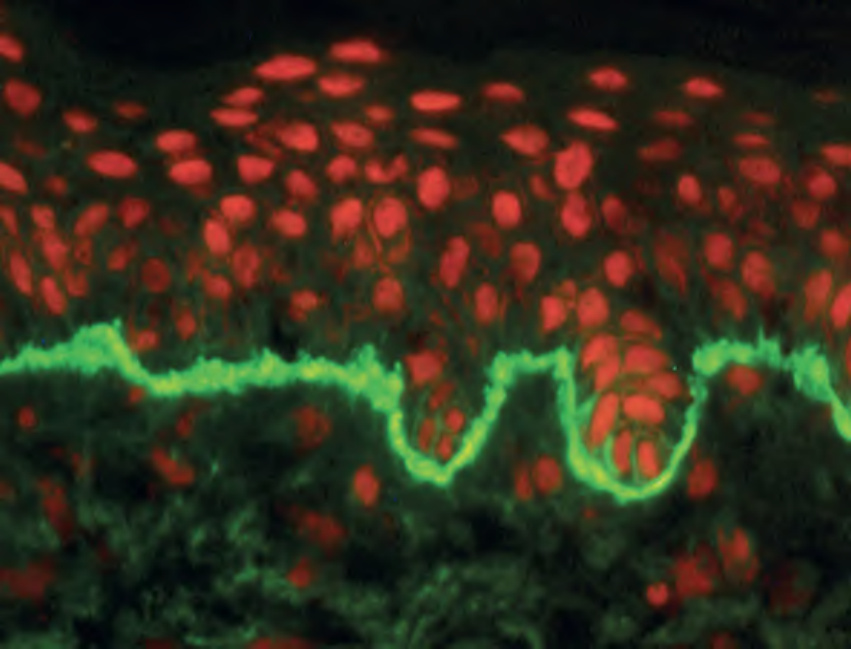
Fig. 27.3 Gestational pemphigoid – direct immunofluorescence (IF)
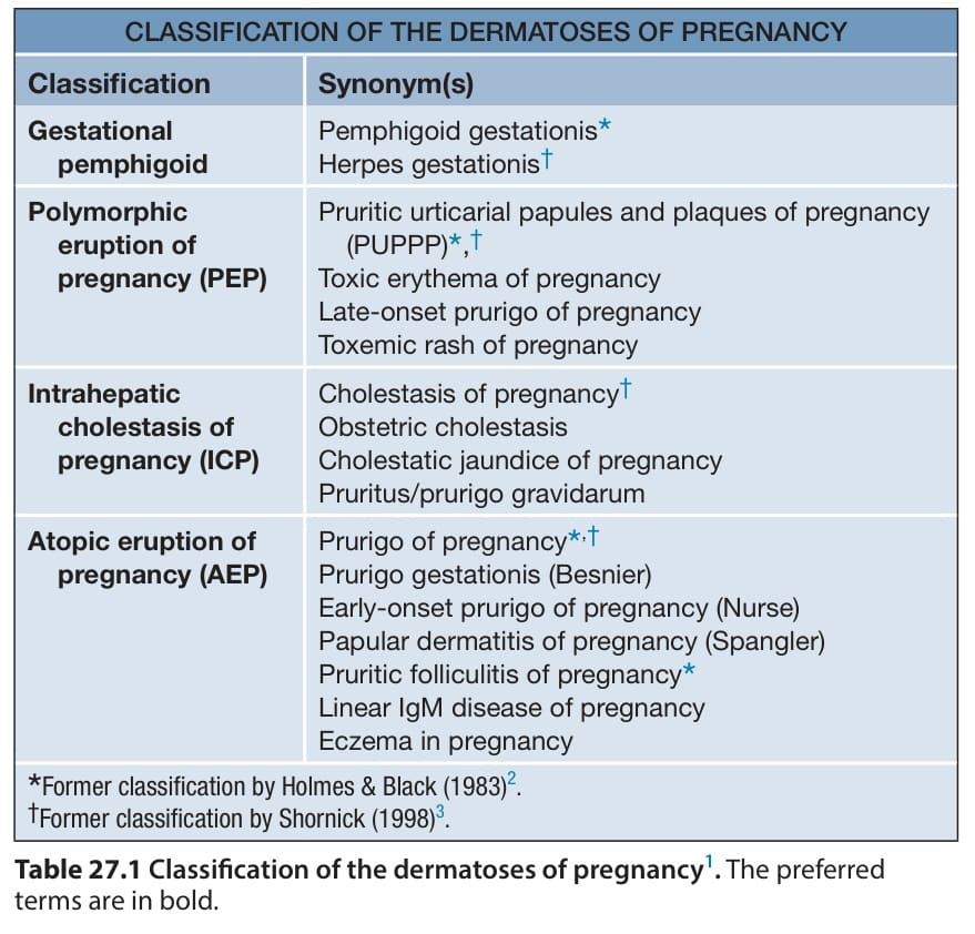
Table 27.1 Classification of the dermatoses of pregnancy. The preferred terms are in bold.
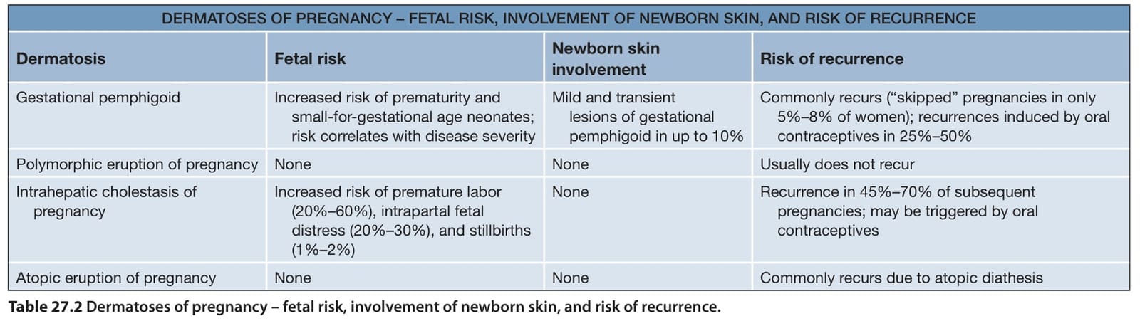
Table 27.2 Dermatoses of pregnancy – fetal risk, involvement of newborn skin, and risk of recurrence.
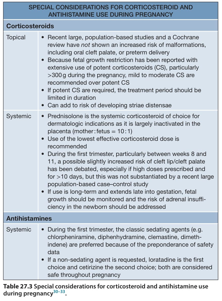
Table 27.3 Special considerations for corticosteroid and antihistamine use during pregnancy.