POLYMORPHIC ERUPTION OF PREGNANCY
Synonyms: Pruritic urticarial papules and plaques of pregnancy (PUPPP) Bourne’s “toxaemic rash of pregnancy” Nurse’s “late-onset prurigo” of pregnancy Toxic erythema of pregnancy
Key features
Urticarial papules and plaques that usually first appear within striae distensae during the latter portion of the third trimester or immediately postpartum
Development of polymorphous features (vesicles, erythema, target and eczematous lesions) with disease progression
Most frequent in primiparous women
Nonspecific histologic features, negative IF, and normal routine laboratory evaluation
No maternal or fetal risks; usually does not recur
Introduction
Polymorphic eruption of pregnancy (PEP), formerly known as PUPPP, is a common gestational dermatosis. It is characterized by a typical clinical presentation, normal laboratory tests, and negative IF or ELISA.
History
The term PUPPP, introduced by Lawley et al. in 1979, focuses on the initial clinical findings in this disorder but overlooks its later polymorphous features. In order to encompass the entire clinical spectrum, the term “polymorphic eruption of pregnancy” was introduced and is now generally accepted.
Epidemiology
The incidence is ~1 in 160 deliveries. It is seen predominantly in primiparous women and tends not to recur in subsequent pregnancies. There is neither an autoimmune diathesis nor an association with a specific HLA type.
Pathogenesis
The cause of PEP is unknown. Reference has been made to increased maternal weight gain and an increased frequency of multiple-gestation pregnancies. It has therefore been suggested that rapid, late stretching of abdominal skin may lead to damage of connective tissue and elicitation of an allergic-type reaction, resulting in the initial appearance of the eruption within striae. The lesions then become generalized as the inflammatory response develops cross-reactivity to collagen in otherwise normal-appearing skin. Immune tolerance during subsequent pregnancies might prevent recurrence. Additional theories include increased levels of progesterone in association with multiple gestations and peripheral chimerism (deposition of fetal DNA) that favors skin with increased vascularity and damaged collagen.
Clinical Features
Pruritic erythematous and edematous papules and plaques usually first appear within the abdominal striae, typically with periumbilical sparing (Fig. 27.4). Onset is most often during the latter part of the third trimester (85%) or in the immediate postpartum period (15%). The eruption typically spreads over a matter of days, but in general spares the face, palms, and soles. While pruritic urticarial papules are the initial lesions in almost all patients, approximately half will develop more polymorphic features as the disease evolves, including widespread erythema, target lesions, tiny vesicles, and eczematous plaques (Fig. 27.5). Irrespective of whether the eruption starts during pregnancy or postpartum, lesions resolve over an average of 4 weeks.
There are no maternal or fetal morbidities, and recurrences are unusual except for subsequent multiple-gestation pregnancies. To date, only a single possible case of newborn skin involvement has been described; however, the possibility of gestational pemphigoid was not excluded by IF studies. Thus, it is generally agreed that neonatal skin is not affected by PEP.
Pathology
Skin biopsy specimens reveal nonspecific findings. Epidermal changes vary from modest spongiosis to acanthosis with hyperkeratosis and parakeratosis, depending upon the stage of the disease. The dermis shows a nonspecific perivascular lymphocytic infiltrate with a variable degree of dermal edema and a variable number of neutrophils and/or eosinophils. Early lesions may resemble arthropod bite reactions, with a deeper dermal infiltrate and an absence of epidermal changes (Fig. 27.6). The histologic correlate of microvesiculation is severe epidermal spongiosis and/or dermal edema. Direct IF reveals no relevant abnormalities and indirect IF is negative. Routine laboratory evaluation is normal.
Differential Diagnosis
Since lesions of PEP may show microvesiculation, contact dermatitis must be considered. Drug eruptions, urticaria, or viral exanthems may also be in the clinical differential diagnosis. The most important entity to exclude is urticarial gestational pemphigoid, whose lesions tend to appear earlier during gestation, have no association with abdominal striae and often involve the umbilicus, along with positive IF of perilesional skin.
A, B On the abdomen, the edematous urticarial lesions favor the striae while sparing the umbilicus; note how skin phototype influences the color of the lesions. C Although there is periumbilical involvement, the umbilicus itself is spared and the edematous striae point to PEP.
Treatment
The majority of patients benefit from topical corticosteroids and oral antihistamines. More severe disease with a distressing degree of pruritus can be safely treated with a short course of systemic corticosteroids (see Table 27.3). Since the disease is self-limited and without serious sequelae, a conservative approach is justified.
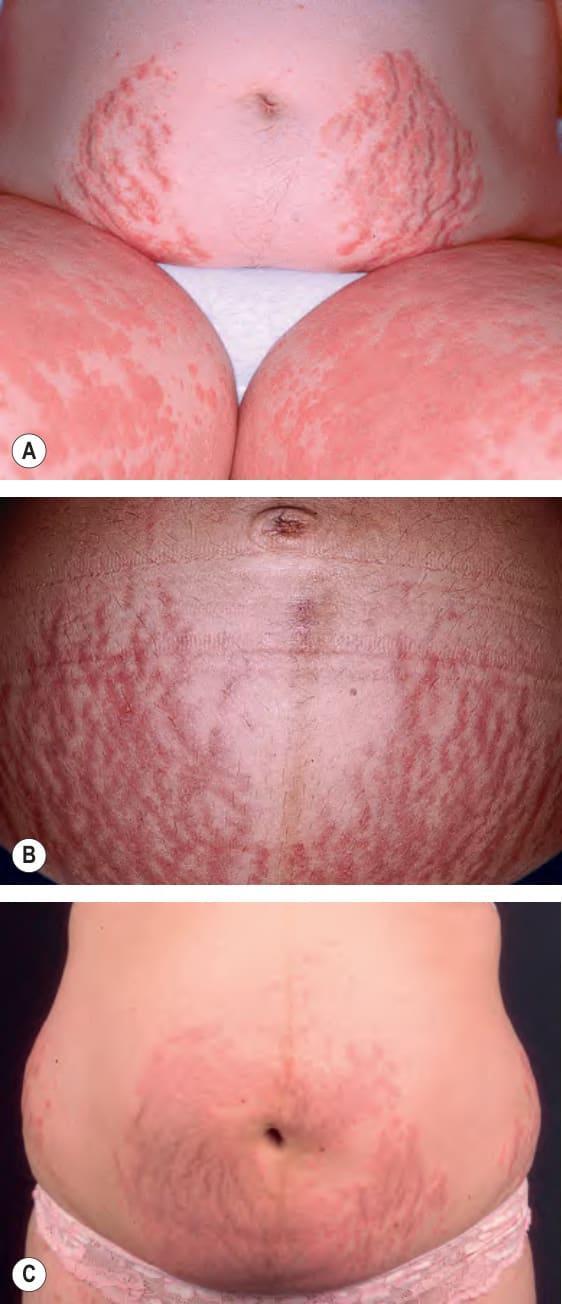
Fig. 27.4 Polymorphic eruption of pregnancy.
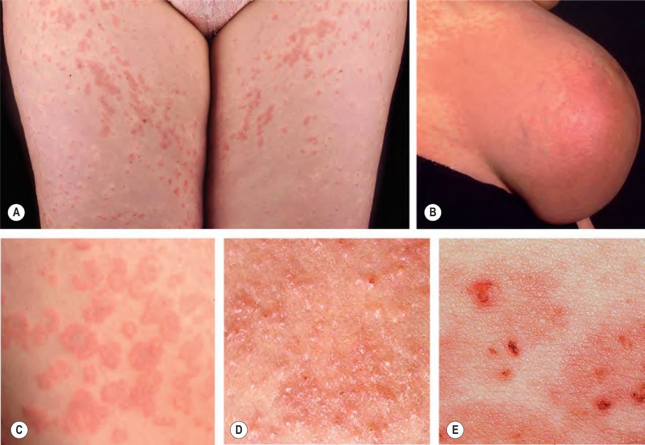
Fig. 27.5 Polymorphic eruption of pregnancy. The clinical spectrum includes: A urticarial papules and plaques, some of which demonstrate a steal phenomenon; B macular erythema, which can be widespread; C targetoid lesions; D tiny vesicles due to marked epidermal spongiosis or dermal edema; and E eczematous plaques, especially as lesions age.
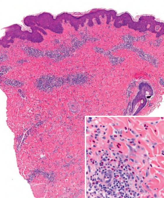
Fig. 27.6 Polymorphic eruption of pregnancy – histopathologic features. In this early lesion, a superficial and deep perivascular and interstitial lymphohistiocytic infiltrate is seen within the dermis. Numerous eosinophils are also present (inset).
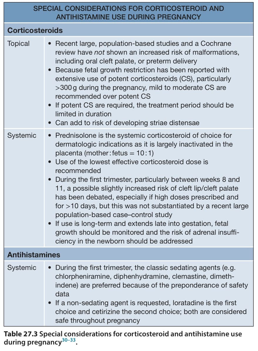
Table 27.3 Special considerations for corticosteroid and antihistamine use during pregnancy.