ATOPIC ERUPTION OF PREGNANCY
Synonyms: Prurigo of pregnancy Besnier’s “prurigo gestationis” Nurse’s “early-onset prurigo” of pregnancy Spangler’s “papular dermatitis of pregnancy” Pruritic folliculitis of pregnancy Eczema in pregnancy
Key features
Eczematous and/or papular skin lesions in a patient with an atopic diathesis in whom other specific dermatoses have been excluded
Most common pruritic disorder during pregnancy
Generally appears earlier than other pregnancy-related dermatoses
(75% before the third trimester)
Nonspecific histology; negative direct IF; elevated serum IgE levels in up to 70% of patients
No maternal or fetal risks; commonly recurs in subsequent pregnancies
Introduction
Atopic eruption of pregnancy (AEP) is defined as either an exacerbation or the first occurrence of eczematous and/or papular skin changes during pregnancy in atopic individuals. As the majority of patients belong to the second group, the atopic link is often overlooked, leading to a number of different diagnoses, as evidenced by the many synonyms.
History
In retrospect, an association with atopy dates back to the first reports. When Besnier described the disorder “prurigo gestationis” in 1904, “prurigo” was the term dermatologists used for atopic dermatitis (Besnier was the first to note the association between atopic dermatitis, allergic rhinitis, and asthma). Nurse, in 1968, described accompanying eczematous features in most of the 31 patients in his “early-onset” prurigo group. In 1983, Holmes and Black were the first to suggest that “prurigo of pregnancy” could simply result from pregnancy-related pruritus in women with an atopic diathesis rather than being a distinct entity.
Epidemiology
AEP is by far the most common pruritic disorder in pregnant women and it tends to appear earlier than the other pregnancy-related dermatoses. Its incidence is not known but may be as high as 1 in 5 to 1 in 20.
Pathogenesis
To prevent fetal rejection, a normal pregnancy is characterized by a lack of strong maternal cell-mediated immune function and reduced Th1 cytokine production (e.g. IL-12, interferon-γ) as well as a dominant humoral immune response with increased Th2 cytokine production (e.g. IL-4, IL-10). This natural switch towards a dominant Th2 response, which worsens the imbalance already present in most atopic patients, is thought to favor the development of AEP.
Clinical Features
In contrast to the other specific dermatoses of pregnancy, AEP appears earlier, often during the first trimester, with 75% of patients presenting before the third trimester. Approximately 20% of women experience an exacerbation of pre-existing atopic dermatitis, while the remaining 80% develop atopic skin changes for the first time during pregnancy. Two-thirds of patients present with eczematous lesions (Fig. 27.8), often involving “atopic sites” such as the face, neck, and flexural aspects of the extremities. One-third develop a papular eruption on the trunk and extremities, composed of either classic prurigo lesions or small erythematous papules (Fig. 27.9). Findings typically include xerosis (often marked) and other signs of the underlying atopic diathesis (see Ch. 12). Fetal and maternal prognoses are excellent and recurrences in subsequent pregnancies are common.
Pathology
Depending on the stage of the lesion, histologic features can vary. Epidermal changes include spongiosis, acanthosis, and erosions; the dermal infiltrate is composed of lymphocytes and usually admixed eosinophils. If the histologic section includes a hair follicle, there may be sterile follicular inflammation. Direct IF is negative. Serum IgE levels may be elevated in up to 70% of patients, usually to a mild degree.
Differential Diagnosis
Of the specific pregnancy dermatoses, PEP and ICP are the ones that in particular need to be excluded. In AEP, the eruption starts significantly earlier during gestation and has no association with striae; serum bile acid levels are also normal. Furthermore, other pruritic dermatoses not
specifically associated with pregnancy (e.g. scabies, viral exanthems, drug eruptions, contact dermatitis) must be considered.
Treatment
Cutaneous lesions respond rapidly to topical corticosteroids with or without systemic antihistamines. Topical tacrolimus may be used for sites such as face and intertriginous zones. Emollients, humectants, and topical antipruritic agents also play a role, as they do in non-pregnant patients with atopic dermatitis. Topical urea (10%), polidocanol, pramoxine, and menthol are considered safe during pregnancy. UVB irradiation is very helpful for severe disease. Secondary bacterial infection may require systemic antibiotics (e.g. penicillins, cephalosporins).
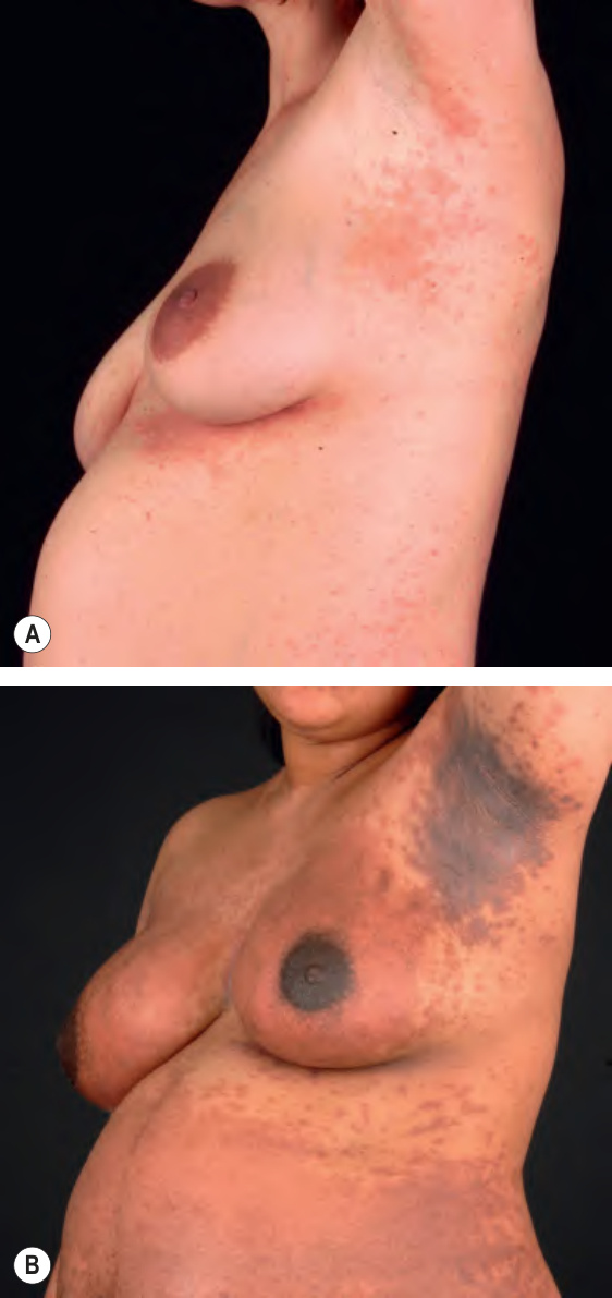
Fig. 27.8 Atopic eruption of pregnancy – eczematous lesions. The eczematous lesions often involve flexural areas and friction sites (A,B), as well as the breasts and abdomen (B). These changes are seen in approximately two-thirds of patients.
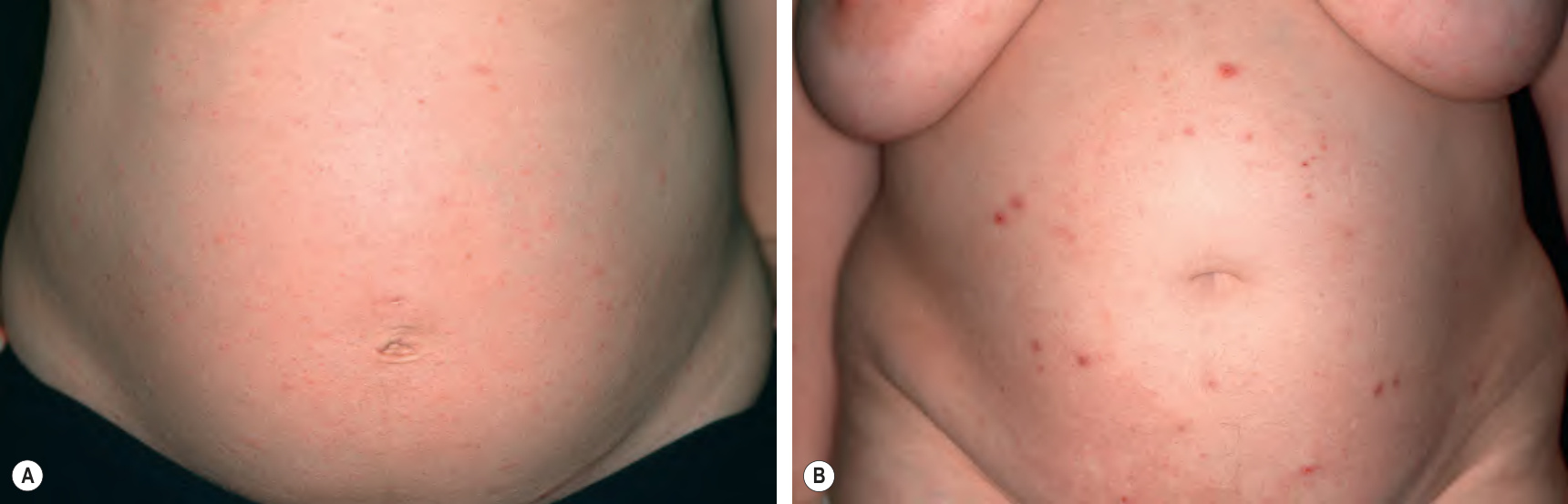
Fig. 27.9 Atopic eruption of pregnancy – papular eruption. Scattered small erythematous papules (A) or excoriated prurigo lesions (B) favor the abdomen and extremities. These changes are seen in approximately one-third of patients. Note the absence of striae distensae.