HISTORY
The modern history of pemphigus began with the discovery by Beutner and Jordon in 1964 of circulating antibodies directed against the cell surface of keratinocytes in the sera of pemphigus vulgaris patients (Fig. 29.1). This was followed by the finding of in vivo IgG deposition on the cell surface of keratinocytes in patients’ skin. These discoveries formed the basis of our understanding of pemphigus as a tissue-specific autoimmune disease of the skin and mucous membranes. In the late 1970s and early 1980s, pemphigus autoantibodies were shown to be pathogenic, i.e. they could induce blister formation in skin organ-culture systems as well as by passive transfer of patients’ IgG to neonatal mice. In the mid and late 1980s, the target antigens of pemphigus were characterized by immunochemical methods, such as immunoprecipitation and immunoblotting. In the early 1990s, the isolation of cDNA for pemphigus antigens demonstrated that pemphigus is an anti- cadherin autoimmune disease.
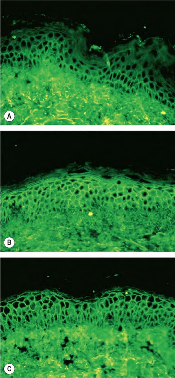
Fig. 29.1 Indirect immunofluorescence of pemphigus sera with normal human epidermis as a substrate. The hallmark of pemphigus is the finding of IgG autoantibodies directed against the cell surface of keratinocytes. A Pemphigus vulgaris sera containing anti-desmoglein 3 (anti- Dsg3) IgG alone stain predominantly the cell surfaces in the lower epidermis. B Pemphigus vulgaris sera containing both anti-Dsg3 IgG and anti-Dsg1 IgG stain the cell surfaces throughout the epidermis. C Pemphigus foliaceus sera, which contain only anti-Dsg1 IgG, stain the cell surfaces throughout the epidermis, but more intensely in the superficial layers.