歷史(HISTORY)
天疱瘡的現代歷史始於 1964 年 Beutner 與 Jordon 的發現,他們在 pemphigus vulgaris 病人的血清中找到針對角質形成細胞(keratinocytes)細胞表面的循環抗體(圖 29.1)。隨後又在病人皮膚中發現角質形成細胞細胞表面有體內(in vivo)IgG 沉積。這些發現構成了我們理解天疱瘡為一種皮膚與黏膜之組織特異性自體免疫疾病的基礎。在 1970 年代後期與 1980 年代初期,天疱瘡自體抗體被證實具有致病性,亦即它們可在皮膚器官培養系統中誘發水疱形成,也可藉由將病人 IgG 被動轉移至新生小鼠而誘發。在 1980 年代中後期,天疱瘡的標的抗原以免疫化學方法被鑑定出來,例如免疫沉澱法(immunoprecipitation)與免疫墨點法(immunoblotting)。在 1990 年代初期,天疱瘡抗原 cDNA 的分離證明了天疱瘡是一種抗 cadherin 的自體免疫疾病。
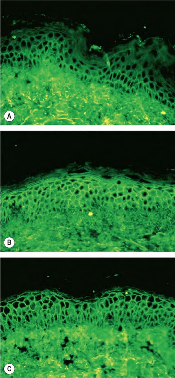
圖 29-1:以正常人類表皮為受質,對天疱瘡血清進行間接免疫螢光染色
Fig. 29.1 Indirect immunofluorescence of pemphigus sera with normal human epidermis as a substrate. The hallmark of pemphigus is the finding of IgG autoantibodies directed against the cell surface of keratinocytes. A Pemphigus vulgaris sera containing anti-desmoglein 3 (anti- Dsg3) IgG alone stain predominantly the cell surfaces in the lower epidermis. B Pemphigus vulgaris sera containing both anti-Dsg3 IgG and anti-Dsg1 IgG stain the cell surfaces throughout the epidermis. C Pemphigus foliaceus sera, which contain only anti-Dsg1 IgG, stain the cell surfaces throughout the epidermis, but more intensely in the superficial layers.
(圖 29.1 說明:天疱瘡的標誌是發現針對角質形成細胞細胞表面的 IgG 自體抗體。A 僅含抗 desmoglein 3(anti-Dsg3)IgG 的 pemphigus vulgaris 血清,主要染色表皮下部的細胞表面。B 同時含抗 Dsg3 IgG 與抗 Dsg1 IgG 的 pemphigus vulgaris 血清,染色整層表皮的細胞表面。C 僅含抗 Dsg1 IgG 的 pemphigus foliaceus 血清,染色整層表皮的細胞表面,但在表淺層更為強烈。)