CLINICAL FEATURES
Pemphigus Vulgaris
Essentially all patients with pemphigus vulgaris develop painful erosions of the oral mucosa. More than half of the patients also develop flaccid blisters and widespread cutaneous erosions. Pemphigus vulgaris is therefore divided into two subgroups: (1) the mucosal-dominant type with mucosal erosions but minimal skin involvement; and (2) the mucocutaneous type with extensive skin blisters and erosions in addition to mucosal involvement (see Fig. 29.4).
Mucous membrane lesions usually present as painful erosions (Fig. 29.5). Intact blisters are rare, probably because they are fragile and break easily. Although scattered or extensive erosions may be seen anywhere in the oral cavity, the most common sites are the buccal and palatine mucosae. The erosions are of different sizes with an irregular and ill-defined border, which, when extensive or painful, may result in decreased oral intake of food or liquids. The diagnosis of pemphigus vulgaris tends to be delayed in patients presenting with only oral involvement, as compared to patients with skin lesions.
The lesions may extend out onto the vermilion lip and lead to thick, fissured hemorrhagic crusts. Involvement of the throat produces hoarseness and difficulty in swallowing. The esophagus also may be involved and sloughing of its entire lining in the form of a cast has been reported. The conjunctivae, nasal mucosa, vagina, labia, penis and anus
Essentially all patients develop painful oral mucosal erosions. The most common sites are the buccal (A) and palatine mucosae, but lesions can also develop on the gingivae (B) and tongue (C). A, Courtesy Lorenzo Cerroni, MD; B,C, Courtesy Jeffrey P. Callen, MD.
can develop lesions as well. Cytology of vaginal cells may be misread as a malignancy when vaginal lesions are present.
The primary skin lesions of pemphigus vulgaris are flaccid, thinwalled, easily ruptured blisters (Fig. 29.6). They can appear anywhere on the skin surface and arise on either normal-appearing skin or erythematous bases. The fluid within the bullae is initially clear but may become hemorrhagic, turbid, or even seropurulent. The blisters are fragile and soon rupture to form painful erosions that ooze and bleed easily. These erosions often attain a large size and can become generalized. The erosions soon become partially covered with crusts that have little or no tendency to heal. Those lesions that do heal often leave hyperpigmented patches with no scarring. Associated pruritus is uncommon. Table 29.3 outlines more unusual clinical presentations of pemphigus vulgaris.
Because of an absence of cohesion within the epidermis, its upper layers easily move laterally with slight pressure or rubbing in patients with active disease (Nikolsky sign). The lack of cohesion of the skin may also be demonstrated with the “bulla-spread phenomenon” – gentle pressure on an intact bulla forces the fluid to spread under the skin away from the site of pressure (Asboe–Hansen sign, also referred to as the “indirect Nikolsky” or “Nikolsky II” sign). Without appropriate treatment, pemphigus vulgaris can be fatal because a large portion of the skin loses its epidermal barrier function, leading to the loss of body fluids or to secondary bacterial infections.
Pemphigus Vegetans
Pemphigus vegetans is a rare vegetative variant of pemphigus vulgaris, and it is thought to represent a reactive pattern of the skin to the autoimmune insult of pemphigus vulgaris.
Pemphigus vegetans is characterized by flaccid blisters that become erosions and then form fungoid vegetations or papillomatous proliferations, especially in intertriginous areas and on the scalp or face (Fig. 29.7). Pustules rather than vesicles characterize early lesions, but these soon progress to vegetative plaques. The tongue may show cerebriform-like changes. Two subtypes are recognized: the severe Neumann type and the mild Hallopeau type. Occasionally, a vegetative response may also be seen in lesions of pemphigus vulgaris that tend to be resistant to therapy and remain for long periods of time in one location (see Fig. 29.6D).
Pemphigus Foliaceus
Patients with pemphigus foliaceus develop scaly, crusted cutaneous erosions, often on an erythematous base, but they do not have clinically apparent mucosal involvement even with widespread disease.
Flaccid blisters and an erosion due to rupture of a bulla. B, C Multiple erosions and hemorrhagic crusts of the back that can become extensive. Secondary bacterial infections are a potential complication. D A vegetative response can occasionally be seen in chronic recalcitrant lesions; the patient has a Cushingoid appearance due to use of chronic systemic corticosteroids. E The dyshidrosiform variant is uncommon. A, D, Courtesy Luis Requena, MD; B, C, Courtesy Lorenzo Cerroni, MD; E, Courtesy Louis A. Fragola, Jr, MD.
The onset of disease is often subtle, with a few scattered crusted lesions that are transient and are frequently mistaken for impetigo. They are usually well demarcated and have a seborrheic distribution, i.e. they favor the face, scalp, and upper trunk (Fig. 29.8A–C). Because the vesicle is so superficial and fragile, often only the resultant crust and scale are seen (Fig. 29.8D, E). The disease may stay localized for years or it may rapidly progress, in some cases to generalized involvement and an exfoliative erythroderma (see Ch. 10). The Nikolsky sign is present. In contrast to the extensive oral lesions in pemphigus vulgaris, it is extremely rare, if ever, for patients with pemphigus foliaceus to develop mucosal involvement. Generally, patients with pemphigus foliaceus are not severely ill. However, they can experience burning and pain in association with the skin lesions.
Pemphigus Erythematosus (Senear–Usher Syndrome)
Pemphigus erythematosus is simply a localized variant of pemphigus foliaceus. Typical scaly and crusted lesions of pemphigus foliaceus appear in the malar region of the face (Fig. 29.9) and in other “seborrheic” areas. Originally, the term “pemphigus erythematosus” was introduced to describe patients with immunologic features of both lupus erythematosus (LE) and pemphigus, i.e. in vivo IgG and C3 deposition on cell surfaces of keratinocytes as well as the basement membrane zone, in addition to circulating anti-nuclear antibodies. However, only a few patients have been reported to actually have the two diseases concurrently.
Herpetiform Pemphigus
Most patients with herpetiform pemphigus have a clinical variant of pemphigus foliaceus and the remainder may have a variant of
pemphigus vulgaris. This disorder is characterized by: (1) erythematous urticarial plaques and tense vesicles that present in a herpetiform arrangement; (2) eosinophilic spongiosis and subcorneal pustules with minimal or no apparent acantholysis histologically; and (3) IgG autoantibodies directed against the cell surfaces of keratinocytes. The target antigen is Dsg1 in most cases and Dsg3 in the remainder. Some patients with herpetiform pemphigus will have features of pemphigus foliaceus or vulgaris during the course of their disease, and some patients will evolve into having pemphigus foliaceus or vulgaris. It is assumed that the pathogenic blister-inducing activity of the IgG autoantibodies in herpetiform pemphigus might be weaker than that seen in classic forms of pemphigus. Although often clinically less severe than pemphigus vulgaris, the course may be more chronic.
Drug-Induced Pemphigus
There are sporadic cases of pemphigus associated with the use of drugs, in particular penicillamine and captopril (see Table 29.1). In patients receiving penicillamine, pemphigus foliaceus is seen more commonly than pemphigus vulgaris, with a ratio of approximately 4:1. Although most patients with drug-induced pemphigus are shown to have autoantibodies against the same molecules involved in sporadic pemphigus, evidence suggests that some drugs may induce acantholysis without the production of antibodies. Both penicillamine and captopril contain sulfhydryl groups that are speculated to interact with the sulfhydryl groups in Dsg1 and Dsg3. This interaction may modify the antigenicity of the desmogleins, which may lead to autoantibody production, or their interaction may directly interfere with the adhesive function of the desmogleins. Imiquimod-induced pemphigus has also been reported as has a flare or recurrence of pemphigus due to immune checkpoint inhibitors. Most, but not all, patients with drug-induced pemphigus go into remission after the offending drug is discontinued.
Paraneoplastic Pemphigus
Paraneoplastic pemphigus is associated with underlying neoplasms, both malignant and benign. The most commonly associated neoplasms are non-Hodgkin lymphoma and chronic lymphocytic leukemia, followed by Castleman disease, malignant and benign thymomas, sarcomas, and Waldenström macroglobulinemia. Non-Hodgkin lymphoma and chronic lymphocytic leukemia together account for two-thirds of patients. Castleman disease, a very rare lymphoproliferative disorder, is the third most commonly associated neoplasm in adults and the most commonly associated tumor in children and adolescents; its association with paraneoplastic pemphigus is strikingly disproportionate to its general occurrence. The absence of common tumors, such as adenocarcinomas of the breast or colon and squamous cell carcinomas, is notable.
The most constant clinical feature of paraneoplastic pemphigus is the presence of intractable stomatitis. Severe stomatitis is usually the earliest presenting sign and, after treatment, it is the one that persists and is extremely resistant to therapy. This stomatitis consists of erosions and ulcerations that affect all surfaces of the oropharynx and characteristically extend onto the vermilion lip (Fig. 29.10). Most patients also have a severe pseudomembranous conjunctivitis, which may progress to scarring and obliteration of the conjunctival fornices. Nasopharyngeal, esophageal, vaginal, labial, penile, and perianal lesions may also be seen.
Cutaneous findings are quite polymorphic and may present as erythematous macules, flaccid blisters and erosions resembling pemphigus vulgaris, tense blisters resembling bullous pemphigoid, erythema multiforme-like lesions, and lichenoid eruptions. The occurrence of blisters and erythema multiforme-like lesions on the palms and soles is often used to differentiate paraneoplastic pemphigus from pemphigus vulgaris, in which lesions on the palms and soles are unusual. In the chronic form of the disease, a lichenoid eruption may predominate over blistering lesions. Some patients with paraneoplastic pemphigus develop bronchiolitis obliterans, which can be fatal as a result of respiratory failure. Although its pathophysiologic mechanism is still unclear, ectopic expression of epidermal antigens in the setting of squamous metaplasia is thought to render the lung a target organ. Of note, a chest X-ray or CT scan obtained at the onset of bronchiolitis obliterans may be normal but pulmonary function tests will show small airway obstruction that does not reverse with bronchodilators.
IgA Pemphigus
IgA pemphigus represents a more recently characterized group of autoimmune intraepidermal blistering diseases presenting with a vesiculo pustular eruption, neutrophilic infiltration of the skin, and in vivo bound and circulating IgA autoantibodies against the cell surface of keratinocytes; no IgG autoantibodies are present. IgA pemphigus usually occurs in middle-aged or elderly persons. Two distinct types of IgA pemphigus have been described: the subcorneal pustular dermatosis type and the intraepidermal neutrophilic type. Patients with both types of IgA
pemphigus present with flaccid vesicles or pustules on either erythematous or normal skin (Fig. 29.11A). In both types, the pustules tend to coalesce to form an annular or circinate pattern with crusts in the center of the lesion (Fig. 29.11B), although a sunflower-like configuration of pustules is a characteristic sign of the intraepidermal neutrophilic type.
The most common sites of involvement are the axilla and groin, but lesions can also develop on the trunk and proximal extremities. Mucous membrane involvement is rare, and pruritus is often a significant symptom. In some patients, there is an underlying associated disorder such as IgA myeloma or ulcerative colitis. Because the sub-corneal pustular dermatosis type of IgA pemphigus is clinically and histologically indistinguishable from classic subcorneal pustular dermatosis (Sneddon–Wilkinson disease; see Ch. 8), immunologic evaluation is essential to differentiate the two diseases.
IgA deposition on cell surfaces of epidermal keratinocytes is present in all cases, as shown by direct immunofluorescence (DIF) microscopy, and many patients have detectable circulating IgA autoantibodies, as shown by indirect immunofluorescence (IIF) microscopy. In the subcorneal pustular dermatosis type, IgA autoantibodies tend to react against upper epidermal surfaces, while in the intraepidermal neutrophilic type, IgA autoantibodies are found throughout the entire epidermis. The subclass of IgA autoantibodies is exclusively IgA1. IgA autoantibodies in the subcorneal pustular dermatosis type were shown to recognize desmocollin 1, while the autoimmune targets of the intraepidermal neutrophilic type remain to be identified. A subset of IgA pemphigus patients have IgA autoantibodies directed against Dsg1 or Dsg3, making the autoimmune targets of IgA pemphigus more heterogeneous. Besides the two major forms of IgA pemphigus, some authors include three additional forms of IgA pemphigus – IgA pemphigus foliaceus, IgA pemphigus vulgaris, and IgA pemphigus vegetans. The exact pathogenic role of IgA autoantibodies in inducing pustular formation in IgA pemphigus remains to be elucidated.
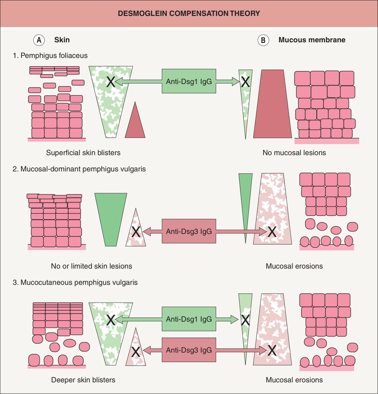
Fig. 29.4 Logical explanation for the localization of blister formation in classic pemphigus by desmoglein compensation theory. The colored triangles represent the distribution of desmoglein 1 (Dsg1, green) and desmoglein 3 (Dsg3, pink) in the skin (A) and mucous membranes (B). Pemphigus foliaceus sera contain only anti-Dsg1 IgG, which causes superficial blisters in the skin because Dsg3 functionally compensates for the impaired Dsg1 in the lower part of the epidermis (A1), whereas those antibodies do not cause blisters in the mucous membranes because cell–cell adhesion is mainly mediated by Dsg3 (B1). Sera containing only anti-Dsg3 IgG cause no or only limited blisters in the skin because Dsg1 compensates for the loss of Dsg3- mediated adhesion (A2); however, these sera induce separation in the mucous membranes, where the low expression of Dsg1 will not compensate for the loss of Dsg3-mediated adhesion (B2). When sera contain both anti- Dsg1 and anti-Dsg3 IgG, the function of both Dsgs is compromised and blisters occur in both the skin and mucous membranes (A3, B3). In neonatal skin, the situation is similar to that shown here for mucous membranes.
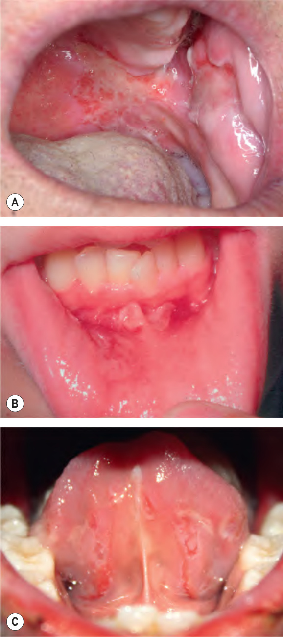
Fig. 29.5 Pemphigus vulgaris – oral involvement.
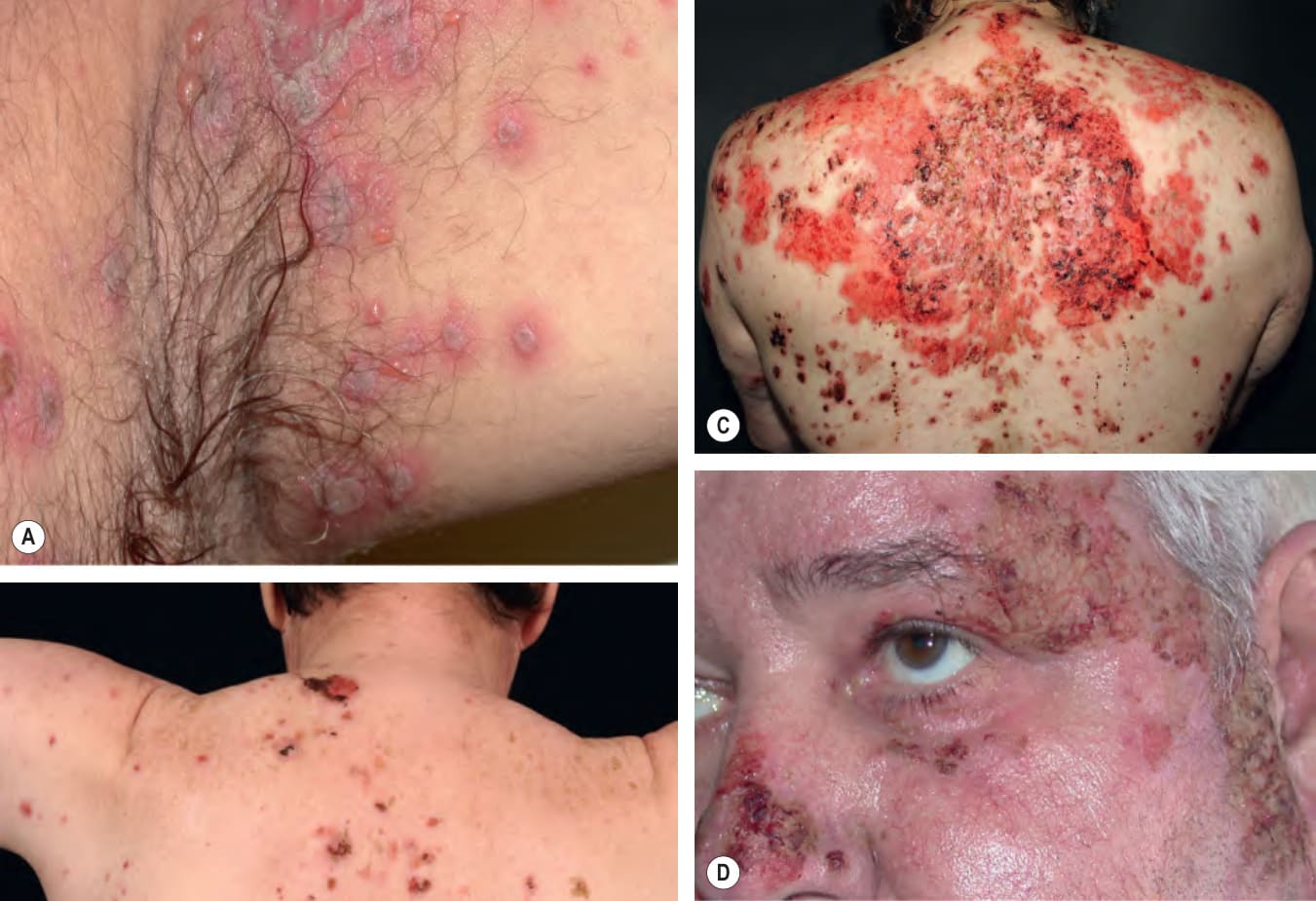
Fig. 29.6 Pemphigus vulgaris – cutaneous involvement.A
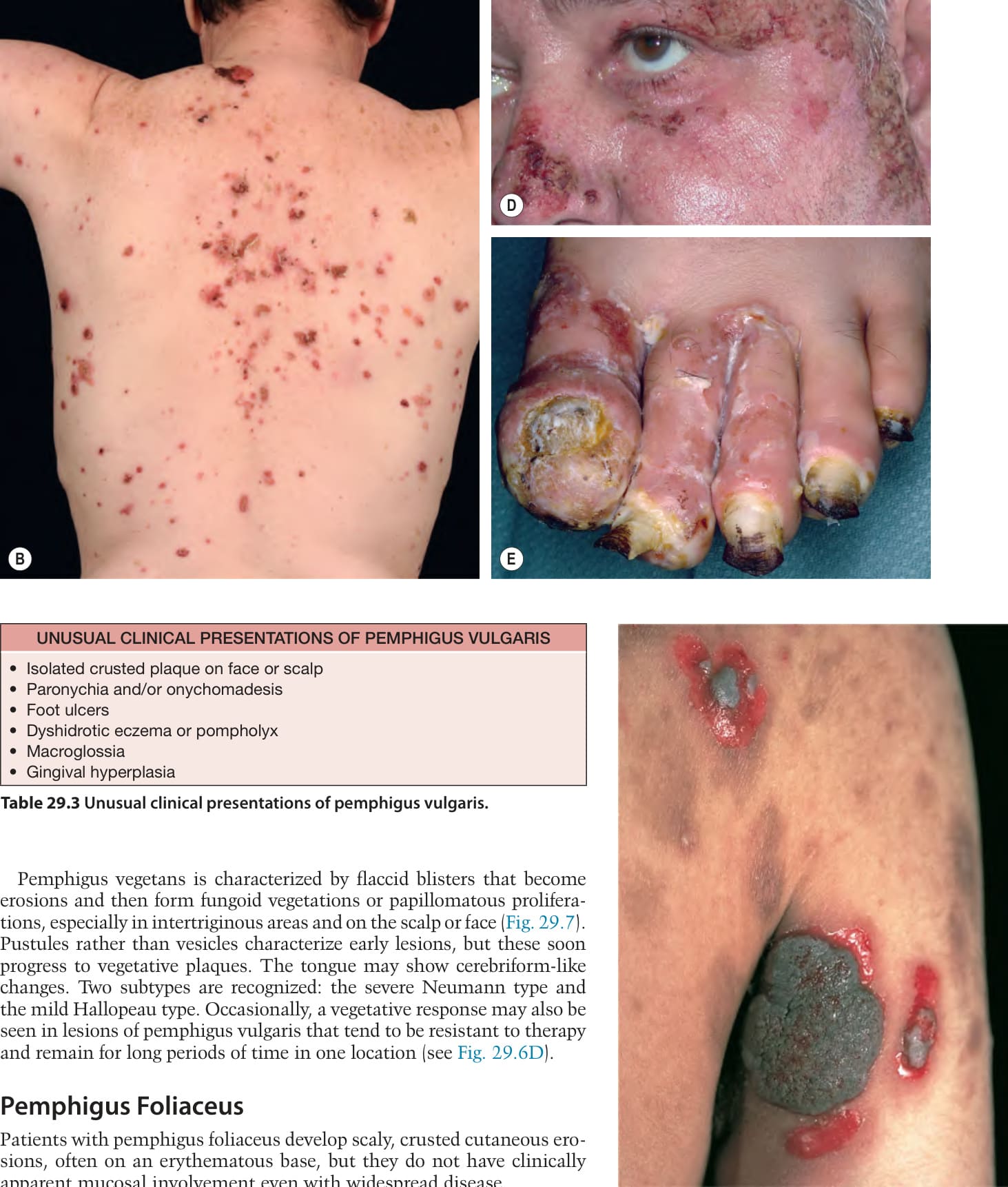
Fig. 29.7 Pemphigus vegetans. Large, thick, vegetating, papillomatous plaques arising in conjunction with erosions. Healed lesions have residual postinflammatory hyperpigmentation.
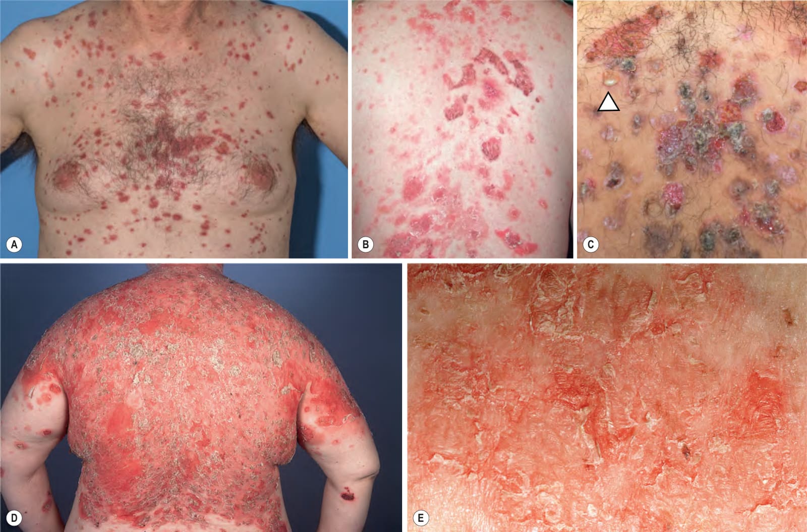
Fig. 29.8 Pemphigus foliaceus.A, B Multiple superficial erosions arising on an erythematous base on the trunk, a common site of involvement. Larger erosions as well as the characteristic scale-crusts are seen in B. C Thicker scale-crusts in addition to large erosions and one flaccid vesicle with seropurulent fluid where pus has settled in the lower half due to gravity similar to hypopyon (“half-half” blisters) (arrowhead). D As the disease progresses, the lesions become confluent, but because the vesicles are fragile and rupture easily, only erosions with scale-crust are observed. E The scales have been likened to cornflakes. A, Courtesy Edward Cowen, MD; C, Courtesy Kalman Watsky, MD.
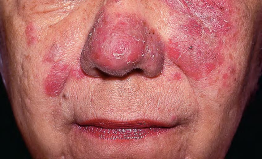
Fig. 29.9 Pemphigus erythematosus. Erythematous plaques with scale-crust and erosions on the nose and malar area of the face. Courtesy Ronald P. Rapini, MD.
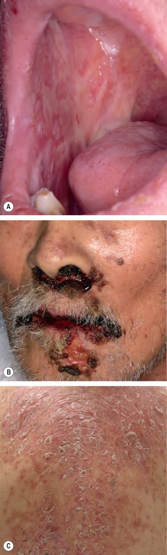
Fig. 29.10 Paraneoplastic pemphigus.A The characteristic clinical feature is severe intractable stomatitis with multiple erosions; there may be a resemblance to erosive oral lichen planus. B The erosions, along with hemorrhagic crusts, can extend onto the vermilion lip and involve the nasal mucosa. C Papulosquamous lichenoid lesions, reflecting an interface dermatitis, can also be present. A, Courtesy Luis Requena, MD; C, Courtesy Jennie Clarke, MD.
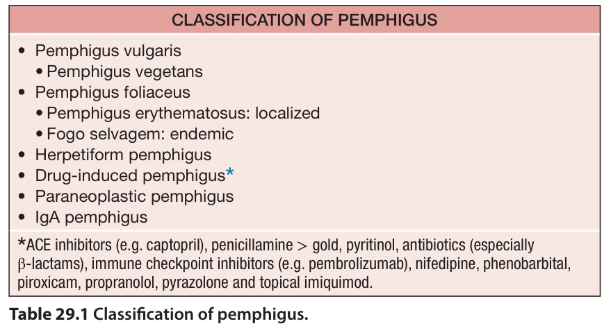
Table 29.1 Classification of pemphigus.
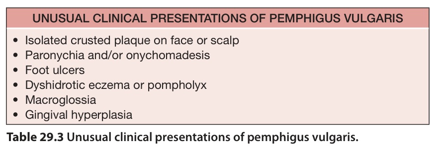
Table 29.3 Unusual clinical presentations of pemphigus vulgaris.