PATHOLOGY
For routine histology, it is best to perform biopsies of early lesions. If the vesicle or edematous papule is small enough, the entire lesion can be removed. Otherwise, the biopsy specimen should include the edge of a fresh vesicle or bulla plus the inflammatory rim (Fig. 29.12). Examination of perilesional, rather than lesional, skin is recommended for DIF in order to avoid negative staining due to secondary degeneration of target antigens and immunoreactants. In patients with only mucosal lesions, the biopsy specimen should consist of the active border of a denuded area, since intact blisters are rarely encountered. Cytologic examination (Tzanck smear) is useful for the rapid demonstration of acantholytic epidermal cells within the blister cavity. However, this bedside test merely represents a preliminary diagnostic tool and it should not supplant histologic examination. This is because acantholytic keratinocytes are occasionally seen in various non-acantholytic vesiculobullous or pustular diseases as a result of secondary acantholysis.
Pemphigus Vulgaris
The characteristic histologic finding in this form of pemphigus is intraepidermal blister formation due to a loss of cell–cell adhesion of keratinocytes (acantholysis) without keratinocyte necrosis (Fig. 29.13A). Whereas acantholysis usually occurs just above the basal cell layer (suprabasilar acantholysis), intraepithelial separation may occasionally be higher in the stratum spinosum. A few rounded-up (acantholytic) keratinocytes as well as clusters of epidermal cells are often seen within the blister cavity. Although the basal cells lose lateral desmosomal contact with their neighbors, they maintain their attachment to the basement membrane via hemidesmosomes, thus giving the appearance of a “row of tombstones” (Fig. 29.13B). The acantholytic process may involve hair follicles.
(SPD) type.A Numerous superficial pustules arising within areas of erythema; these pustules rupture easily. The desquamation has a figurate configuration and overall there is a resemblance to pustular psoriasis. B Pustules tend to coalesce to form an annular or figurate pattern with crusts present centrally. Note the accumulation of the pustular component in the dependent portion of the vesiculopustule. A, Courtesy Luis Requena, MD.
The dermal papillary outline is usually maintained and, frequently, the papillae protrude into the blister cavity. The blister cavity may contain a few inflammatory cells, notably eosinophils, and in the dermis there is a moderate perivascular mononuclear cell infiltrate with conspicuous eosinophils. In rare instances, the earliest histologic finding consists of eosinophilic spongiosis (Table 29.4), in which eosinophils infiltrate a spongiotic epidermis with little or no evidence of acantholysis.
In pemphigus vegetans, suprabasilar acantholysis is seen, in addition to considerable papillomatosis and acanthosis. Characteristically, there is an intense inflammatory cell infiltrate containing numerous eosinophils, and intraepidermal microabscesses of eosinophils are often seen.
Pemphigus Foliaceus
The histologic changes of pemphigus foliaceus, pemphigus erythematosus, and fogo selvagem are identical. Early blisters in pemphigus foliaceus have acantholysis in the upper epidermis, within or adjacent to the granular layer (Fig. 29.14). Because the blisters are superficial and fragile, it is often difficult to obtain an intact lesion for histologic examination. As a result, acantholysis is sometimes difficult to detect, but usually a few acantholytic keratinocytes can be found attached to the roof or floor of the blister. Of note, these cells are often almond- or spindle-shaped rather than round. When the blisters form within the granular layer, a “missing” horny layer with a few acantholytic keratinocytes
may be the only histopathologic finding (the horny layer detaches as a consequence of superficial acantholysis). Although the deeper epidermis usually remains intact, secondary clefts may develop, leading to detachment of the epidermis in its midlevel, but rarely giving rise to limited areas of separation right above the basal layer.
These superficial blisters are histologically indistinguishable from those seen in staphylococcal scalded skin syndrome or bullous impetigo, because Dsg1 is targeted in both of these diseases. Sometimes the blister cavity contains numerous acute inflammatory cells, particularly neutrophils. Eosinophilic spongiosis can be also seen in very early lesions of pemphigus foliaceus (see Table 29.4). The dermis shows a moderate number of inflammatory cells, among which eosinophils are often present.
Paraneoplastic Pemphigus
The histologic findings of cutaneous lesions in paraneoplastic pemphigus show considerable variability, reflecting the polymorphism seen clinically. The lesions show a unique combination of pemphigus vulgaris-like, erythema multiforme-like, and lichen planus-like histologic features, sometimes in the same specimen (Fig. 29.15). Intact cutaneous blisters demonstrate suprabasilar acantholysis and individual keratinocyte necrosis as well as lymphocytes within the epidermis. In addition, features of an interface dermatitis can be seen, including basal cell liquefactive degeneration or a dense band-like lymphocytic infiltrate in the upper dermis. Eosinophils are rare. Biopsy specimens of the severe ulcerative stomatitis usually yield only nonspecific changes of inflammation, but the perilesional oral epithelium should show suprabasilar acantholysis.
IgA Pemphigus
The characteristic histologic feature in IgA pemphigus is formation of an intraepidermal pustule or vesicle (Fig. 29.16). The contents of the pustules consist predominantly of neutrophils. Acantholysis is usually not seen. IgA pemphigus is divided into two subtypes depending on the
A Blisters in the skin show suprabasilar acantholysis with a few acantholytic cells in the blister cavity (inset). B Because blisters in the mouth rarely remain intact, the blister roof is often not seen in oral biopsy specimens. Nonetheless, the diagnosis can be made by the location of the split within the epithelium, the presence of acantholytic cells, and the “tombstones” appearance of the basal cells. Courtesy Lorenzo Cerroni, MD.
level of intraepidermal pustule; in the subcorneal pustular dermatosis type, pustules are located subcorneally in the upper epidermis, while in the intraepidermal neutrophilic type, suprabasilar pustules involving the lower or entire epidermis are present.
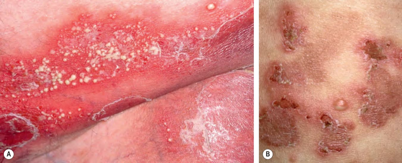
Fig. 29.11 IgA pemphigus – subcorneal pustular dermatosis
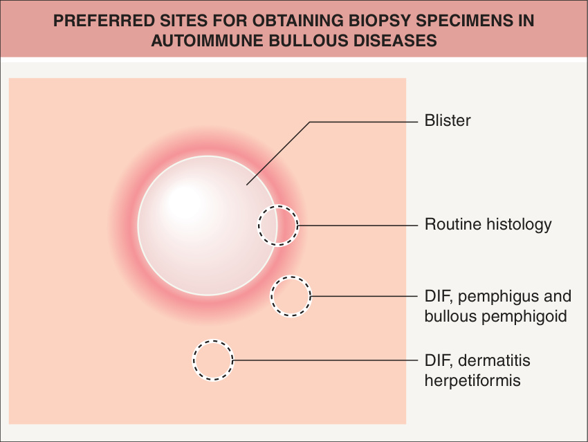
Fig. 29.12 Preferred sites for obtaining biopsy specimens in autoimmune bullous diseases. If the lesion is small enough, the entire vesicle can be removed for routine histology. If the lesions are not small, the edge of a fresh vesicle or bulla plus the inflammatory rim is recommended. For direct immunofluorescence (DIF) for various forms of pemphigus and bullous pemphigoid, perilesional skin is preferred, whereas nearby normal skin is recommended in dermatitis herpetiformis.
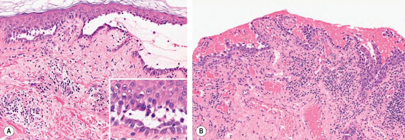
Fig. 29.13 Pemphigus vulgaris – histopathologic features.
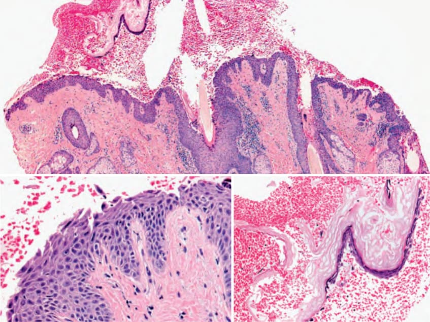
Fig. 29.14 Pemphigus foliaceus – histopathologic features. Because of the fragility of the blisters, the blister roof may become detached (right inset). The split is in the upper spinous layer of the epidermis and acantholytic cells are readily apparent (left inset). Courtesy Lorenzo Cerroni, MD.
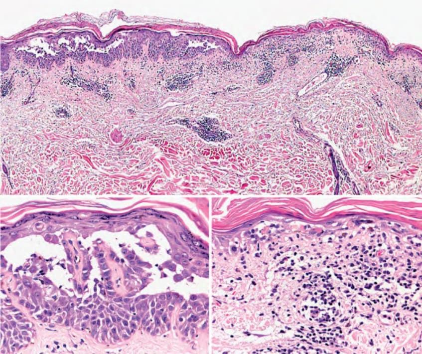
Fig. 29.15 Paraneoplastic pemphigus – histopathologic features. In the same biopsy specimen, there are areas of suprabasilar acantholysis (left inset) as well as an interface dermatitis with basal cell vacuolar change, necrotic keratinocytes, and lymphocytes within the epidermis (right inset). Courtesy Lorenzo Cerroni, MD.
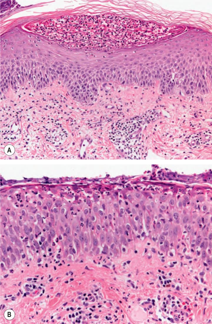
Fig. 29.16 IgA pemphigus – histopathologic features. Intraepidermal pustule with neutrophils present subcorneally in the subcorneal pustular dermatosis type (A), and within the entire epidermis in the intraepidermal neutrophilic type (B). Courtesy Lorenzo Cerroni, MD.
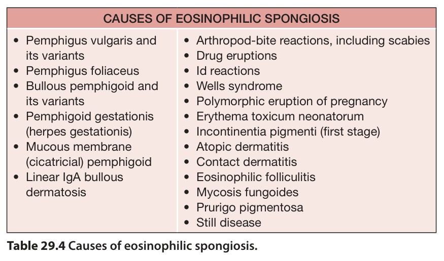
Table 29.4 Causes of eosinophilic spongiosis.