DIFFERENTIAL DIAGNOSIS
Demonstration of IgG (or IgA for IgA pemphigus) autoantibodies directed against the cell surface of keratinocytes is the gold standard for the diagnosis of pemphigus. By detection of these autoantibodies, pemphigus can be differentiated from other vesiculobullous or pustular diseases. Methods for demonstrating pemphigus autoantibodies include DIF, IIF, immunoprecipitation, immunoblotting, and enzyme-linked immunosorbent assay (ELISA).
DIF examines patients’ skin or mucous membranes in order to demonstrate in vivo bound IgG deposition on the keratinocyte cell surfaces (Fig. 29.17A). The biopsy specimen should be taken from perilesional normal skin or mucous membrane (see Fig. 29.12). DIF is the most reliable and sensitive diagnostic test for all forms of pemphigus. However, on occasion, nonspecific cell surface staining may occur in other skin diseases, such as spongiotic dermatitis, burns, toxic epidermal necro lysis, systemic LE and lichen planus, or in patients with antibodies directed against blood groups A and B. If the DIF is negative, the diagnosis of pemphigus should be seriously questioned. IgG deposition is seen in up to 100% of patients with active pemphigus vulgaris and pemphigus foliaceus. IgM deposition is not found, but occasionally IgA deposition may also be seen. Complement (C3) deposition is not necessarily observed, probably because the dominant subclass of IgG is IgG4, which does not fix complement. The staining pattern of paraneoplastic pemphigus shows deposition of IgG and C3 on epidermal cell surfaces as well as variably along the basement membrane zone. In IgA pemphigus, IgA (but not IgG) deposition is detected on keratinocyte cell surfaces.
IIF examines patients’ sera in order to demonstrate circulating IgG autoantibodies directed against epithelial cell surfaces (Fig. 29.17B). In order to optimize sensitivity, there are recommended substrates based upon the particular autoantibody (Table 29.5). Except for a few patients with early localized disease or those in remission, most patients with pemphigus have circulating anti-epithelial cell surface IgG as determined by IIF. Despite the different antigens involved in pemphigus vulgaris and foliaceus, the staining pattern obtained by DIF or IIF is similar, which makes it difficult to serologically distinguish the two diseases (compare Fig. 29.1B & C). Immunoprecipitation, immuno blotting, and ELISA can be used to demonstrate the target molecules of these autoantibodies.
Immunoprecipitation and immunoblotting detect the target anti-gens as protein bands of a particular molecular weight that have been separated by electrophoresis (see Ch. 30). Immunoblotting requires denaturation of the protein substrates, but immunoprecipitation does not. Therefore, immunoprecipitation is superior to immunoblotting in detecting antibodies that react with epitopes dependent on threedimensional structure (conformational epitopes), while immunoblots detect antibodies that react with epitopes that are preserved even after denaturation (linear epitopes). However, an immunoblot is easier to perform because immunoprecipitation requires radioisotopes to label protein substrates.
ELISA is increasingly being used to detect antigen-specific autoantibodies for the diagnosis of pemphigus (Table 29.6). The patient’s serum is tested on ELISA plates pre-coated with relevant recombinant proteins, e.g. Dsg1, Dsg3. Thus, specific antibodies directed against Dsg1 or Dsg3 can be detected. This assay has enabled investigators to serologically distinguish subtypes of pemphigus vulgaris and foliaceus. If a serum is positive against Dsg1 but negative against Dsg3, this suggests a diagnosis of pemphigus foliaceus. If negative against Dsg1 but positive against Dsg3, this suggests a diagnosis of the mucosaldominant type of pemphigus vulgaris. If positive against both Dsg1 and Dsg3, this suggests a diagnosis of mucocutaneous type of pemphigus vulgaris (see Fig. 29.4). Furthermore, ELISA scores demonstrate parallel fluctuations with disease activity and are useful in monitoring disease activity, planning schedules for tapering corticosteroids, and predicting flares or relapses before there is clinical evidence. The chemiluminescent enzyme immunoassay (CLEIA) is an even more sensitive and more rapid technique for detecting IgG autoantibodies in patients with pemphigus.
Differential Diagnosis of Pemphigus Vulgaris
Establishing the diagnosis in patients with only oral lesions is more difficult than in those with cutaneous blisters and erosions. The differential diagnosis of mucosal lesions of pemphigus vulgaris includes acute herpetic stomatitis, aphthous stomatitis, erythema multiforme major or Stevens–Johnson syndrome, lichen planus, systemic LE, and mucous membrane (cicatricial) pemphigoid. For cutaneous lesions of pemphigus vulgaris, the differential diagnosis includes other forms of pemphigus, bullous pemphigoid, linear IgA bullous dermatosis, erythema multiforme, Hailey–Hailey disease, and transient acantholytic dermatosis (Grover disease). The differential diagnosis of pemphigus vegetans is outlined in Table 29.7.
The demonstration of IgG autoantibodies against keratinocyte cell surfaces and anti-Dsg3 IgG, as mentioned above, will exclude these diseases (with the exception of drug-induced pemphigus vulgaris and paraneoplastic pemphigus). Fresh blisters of bullous pemphigoid are tense due to a subepidermal split. Hailey–Hailey disease has full-thickness acantholysis (“dilapidated brick wall”) with epidermal hyperplasia and an impetiginized scale, and the acantholysis does not extend down follicles as it does in pemphigus. Transient acantholytic dermatosis exhibits only small foci of intraepidermal acantholysis in addition to dyskeratosis; the latter is not present in pemphigus.
Differential Diagnosis of Pemphigus Foliaceus
This differential includes other forms of pemphigus, bullous impetigo, subcorneal pustular dermatosis, subacute cutaneous LE, and seborrheic dermatitis. As discussed earlier for pemphigus vulgaris, the demonstration of IgG autoantibodies against epidermal cell surfaces is essential for separating these disorders from the pemphigus
family. A complete review of medications should be done to exclude the possibility of drug-induced pemphigus foliaceus. Because the lesions of pemphigus foliaceus may become secondarily infected, the finding of bacteria does not confirm a diagnosis of bullous impetigo. Likewise, a clinical flare or recalcitrant disease may represent a super-imposed disorder, e.g. tinea corporis, especially in patients on systemic corticosteroids.
Differential Diagnosis of Paraneoplastic Pemphigus
This differential includes pemphigus vulgaris, mucous membrane pemphigoid, erythema multiforme or Stevens–Johnson syndrome, lichen planus, GVHD, persistent HSV infection, and other viral infections;
stomatitis due to chemotherapy will be limited in duration (7–14 days) (Table 29.8). In two-thirds of patients with paraneoplastic pemphigus, it arises in the context of a known underlying neoplasm, usually a lymphoproliferative disorder, which may have been treated for years before the mucocutaneous lesions appear. In approximately one-third of patients, the underlying neoplasm is undiagnosed at the time the mucocutaneous disease develops. In these latter patients, examination for an occult neoplasm is required, and, given the most commonly associated ones (see earlier), a CT scan of the chest, abdomen, and pelvis should accompany a complete blood count, flow cytometry of peripheral blood, LDH, and serum protein and immunofixation electrophoreses. Severe intractable stomatitis that extends onto the vermilion lip is a key clinical feature in differentiating paraneoplastic pemphigus from
most cases of pemphigus vulgaris or mucous membrane pemphigoid. The gold standard for the diagnosis of paraneoplastic pemphigus is the demonstration of the characteristic combination of IgG autoantibodies – anti-plakin autoantibodies by immunoprecipitation, immunoblotting or ELISA and anti-desmoglein autoantibodies by ELISA (see Table 29.6).
Differential Diagnosis of IgA Pemphigus
The differential diagnosis includes subcorneal pustular dermatosis, pemphigus foliaceus, bullous impetigo, dermatitis herpetiformis, linear IgA bullous dermatosis, and pustular psoriasis. The clinical and histologic features of the SPD type of IgA pemphigus and classic subcorneal pustular dermatosis are indistinguishable. The demonstration of IgA autoantibodies is essential for separating these other disorders from IgA pemphigus.
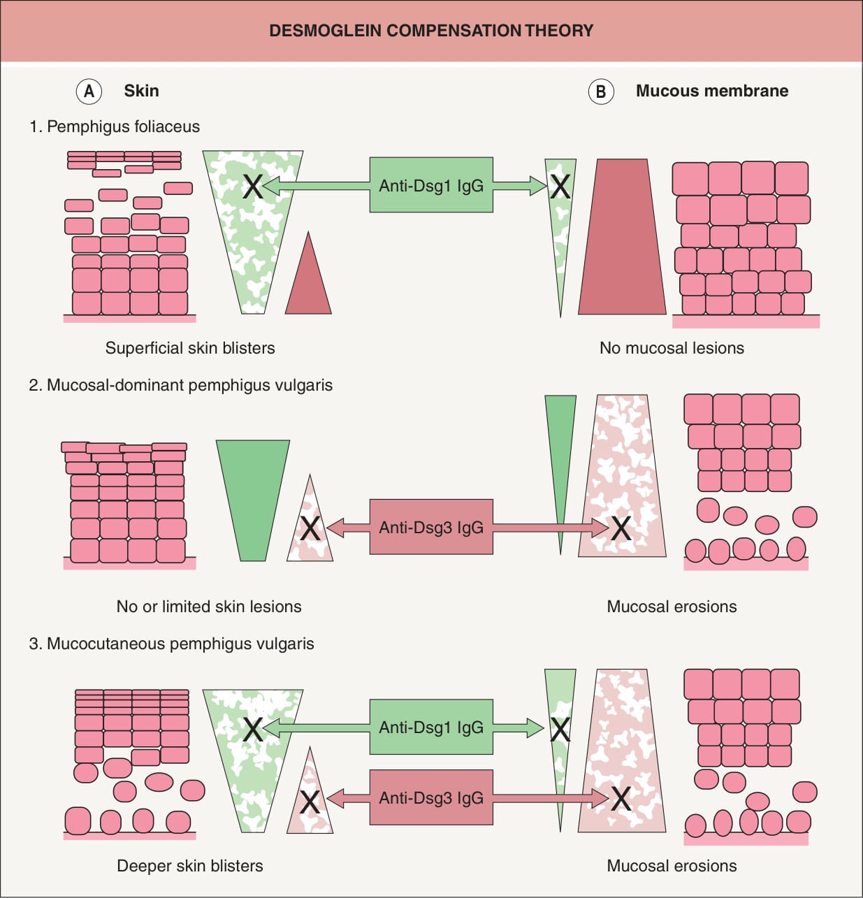
Fig. 29.4 Logical explanation for the localization of blister formation in classic pemphigus by desmoglein compensation theory. The colored triangles represent the distribution of desmoglein 1 (Dsg1, green) and desmoglein 3 (Dsg3, pink) in the skin (A) and mucous membranes (B). Pemphigus foliaceus sera contain only anti-Dsg1 IgG, which causes superficial blisters in the skin because Dsg3 functionally compensates for the impaired Dsg1 in the lower part of the epidermis (A1), whereas those antibodies do not cause blisters in the mucous membranes because cell–cell adhesion is mainly mediated by Dsg3 (B1). Sera containing only anti-Dsg3 IgG cause no or only limited blisters in the skin because Dsg1 compensates for the loss of Dsg3- mediated adhesion (A2); however, these sera induce separation in the mucous membranes, where the low expression of Dsg1 will not compensate for the loss of Dsg3-mediated adhesion (B2). When sera contain both anti- Dsg1 and anti-Dsg3 IgG, the function of both Dsgs is compromised and blisters occur in both the skin and mucous membranes (A3, B3). In neonatal skin, the situation is similar to that shown here for mucous membranes.
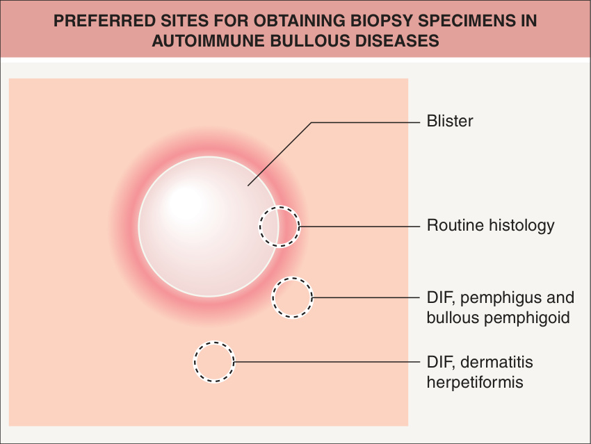
Fig. 29.12 Preferred sites for obtaining biopsy specimens in autoimmune bullous diseases. If the lesion is small enough, the entire vesicle can be removed for routine histology. If the lesions are not small, the edge of a fresh vesicle or bulla plus the inflammatory rim is recommended. For direct immunofluorescence (DIF) for various forms of pemphigus and bullous pemphigoid, perilesional skin is preferred, whereas nearby normal skin is recommended in dermatitis herpetiformis.
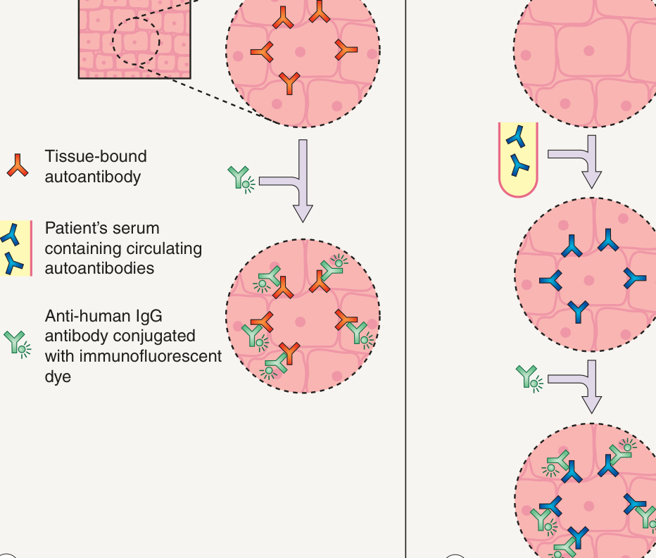
Fig. 29.17 Basic techniques of direct immunofluorescence (DIF) and indirect immunofluorescence (IIF).A DIF is performed on skin biopsy specimens in order to detect in vivo bound IgG. B IIF is performed utilizing patients’ sera in order to detect circulating autoantibodies that bind epithelial antigens.
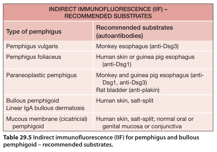
Table 29.5 Indirect immunofluorescence (IIF) for pemphigus and bullous pemphigoid – recommended substrates.
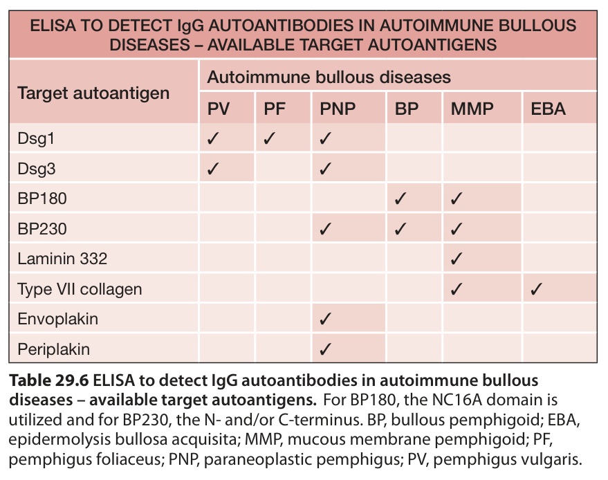
Table 29.6 ELISA to detect IgG autoantibodies in autoimmune bullous diseases – available target autoantigens.For BP180, the NC16A domain is utilized and for BP230, the N- and/or C-terminus. BP, bullous pemphigoid; EBA, epidermolysis bullosa acquisita; MMP, mucous membrane pemphigoid; PF, pemphigus foliaceus; PNP, paraneoplastic pemphigus; PV, pemphigus vulgaris.
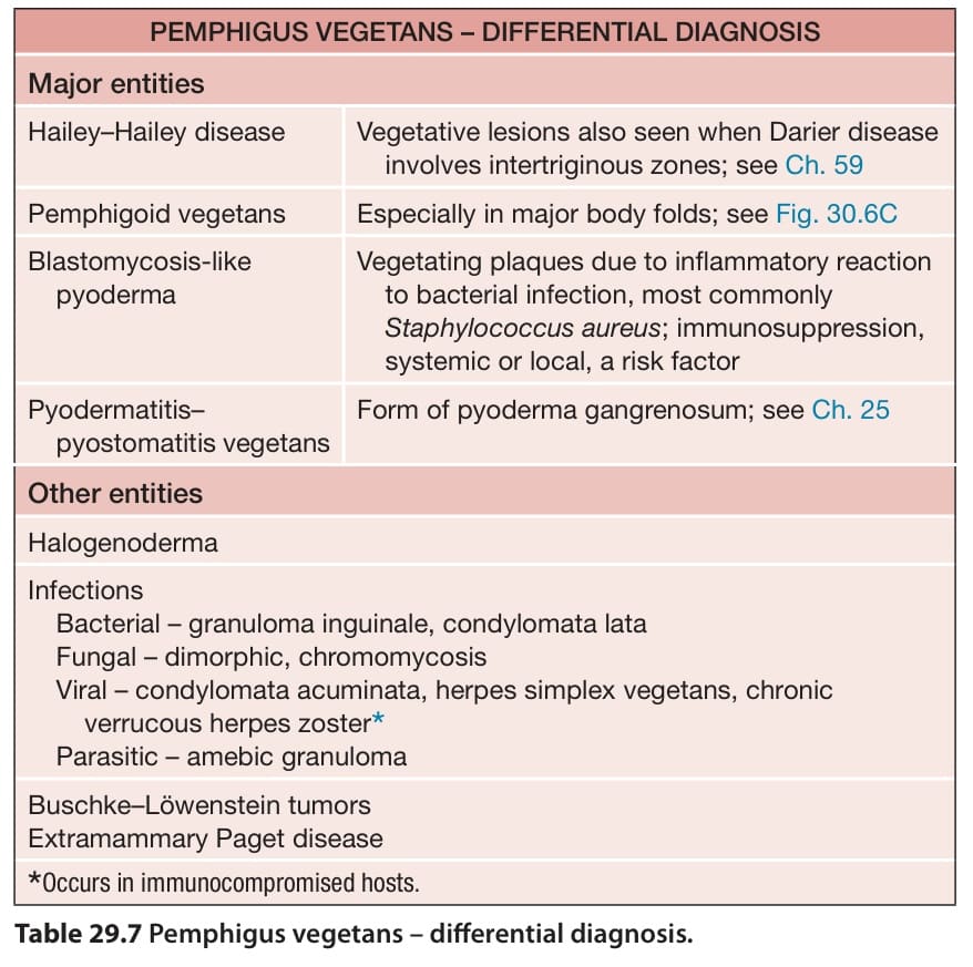
Table 29.7 Pemphigus vegetans – differential diagnosis.
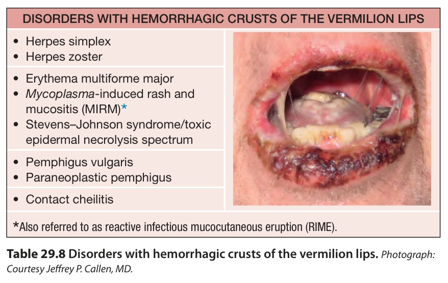
Table 29.8 Disorders with hemorrhagic crusts of the vermilion lips.Photograph: Courtesy Jeffrey P. Callen, MD.