鑑別診斷(DIFFERENTIAL DIAGNOSIS)
證實有針對角質形成細胞細胞表面的 IgG(IgA pemphigus 則為 IgA)自體抗體,是診斷天疱瘡的黃金標準。藉由偵測這些自體抗體,可將天疱瘡與其他水疱性或膿疱性疾病區分。證實天疱瘡自體抗體的方法包括 DIF、IIF、免疫沉澱、免疫墨點法,以及酵素連結免疫吸附分析(enzyme-linked immunosorbent assay, ELISA)。
DIF 檢查病人的皮膚或黏膜,以證實體內結合的 IgG 沉積於角質形成細胞細胞表面(圖 29.17A)。切片檢體應取自病灶周圍的正常皮膚或黏膜(見圖 29.12)。DIF 是所有型式天疱瘡最可靠且最敏感的診斷檢查。然而,偶爾在其他皮膚疾病也可能出現非特異性的細胞表面染色,例如海綿水腫性皮膚炎、燒傷、毒性表皮壞死溶解症、全身性 LE 與扁平苔癬,或出現在具有抗 A、B 血型抗體的病人。若 DIF 為陰性,應嚴重質疑天疱瘡的診斷。在活動性 pemphigus vulgaris 與 pemphigus foliaceus 的病人中,IgG 沉積可見於高達 100% 的病人。不會發現 IgM 沉積,但偶爾也可見到 IgA 沉積。補體(C3)沉積不一定會觀察到,可能是因為主要的 IgG 亞類為 IgG4,而 IgG4 不會固定補體。paraneoplastic pemphigus 的染色模式顯示 IgG 與 C3 沉積於表皮細胞表面,並不定程度地沿著基底膜帶沉積。在 IgA pemphigus 中,可在角質形成細胞細胞表面偵測到 IgA(而非 IgG)沉積。
IIF 檢查病人的血清,以證實循環中針對上皮細胞表面的 IgG 自體抗體(圖 29.17B)。為使敏感度最佳化,會依特定自體抗體而有建議的受質(表 29.5)。除了少數早期局限性疾病或處於緩解期的病人之外,大多數天疱瘡病人以 IIF 測定皆有循環中的抗上皮細胞表面 IgG。儘管 pemphigus vulgaris 與 foliaceus 所涉及的抗原不同,DIF 或 IIF 所得到的染色模式相似,這使得在血清學上難以區分這兩種疾病(比較圖 29.1B 與 C)。免疫沉澱、免疫墨點法與 ELISA 可用來證實這些自體抗體的標的分子。
免疫沉澱與免疫墨點法將標的抗原偵測為經電泳分離、具特定分子量的蛋白條帶(見第 30 章)。免疫墨點法需要蛋白質受質變性,免疫沉澱則不需要。因此,在偵測與依賴三維結構之抗原表位(構形表位,conformational epitopes)反應的抗體上,免疫沉澱優於免疫墨點法;而免疫墨點法則偵測與即使變性後仍保留之抗原表位(線性表位,linear epitopes)反應的抗體。然而,免疫墨點法較易執行,因為免疫沉澱需要放射性同位素來標記蛋白質受質。
ELISA 越來越常被用來偵測抗原特異性自體抗體以診斷天疱瘡(表 29.6)。病人的血清在預先塗覆相關重組蛋白(例如 Dsg1、Dsg3)的 ELISA 盤上進行檢測。因此可偵測到針對 Dsg1 或 Dsg3 的特異性抗體。此分析法使研究者能在血清學上區分 pemphigus vulgaris 與 foliaceus 的亞型。若血清對 Dsg1 陽性而對 Dsg3 陰性,提示 pemphigus foliaceus 的診斷。若對 Dsg1 陰性而對 Dsg3 陽性,提示黏膜主導型 pemphigus vulgaris 的診斷。若對 Dsg1 與 Dsg3 皆陽性,提示黏膜皮膚型 pemphigus vulgaris 的診斷(見圖 29.4)。此外,ELISA 分數與疾病活動度呈平行波動,可用於監測疾病活動度、規劃皮質類固醇減量時程,以及在出現臨床證據之前預測惡化或復發。化學發光酵素免疫分析(chemiluminescent enzyme immunoassay, CLEIA)在偵測天疱瘡病人的 IgG 自體抗體上,是更敏感且更快速的技術。
Pemphigus Vulgaris 的鑑別診斷(Differential Diagnosis of Pemphigus Vulgaris)
在只有口腔病灶的病人建立診斷,比在有皮膚水疱與糜爛的病人更為困難。pemphigus vulgaris 黏膜病灶的鑑別診斷包括急性疱疹性口腔炎、阿弗他口腔炎、重症多形性紅斑或 Stevens–Johnson syndrome、扁平苔癬、全身性 LE,以及黏膜(瘢痕性)類天疱瘡(mucous membrane [cicatricial] pemphigoid)。就 pemphigus vulgaris 的皮膚病灶而言,鑑別診斷包括其他型式的天疱瘡、bullous pemphigoid、linear IgA bullous dermatosis、多形性紅斑、Hailey–Hailey disease,以及暫時性棘層鬆解性皮膚病(transient acantholytic dermatosis, Grover disease)。pemphigus vegetans 的鑑別診斷列於表 29.7。
如前所述,證實有針對角質形成細胞細胞表面的 IgG 自體抗體以及 anti-Dsg3 IgG,可排除這些疾病(藥物誘發 pemphigus vulgaris 與 paraneoplastic pemphigus 除外)。bullous pemphigoid 的新鮮水疱因表皮下裂隙而呈緊繃。Hailey–Hailey disease 有全層棘層鬆解(「破敗磚牆」,dilapidated brick wall)並伴表皮增生與膿痂樣鱗屑,且其棘層鬆解不像天疱瘡那樣向下延伸至毛囊。暫時性棘層鬆解性皮膚病除了角化不良(dyskeratosis)之外,只呈現小灶性的表皮內棘層鬆解;而角化不良在天疱瘡並不存在。
Pemphigus Foliaceus 的鑑別診斷(Differential Diagnosis of Pemphigus Foliaceus)
此鑑別診斷包括其他型式的天疱瘡、大疱性膿痂疹、subcorneal pustular dermatosis、亞急性皮膚型 LE,以及脂漏性皮膚炎。如前面針對 pemphigus vulgaris 所討論的,證實有針對表皮細胞表面的 IgG 自體抗體,對於將這些疾病與天疱瘡家族
區分開來至關重要。應完整回顧用藥,以排除藥物誘發 pemphigus foliaceus 的可能性。由於 pemphigus foliaceus 的病灶可能續發感染,發現細菌並不能確認大疱性膿痂疹的診斷。同樣地,臨床上的惡化或頑固性疾病可能代表合併存在的疾病,例如體癬,特別是在使用全身性皮質類固醇的病人。
Paraneoplastic Pemphigus 的鑑別診斷(Differential Diagnosis of Paraneoplastic Pemphigus)
此鑑別診斷包括 pemphigus vulgaris、mucous membrane pemphigoid、多形性紅斑或 Stevens–Johnson syndrome、扁平苔癬、GVHD、持續性 HSV 感染,以及其他病毒感染;
化學治療所致的口腔炎其持續時間有限(7–14 天)(表 29.8)。在三分之二的 paraneoplastic pemphigus 病人中,此病發生於已知潛在腫瘤的情境下,通常是淋巴增生性疾病,且可能在黏膜皮膚病灶出現之前已治療多年。約三分之一的病人,在黏膜皮膚疾病發生時其潛在腫瘤尚未診斷。在後者這些病人中,必須進行隱匿性腫瘤的檢查;鑑於最常相關的腫瘤(見前述),應在全血球計數、周邊血流式細胞術、LDH,以及血清蛋白與免疫固定電泳之外,加做胸部、腹部與骨盆的電腦斷層掃描。延伸至唇紅的嚴重頑固性口腔炎,是區分 paraneoplastic pemphigus 與
大多數 pemphigus vulgaris 或 mucous membrane pemphigoid 病例的關鍵臨床特徵。診斷 paraneoplastic pemphigus 的黃金標準是證實 IgG 自體抗體的特徵性組合——以免疫沉澱、免疫墨點法或 ELISA 證實 anti-plakin 自體抗體,並以 ELISA 證實 anti-desmoglein 自體抗體(見表 29.6)。
IgA Pemphigus 的鑑別診斷(Differential Diagnosis of IgA Pemphigus)
鑑別診斷包括 subcorneal pustular dermatosis、pemphigus foliaceus、大疱性膿痂疹、dermatitis herpetiformis、linear IgA bullous dermatosis,以及膿疱性乾癬。IgA pemphigus 之 SPD 型與典型 subcorneal pustular dermatosis 的臨床與組織學特徵無法區分。證實有 IgA 自體抗體,對於將這些其他疾病與 IgA pemphigus 區分開來至關重要。
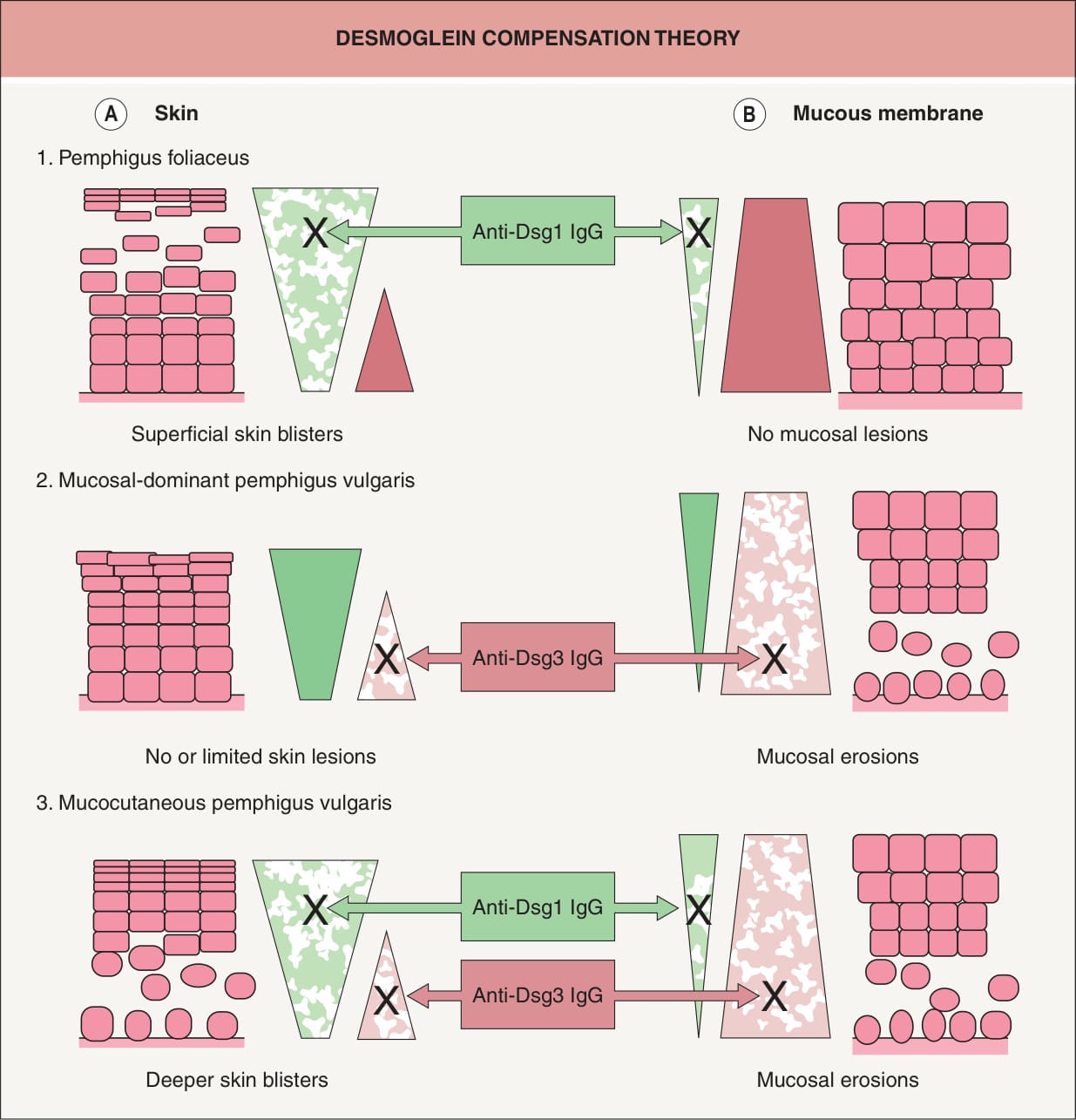
圖 29-4:以 desmoglein 代償理論合理解釋典型天疱瘡水疱形成的部位
Fig. 29.4 Logical explanation for the localization of blister formation in classic pemphigus by desmoglein compensation theory. The colored triangles represent the distribution of desmoglein 1 (Dsg1, green) and desmoglein 3 (Dsg3, pink) in the skin (A) and mucous membranes (B). Pemphigus foliaceus sera contain only anti-Dsg1 IgG, which causes superficial blisters in the skin because Dsg3 functionally compensates for the impaired Dsg1 in the lower part of the epidermis (A1), whereas those antibodies do not cause blisters in the mucous membranes because cell–cell adhesion is mainly mediated by Dsg3 (B1). Sera containing only anti-Dsg3 IgG cause no or only limited blisters in the skin because Dsg1 compensates for the loss of Dsg3- mediated adhesion (A2); however, these sera induce separation in the mucous membranes, where the low expression of Dsg1 will not compensate for the loss of Dsg3-mediated adhesion (B2). When sera contain both anti- Dsg1 and anti-Dsg3 IgG, the function of both Dsgs is compromised and blisters occur in both the skin and mucous membranes (A3, B3). In neonatal skin, the situation is similar to that shown here for mucous membranes.
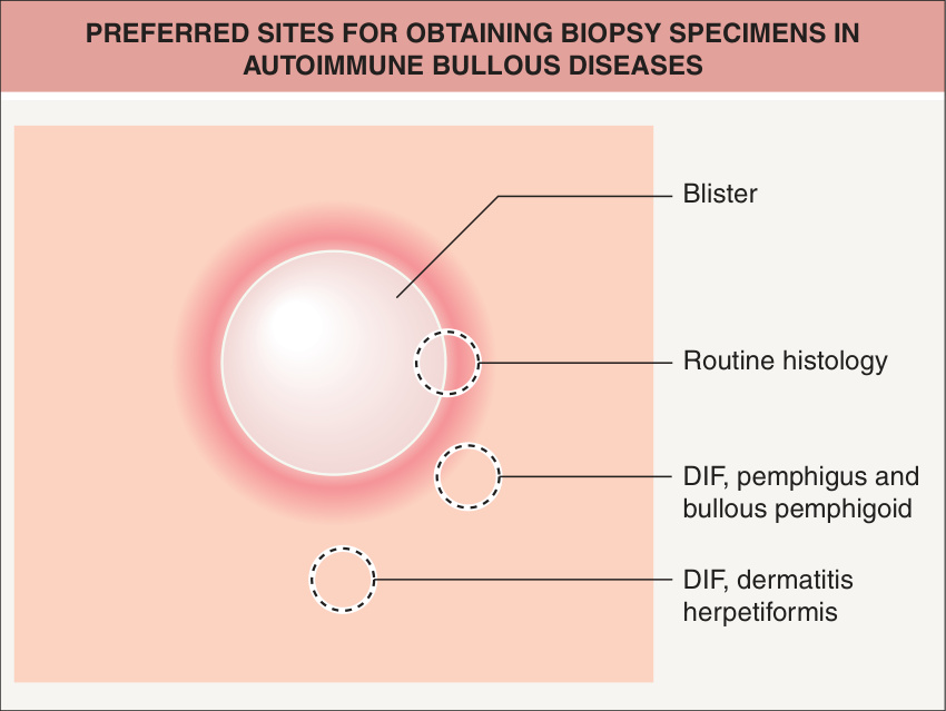
圖 29-12:自體免疫水疱性疾病取得切片檢體的建議部位
Fig. 29.12 Preferred sites for obtaining biopsy specimens in autoimmune bullous diseases. If the lesion is small enough, the entire vesicle can be removed for routine histology. If the lesions are not small, the edge of a fresh vesicle or bulla plus the inflammatory rim is recommended. For direct immunofluorescence (DIF) for various forms of pemphigus and bullous pemphigoid, perilesional skin is preferred, whereas nearby normal skin is recommended in dermatitis herpetiformis.
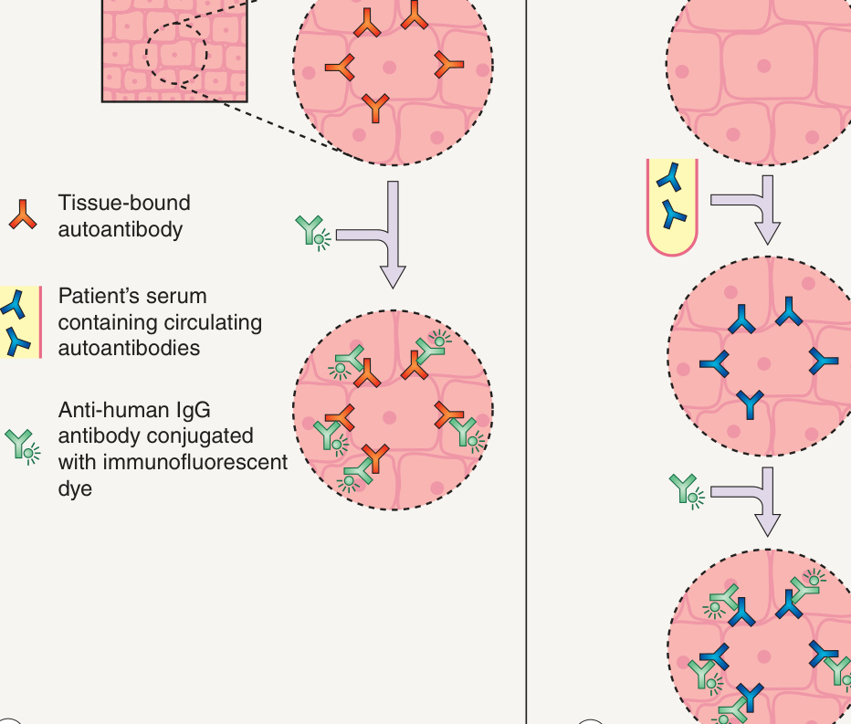
圖 29-17:直接免疫螢光(DIF)與間接免疫螢光(IIF)的基本技術
Fig. 29.17 Basic techniques of direct immunofluorescence (DIF) and indirect immunofluorescence (IIF).A DIF is performed on skin biopsy specimens in order to detect in vivo bound IgG. B IIF is performed utilizing patients’ sera in order to detect circulating autoantibodies that bind epithelial antigens.
(圖 29.17 說明:A DIF 於皮膚切片檢體上進行,以偵測體內結合的 IgG。B IIF 利用病人的血清進行,以偵測結合上皮抗原的循環自體抗體。)
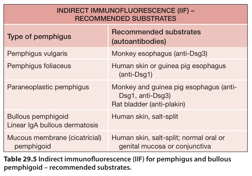
表 29-5:天疱瘡與 bullous pemphigoid 之間接免疫螢光(IIF)——建議的受質
Table 29.5 Indirect immunofluorescence (IIF) for pemphigus and bullous pemphigoid – recommended substrates.
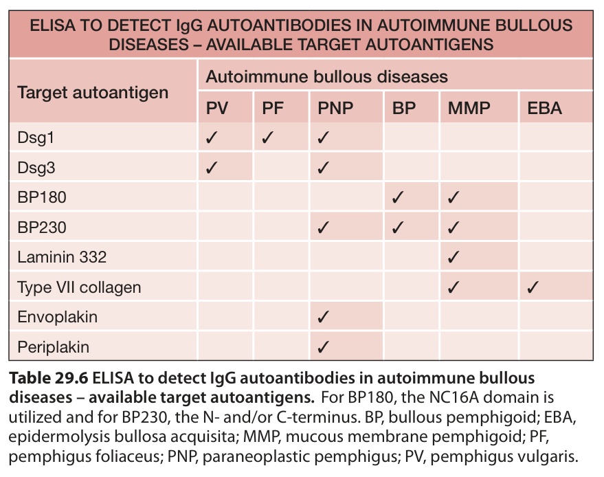
表 29-6:偵測自體免疫水疱性疾病 IgG 自體抗體的 ELISA——可用的標的自體抗原
Table 29.6 ELISA to detect IgG autoantibodies in autoimmune bullous diseases – available target autoantigens.For BP180, the NC16A domain is utilized and for BP230, the N- and/or C-terminus. BP, bullous pemphigoid; EBA, epidermolysis bullosa acquisita; MMP, mucous membrane pemphigoid; PF, pemphigus foliaceus; PNP, paraneoplastic pemphigus; PV, pemphigus vulgaris.
(表 29.6 說明:BP180 使用 NC16A domain,BP230 則使用 N 端及/或 C 端。)
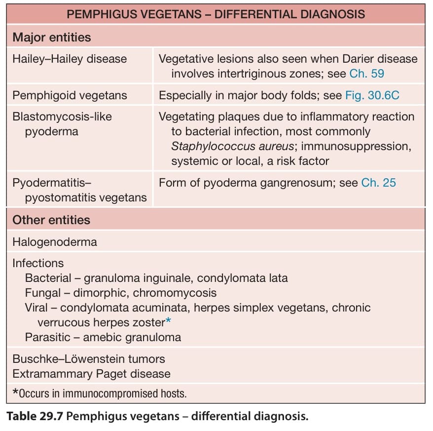
表 29-7:pemphigus vegetans——鑑別診斷
Table 29.7 Pemphigus vegetans – differential diagnosis.
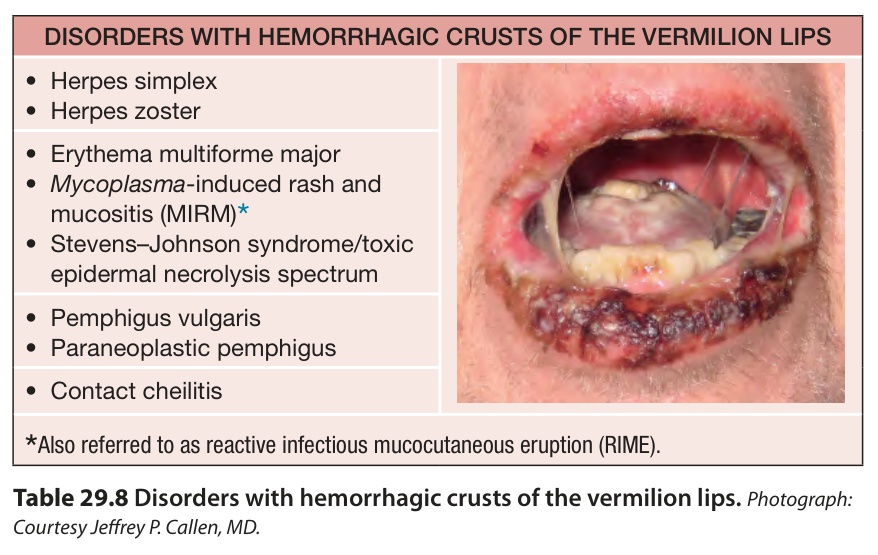
表 29-8:唇紅出現出血性痂皮的疾病
Table 29.8 Disorders with hemorrhagic crusts of the vermilion lips.Photograph: Courtesy Jeffrey P. Callen, MD.