BULLOUS PEMPHIGOID
Synonym: Pemphigoid
Key features
Bullous pemphigoid (BP) is the most common autoimmune subepidermal blistering disease, and its onset is often after 60 years of age
It is usually a chronic disease, with spontaneous exacerbations and remissions, which may be accompanied by significant morbidity
BP is associated with tissue-bound and circulating autoantibodies directed against BP antigen 180 (BP180, BPAG2, or type XVII collagen) and BP antigen 230 (BP230 or BPAG1e), components of junctional adhesion complexes called hemidesmosomes that promote dermal–epidermal cohesion
The spectrum of clinical presentations is extremely broad.
Characteristically, BP is an intensely pruritic eruption with widespread blister formation. In its early stages, or in variants of the disease, only eczematous or urticarial lesions (either localized or generalized) or just excoriations due to intense pruritus are present
Diagnosis relies on immunopathologic examinations, particularly direct and indirect immunofluorescence (IF) microscopy as well as ELISA for anti-BP180/BP230 autoantibodies
Introduction
Bullous pemphigoid (BP) is the most common autoimmune subepidermal blistering disease. It typically presents in older adults as a generalized pruritic bullous eruption and is often associated with significant morbidity. The clinical presentation is quite polymorphic, particularly during the early stages of the disease or in atypical variants, in which full-blown bullous lesions may be absent. In these cases, establishing the diagnosis of BP requires a high degree of suspicion. The anti-gens targeted by patients’ autoantibodies represent two components of hemidesmosomes, the junctional adhesion complexes found in skin and adjacent mucosae (see Fig. 28.3A).
History
In 1953 Lever recognized BP as a disorder distinct from the various types of “true” pemphigus based upon specific clinical and histopathologic features. A decade later, Jordon, Beutner, and colleagues demonstrated that BP patients had tissue-bound and circulating autoantibodies directed against the cutaneous basement membrane zone (BMZ), an observation that suggested that subepidermal dysadhesion was due to autoantibodies directed against structural components of the skin. Further milestones in our understanding of BP included the immunochemical characterization of targeted proteins, the cloning of their genes, and the development of animal models of the disease.
Luca Borradori and Michael Hertl Pemphigoid Group 30
Epidemiology
BP is typically a disease of older adults, with a mean age of presentation between 65 and 85 years. The annual incidence has been estimated to range from 5 to 45 new cases per million population per year. The incidence rate of BP appears to have significantly increased over the past two decades, most likely reflecting an aging population, better recognition of atypical variants, and a rise in drug-induced BP. A relative risk for patients over 90 years of age appears to be ~300-fold higher than for those 60 years of age or younger. The disease also occurs in children, but rarely. Certain HLA class II alleles are more prevalent in patients with BP than in the general population. In Caucasians, a significant association with the allele DQB10301 has been noted, while an increased frequency of the alleles DRB104, DRB11101, and DQB10302 has been observed in Japanese patients.
Pathogenesis
BP is an example of an immune-mediated disease that is associated with a humoral and cellular response directed against two well-characterized self-antigens: BP antigen 180 (BP180, also known as BPAG2 or type XVII collagen) and BP antigen 230 (BP230, also referred to as the epithelial isoform of BPAG1 [BPAG1e]) (Table 30.1). While the former is a transmembrane protein with a large collagenous extracellular domain, the latter is a cytoplasmic protein belonging to the plakin family (see Ch. 28). These two antigens are components of the hemidesmosomes, which are adhesion complexes promoting epithelial–stromal adhesion in stratified and other complex epithelia.
In vitro studies and in vivo animal models have demonstrated the pathogenic role of autoantibodies in BP. Furthermore, in gestational pemphigoid, a disease closely related to BP, the transplacental transfer of anti-BP180 autoantibodies from the mother to the neonate can result in a transient bullous eruption (see Ch. 34).
Humoral and cellular responses
Almost all patients with BP have circulating IgG autoantibodies that bind to BP180. Specifically, it is the non-collagenous NC16A domain, a region of BP180 located extracellularly but close to the transmembrane domain, that constitutes the immunodominant region (see Fig. 31.9). However, additional antigenic sites exist within both the extracellular and intracellular domains of BP180, and they are recognized by up to 70% of BP sera. Patients with BP also exhibit significant autoreactivity to BP230, and BP230-reactive autoantibodies bind predominantly to the COOH-terminal region of this autoantigen. The presence of several antigenic sites throughout BP180 and BP230 most likely results from the phenomenon known as “epitope spreading” (see below). This phenomenon may also account for the finding that patients’ sera rarely contain autoantibodies targeting additional components of the BMZ.
Patients with BP develop an autoreactive T cell response to BP180 and BP230. Likely related to a dysfunction of regulatory T cells, it is crucial for stimulating B cells to produce autoantibodies. The responsiveness of anti-BP180 autoreactive T cells is restricted by certain HLA class II alleles (e.g. HLA-DQB1*0301), which are prevalent in BP patients. These T lymphocytes, whose major relevant epitopes seem to be harbored within the NC16 domain, have a CD4+ phenotype and produce both Th1 (e.g. interferon-γ) and Th2 cytokines (e.g. interleukin [IL]-4, IL-5, IL-13; see Ch. 4). Th2 and Th17 cytokines, which are particularly relevant to the pathophysiology of BP, predominate within lesional tissue and in patients’ sera.
Upon binding of autoantibodies to their target antigens, subepidermal blister formation results from a cascade of events in which Fc
Not an exhaustive list. In the course of these diseases, it is possible to detect autoantibodies directed against additional antigens, the significance of which remains to be established. In certain cases, a so-called “intermolecular epitope spreading” phenomenon is thought to occur.
receptor-mediated mechanisms are critical. The latter involve activation of the complement cascade along with stimulation and recruitment of inflammatory cells including macrophages, neutrophils, mast cells, and eosinophils. These inflammatory cells contribute to tissue damage via liberation of various proteases (e.g. matrix metalloproteinase-9, neutrophil elastase, mast cell proteases), eosinophil toxic granules, and a variety of cytokines (e.g. IL-4, -5, -8, -13, -17, eotaxin).
The latter serve to further amplify the inflammatory response while the proteinases, together with reactive oxygen species, degrade various extracellular matrix proteins as well as BP180. IgG anti- BP180 autoantibodies can also boost the inflammatory response by stimulating keratinocytes to express inflammatory cytokines (Fig. 30.1). Furthermore, IgG autoantibodies are able to directly impair dermal–epidermal adhesion without complement activation.
Finally, BP patients often have circulating IgE anti-BP180 autoantibodies which may bind to the epidermal BMZ. These IgE autoantibodies also seem able to form complexes with shed BP180 fragments, which are prominently detected in the dermis on the surface of mast cells and/ or eosinophils expressing FcεRI. Hence, IgE autoantibodies most likely directly contribute to tissue damage by triggering the degranulation of these FcεRI-expressing cells.
Animal models have provided strong evidence that IgG autoantibodies against BP180 are pathogenic. When passively transferred to neonatal mice (in which BP180 had been fully or partially humanized by genetic engineering), human autoantibodies against the NC16A domain were able to induce a blistering disorder that reproduced all the key features of BP. In addition, passive transfer experiments in mice have indicated that IgE antibodies directed against BP180 contribute to skin inflammation. The prevailing pathomechanism is that antibodies against the ectodomain of BP180 are critical for disease initiation, while the development of antibodies against BP230 represents a secondary event that contributes to tissue damage. However, recent studies utilizing a mouse model demonstrated that anti-BP230 antibodies also had a direct pathogenic effect.
Clinical Features
Non-bullous pemphigoid
The cutaneous manifestations of BP can be extremely polymorphic (Figs. 30.2–30.6). In the prodromal, non-bullous phase of the disease, signs and symptoms are frequently nonspecific, but mild to severe intractable pruritus is a consistent feature. Clinical findings vary from excoriations to eczematous, papular, and/or urticarial lesions that have persisted for several weeks or months (see Figs. 30.3 to 30.5). Importantly, these nonspecific skin findings may remain as the only signs of the disease. At least 20% of patients have neither obvious blisters nor erosions due to ruptured bullae at the time of diagnosis.
Bullous phase
The bullous stage of BP is characterized by the development of vesicles and bullae on apparently normal or erythematous skin, together with urticarial and infiltrated papules and plaques that occasionally assume an annular or figurate pattern. The blisters are tense, up to 1–4 cm in diameter, contain a clear fluid, and may persist for several days, leaving eroded and crusted areas (see Fig. 30.2). Occasionally, the blister fluid becomes blood-tinged. The lesions frequently have a symmetric distribution pattern, and they predominate on the flexural aspects of the extremities and the lower trunk, including the abdomen. Within intertriginous zones, vegetating plaques may be observed (pemphigoid vegetans; see Fig. 30.6C). The extent and degree of disease activity can be assessed by employing the BP disease area index (BPDAI) or a daily blister count.
Residual postinflammatory changes include hyper- and hypopigmentation and, less commonly, milia. Involvement of the oral cavity is observed in 10%–30% of patients, more often in those with moderate to severe disease. The mucosae of the eyes, nose, pharynx, esophagus, and anogenital region are more rarely affected. In ~50% of patients, a peripheral blood eosinophilia is noted.
Clinical variants
Several clinical forms of BP have been described and are outlined in Table 30.2. Gestational pemphigoid (pemphigoid gestationis) is also a variant of BP, which typically occurs during pregnancy (see Ch. 27).
While the individual lesions of BP in infants and in older children (infantile and childhood BP) are similar to those observed in older adults, the sites of involvement can differ (Fig. 30.7). In infants, bullae often first appear in acral locations and then can generalize to other sites, including often the face. Involvement of the genital region (e.g. vulvar childhood pemphigoid) as well as other mucosal sites has been observed in children.
Associated Diseases
The association of internal malignancies with BP is probably related primarily to the older age of the patient. In case–control studies, the trend
towards an increased risk of malignancy was marginal while an English national record linkage study found no overall increased risk for concurrent or subsequent malignancies in BP patients. Nonetheless, individuals with BP should be up to date with age-related cancer screening tests recommended for the general population.
Rarely, BP has been described in patients with inflammatory bowel disease or other autoimmune diseases including Hashimoto thyroiditis,
rheumatoid arthritis, dermatomyositis, and lupus erythematosus (LE). These associations may be fortuitous or they could reflect a genetically determined susceptibility to develop autoimmune disorders. While one case–control study did not find an increased risk for autoimmune diseases, a sample analysis of US hospitalized patients with BP reported a slight risk for other autoimmune disorders.
In some patients, BP appears to be triggered by trauma, burns, radiotherapy or UV irradiation (including PUVA). BP has also been observed in association with certain dermatoses, in particular lichen planus and psoriasis (Fig. 30.8). In lichen planus pemphigoides, BP coexists with lichen planus (see Ch. 11). Theoretically, a dysfunction of T regulatory cells leads to both a lichenoid interface dermatitis and an autoantibody response to BP180 and BP230. In the setting of chronic inflammation at the dermal–epidermal junction, exposure of antigens to autoreactive T lymphocytes could result in a secondary immune response.
Finally, BP has been strongly associated with psychiatric and neurologic disorders; the latter include Parkinson disease, dementia, multiple sclerosis, and stroke. It should be noted that a neuronal variant of BP230 is expressed in the central and peripheral nervous systems, while available data regarding the expression of BP180 are contradictory. Whether exposure to neuronal antigens, via a compromised blood–brain barrier, plays a role in the development of BP remains to be determined.
Drug-induced bullous pemphigoid
In certain patients, systemic medications can lead to the development of BP (Fig. 30.9). There are multiple implicated drugs, in particular diuretics (e.g. furosemide), NSAIDs, antibiotics (e.g. amoxicillin, ciprofloxacin), ACE inhibitors, and TNF inhibitors. More recently, dipeptidyl peptidase-4 inhibitors (e.g. vildagliptin, linagliptin) and immune checkpoint inhibitors (e.g. nivolumab, pembrolizumab) have been strongly associated with BP. A meta-analysis suggested that exposure to aldosterone antagonists, dipeptidyl peptidase-4 inhibitors, anticholinergics, and dopaminergic medications were significantly associated with BP. Therefore, a detailed history is essential. Discontinuation of the drug, or a drug holiday in the case of immune checkpoint inhibitors, may or may not lead to improvement22,29a. As a result, initiation of standard therapies may be required.
A number of hypotheses have been put forth to explain drug-induced BP – an underlying genetic predisposition, medication-related alteration of the antigenic properties of the BMZ, and a breakdown of immune tolerance in the case of immune checkpoint inhibitors. In dipeptidyl peptidase-4 inhibitor-induced BP, autoantibodies also recognize BP180 epitopes other than the NC16A domain.
Diagnosis
The diagnosis of BP is based upon the typical clinical presentation, compatible histologic features, and, most importantly, positive direct immunofluorescence (DIF) microscopy studies (see Fig. 29.17). Further support for the diagnosis of BP requires the detection of specific circulating IgG autoantibodies (anti-BP180, anti-BP230) via either indirect immunofluorescence (IIF) microscopy or ELISA. In the vast majority of patients, these tests allow for the correct classification of patients. However, in a minority of patients (~10%) in whom both IIF microscopy and ELISA are negative, additional immunopathological studies, e.g. n-serration versus u-serration pattern analysis (see below), can be employed to demonstrate indirectly an autoantibody response to BP180 and/or BP230 and thereby exclude other autoimmune bullous diseases (see Table 30.1).
Light microscopy
In the non-bullous phase or in atypical variants of BP, routine histology may provide less specific information, since only eosinophilic spongiosis and/or dermal inflammatory infiltrates admixed with numerous eosinophils may be seen (Fig. 30.10). In biopsy specimens of an early bulla, a subepidermal blister accompanied by a dermal inflammatory infiltrate composed of eosinophils and mononuclear cells is typically observed. The infiltrate favors the uppermost dermis, and the cavity of the bulla contains a net of fibrin with a variable inflammatory
infiltrate (Fig. 30.11). Conventional electron microscopy studies have shown that subepidermal blister formation usually occurs at the level of the lamina lucida.
Direct immunofluorescence (DIF) microscopy
In almost all patients, DIF microscopy of perilesional skin, either normal-appearing or if inflamed 1–2 cm from a bulla, will characteristically demonstrate the presence of fine, linear, continuous deposits of IgG and/ or C3 (and, more rarely, other Ig classes) along the epidermal basement membrane (see Fig. 1.9). IgG4 and IgG1 are the predominant IgG subclasses. Two additional studies are helpful in distinguishing
BP from other autoimmune blistering disorders: (1) close analysis of the linear fluorescence pattern at the BMZ in order to determine if it is n-serrated (BP and linear IgA bullous dermatosis) versus u-serrated (epidermolysis bullosa acquisita); and (2) the salt-split skin assay in which perilesional skin is examined after treatment with 1 M NaCl (see Fig. 28.7). In BP, immune deposits are found in the epidermal side (roof) or in both the epidermal and dermal sides of the split. Although not routinely available, the computer-aided fluorescence overlay anti-gen mapping (FOAM) technique allows for more precise localization of the deposited immunoreactants.
Indirect immunofluorescence (IIF) microscopy
For IIF studies in subepidermal blistering diseases, salt-split normal human skin is the substrate of choice, rather than intact normal human skin or monkey esophagus (see Table 29.5). Circulating anti-basement membrane autoantibodies of the IgG class, and less frequently of the IgA and IgE classes, are detectable in 60%–80% of patients. These autoantibodies typically bind to the epidermal side or, less frequently, to both the epidermal and dermal sides of saline-separated normal human skin (Fig. 30.12). In a single miniature incubation field, multiplex tests that combine IIF with BIOCHIP technology have been used to screen for autoantibodies against multiple target antigens.
ELISA
An increasing number of autoantibodies associated with autoimmune bullous diseases can be detected via ELISA (see Table 29.6). Target anti-gens include the NC16A domain of BP180 and the COOH-terminus (± the NH2-terminus) of BP230. In the case of BP, these tests have been found to be fairly specific (≥90%); occasionally, low-titer, false-positive results are observed in healthy subjects and elderly patients with pruritic cutaneous eruptions. When performed in unselected BP patients, the overall sensitivity of the BP180–NC16A ELISA is comparable to that of IIF (with salt-split human skin as a substrate). Combining the ELISA for BP230 with the BP180– NC16A ELISA increases the overall sensitivity by ~10% so the former
is only recommended in the setting of a negative BP180 ELISA. A multivariant ELISA that contains multiple autoantigens and utilizes BIOCHIP technology has recently been introduced.
Other immunopathological studies
In immunoblot and immunoprecipitation studies of keratinocyte extracts, which are not commercially available, 60%–100% of patients’ sera contain IgG autoantibodies that bind to BP180 and BP230, respectively (Fig. 30.13A,B). Patients’ sera also frequently contain specific IgA and IgE autoantibodies. In general, recombinant forms of BP180 and BP230 expressed in prokaryotic or eukaryotic systems are used for the detection of autoantibodies (Fig. 30.13C).
Differential Diagnosis
Because the clinical findings in the non-bullous forms of BP may be nonspecific, they can resemble a variety of dermatoses, including drug
reactions, contact dermatitis, prurigo (simplex and nodularis), urticarial dermatoses, arthropod reactions, and scabies. These disorders are usually distinguished on the basis of the clinical history and setting, pathologic features, and the negative findings with IF microscopy. The presence of bullae raises the possibility of bullous arthropod bites and the eosinophilic dermatosis of hematologic malignancy, allergic contact dermatitis, bullous drug eruptions (see Ch. 33), dyshidrotic eczema, pseudoporphyria, or porphyria cutanea tarda. In children, bullous impetigo, inherited epidermolysis bullosa, and bullous mastocytosis must also be considered.
The pemphigus group, paraneoplastic pemphigus, and dermatitis herpetiformis can be differentiated on the basis of distinctive immunopathologic findings and clinical context. In patients with a subepidermal blistering disorder associated with linear deposits of IgG or C3 along the BMZ, the presence of the following clinical criteria point to the diagnosis of BP: (1) absence of skin atrophy; (2) absence of mucosal involvement; (3) absence of head and neck involvement; and (4) age greater than 70 years. Nevertheless, the distinction of BP from the following disorders can sometimes prove challenging (see Table 30.1).
●Epidermolysis bullosa acquisita (EBA) has a wide spectrum of clinical presentations (see below). While the classic “non-inflammatory” form of EBA is sufficiently distinctive, the “inflammatory” form closely mimics BP. As in BP, mucosal involvement may be present.
●Linear IgA bullous dermatosis (LABD) represents a group of subepidermal blistering diseases rather than a single entity (see Ch. 31). While features of LABD are polymorphic in adults, during childhood the condition is frequently associated with annular and polycyclic lesions, often with peripheral blisters, as well as involvement of the genital and perioral region. However, identical features are also observed in childhood BP (see Fig. 30.7B).
●Anti-p200 pemphigoid (anti-laminin γ1 pemphigoid). Although patients often have features similar to BP, they tend to be younger and are more likely to have involvement of palmoplantar and cephalic regions as well as the oral mucosa. Neutrophils are usually the predominant cells within the inflammatory infiltrate. These patients have circulating autoantibodies that bind to: (1) the dermal side of salt-split human skin; and (2) a 200 kDa protein in immunoblots of human dermal extracts. The major target antigen, detected by ~90% of sera, is laminin γ1 (see Table 30.1); additional
Eosinophils are present within the dermis as well as the epidermis (eosinophilic spongiosis). Some of the eosinophils have lined up at the dermal–epidermal junction, a typical finding in the urticarial stage of BP. Courtesy Lorenzo Cerroni, MD.
target antigens, which may be pathologically important, have also been identified. Of note, anti-p200 pemphigoid and psoriasis often co-exist, especially in Japanese patients.
●Mucous membrane pemphigoid(MMP) is a heterogeneous group of diseases which have in common the predominant involvement of the mucosae, a chronic course, and a scarring tendency (see below). Skin lesions occur in up to 30% of patients and favor the head and upper trunk. When there is both mucosal and cutaneous involvement, distinction from BP may be difficult, but a predominance of mucosal lesions with a scarring tendency and limited skin involvement point to MMP.
●Chronic pruritic dermatoses of the elderly and early-stage BP. In the elderly, pruritus, alone or in combination with nonspecific skin lesions, is a common complaint. There are myriad underlying causes and a thorough assessment is often required (see Ch. 6). A dilemma arises when patients with pruritus have circulating autoantibodies to the epidermal BMZ and reactivity with BP180 and/or BP230, but routine DIF microscopy remains negative. A small minority of these patients, with initially negative DIF microscopy findings, eventually develop BP. With few exceptions, DIF remains a more definitive diagnostic test than either IIF or ELISA.
A Scattered pruritic urticarial papules and plaques, some of which have a central hemorrhagic crust, in a patient receiving nivolumab, an anti-PD-1 antibody. B Dull violet-brown plaques with a thin inflammatory figurate border and crusting in a patient receiving furosemide. A, Courtesy Edward Cowen, MD; B, Courtesy Jeff Gehlhausen, MD, PhD.
Prognosis
BP is a chronic disease characterized by spontaneous exacerbations. Approximately 30% of BP patients have a relapse during their first year of treatment, with extensive disease and associated dementia representing independent risk factors for relapse. Furthermore, after cessation of therapy, ~50% of patients experience a relapse, most often within the first 3 months.
Mortality is considerable among elderly patients. The estimated death rate during the first year varies between 10% and 40%, depending on the series. Older age, dementia, history of a cerebrovascular accident (CVA), and low Karnofsky score have a negative impact on survival. The use of systemic corticosteroids or immunosuppressive drugs is also likely to influence overall mortality. Impaired quality of life is obviously an issue for many patients given the associated symptoms.
Monitoring
The practicality of using the results of quantitative serologic tests, such as ELISA-BP180 or perhaps distinct inflammatory biomarkers, as a means of guiding treatment remains to be established. That said, serum
levels of IgG autoantibodies to BP180 correlated with disease severity in several ELISA-based studies. Furthermore, determination of anti-BP180 IgG antibody levels by ELISA at days 0, 60, and 150 appeared to help predict disease relapse – a small (<20%) decrease in serum autoantibody levels between days 0 and 60 was associated with relapse during the first year of therapy. Finally, a high BP180–NC16A ELISA score (>27 U/ml) and, to a lesser degree, positive DIF findings at cessation of therapy are both good indicators of future relapse of BP.
As noted previously, the extent and degree of disease activity can be assessed by employing the BP disease area index (BPDAI) or a daily blister count.
Treatment
The choice of treatment depends on the severity of the disease and comorbidities (Tables 30.3 and 30.4). For extensive disease, often defined as >10 new blisters/day or inflammatory lesions involving large areas or multiple body sites, either systemic or widespread ultrapotent corticosteroids are employed. Oral prednisone (0.5–0.75 mg/kg/ day; 1 mg/kg may result in significant side effects and increased mortality) usually controls the disease within 1–2 weeks. If there are no new lesions or pruritus for at least 2 weeks, then the dosage is progressively tapered to a low dose over a period of 4–6 months, or occasionally longer. Low-dose prednisone is then continued for an additional 3–6 months. Rarely, pulse therapy with methylprednisolone is required for rapid control of the disease. In controlled trials, ultrapotent topical corticosteroids, e.g. clobetasol propionate 0.05% cream, had the same efficacy as oral corticosteroids, with fewer systemic side effects and reduced mortality. Two topical regimens have been described: standard and mild, in which the starting doses are clobetasol 40 g/day and clobetasol 10–30 g/day, respectively (see Table 30.3). While ultrapotent topical corticosteroids can be tried first, cost, compliance, and ease of application in the elderly may be limiting factors.
Immunosuppressive therapies can serve as steroid-sparing agents and are employed when corticosteroids alone fail to control the disease, there are contraindications to the use of systemic corticosteroids, and/ or comorbidities exist that limit the dosage of corticosteroid (e.g. diabetes mellitus, osteoporosis, psychosis). The most frequently employed agents are azathioprine, mycophenolate mofetil (1.5–3 g/day), methotrexate (7.5–15 mg/week), and cyclophosphamide (see Table 30.4). The dosage of azathioprine (0.5–2.5 mg/kg/day) should be adjusted according to the level of thiopurine methyltransferase, in order to increase efficacy and reduce myelosuppression (see Ch. 130). In the elderly, the dose of methotrexate requires careful monitoring as renal function is often
Immunoprecipitation studies utilizing radiolabeled human keratinocyte extracts: lanes 1–3, serum samples from BP patients immunoprecipitate proteins of 230 kDa (BP230) and 180 kDa (BP180); lane 4, reactivity of normal human serum. B Immunoblot studies of keratinocyte extracts: lane 1, reactivity of monoclonal antibodies directed against BP230; lane 2, reactivity of monoclonal antibodies directed against BP180; lane 3, reactivity of a BP serum sample with a protein of approximately 230 kDa; lane 4, reactivity of normal human serum. C Reactivity with the extracellular domain of BP180, which was expressed by transfection of COS-7 cells: lanes 1–3, serum samples from BP patients; lane 4, normal human serum.
significantly reduced. The choice of a particular immunosuppressive drug depends on its side-effect profile, the patient’s overall condition, and the experience of the physician.
For localized involvement or mild disease with a limited number of non-bullous inflammatory lesions, potent topical corticosteroids represent first-line therapy. Tetracycline, doxycycline or minocycline, alone or in combination with nicotinamide (500–2000 mg/day), can lead to clinical improvement and may be considered as a therapeutic alternative when contraindications to corticosteroids exist. A trial comparing doxycycline with prednisolone reported acceptable blister control and a better safety profile with doxycyline, but the latter often needed to be combined with corticosteroids during the course of the disease. Unless there is glucose-6-phosphate dehydrogenase deficiency, the use of dapsone may also be warranted, particularly in the presence of mucosal involvement; topical immunomodulators (e.g. tacrolimus) are an additional option.
In case series, both dupilumab and omalizumab have been used as monotherapy or as steroid-sparing agents with favorable responses and safety profiles (see Ch. 128). The pathogenic roles of IL-4 and IL-13 signaling pathways as well as IgE autoantibodies suggest that they are potential therapeutic targets in BP. In treatment-resistant cases, anti- CD20 immunotherapy (rituximab) and IVIg can also be considered. Therapies under investigation include inhibitors of IL-17, IL-12/-23, IL-5Rα, eotaxin, and complement molecules (e.g. C5aR).
While the optimal duration of therapy has not been established, BP patients usually need to be treated for a range of 9–12 months, depending upon the severity of the disease and therapeutic response. The exception is steroid-resistant or steroid-dependent disease. This time period includes a maintenance phase in which low-dose oral prednisone (<10 mg/day) or topical clobetasol propionate (10 g/week) is continued for 1 to 6 months after cessation of clinically active disease. Finally, in all patients with BP, it is important to minimize the complications of both the cutaneous lesions and the systemic treatment, including using osteoporosis prophylaxis and gastric protection (see Ch. 125).
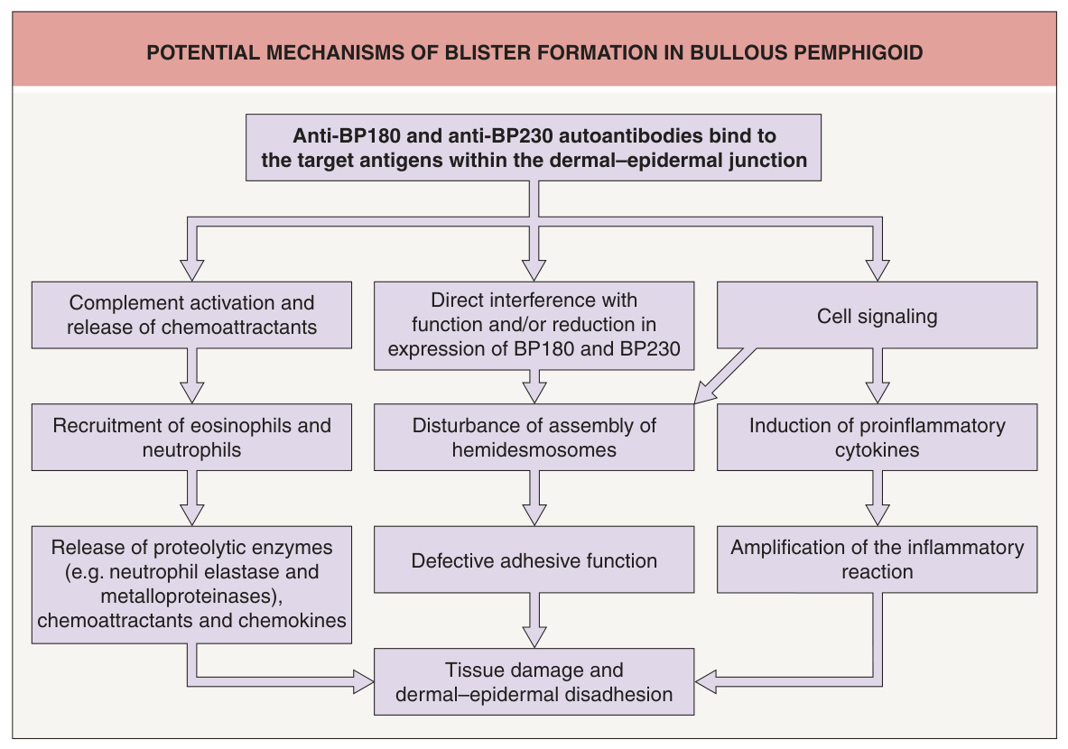
Fig. 30.1 Potential mechanisms of blister formation in bullous pemphigoid. IgG autoantibodies are classically involved in the pathogenesis, but more recently IgE autoantibodies have also been described. The latter may directly trigger the degranulation of FcεRI-expressing cells, such as eosinophils and mast cells.
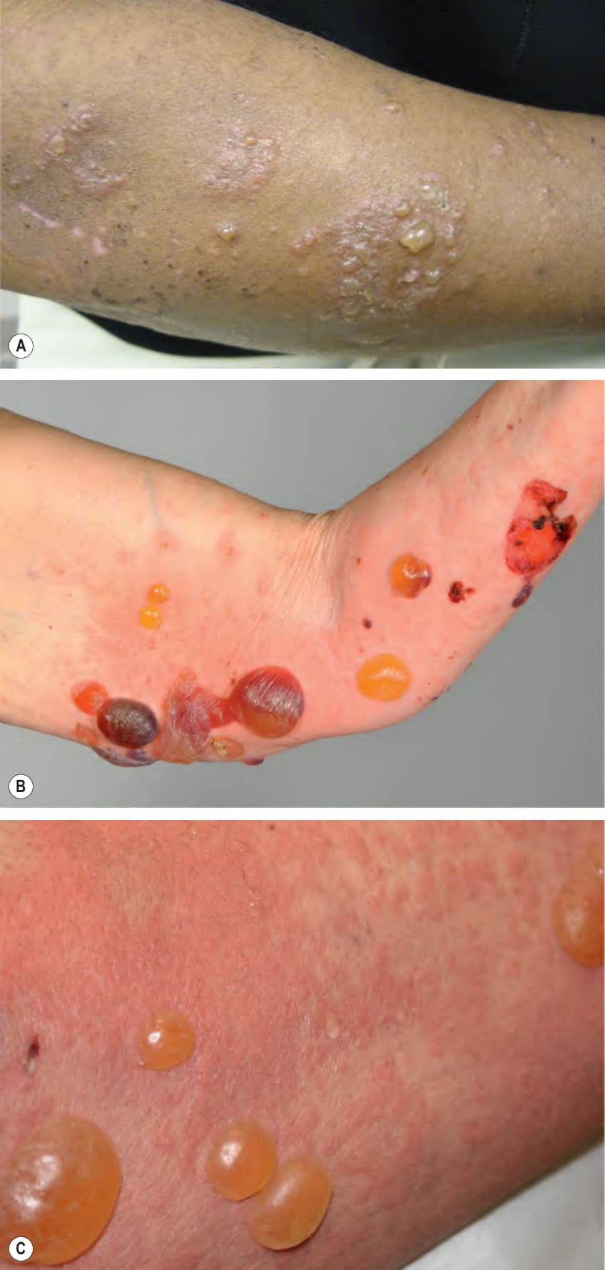
Fig. 30.2 Bullous pemphigoid – classic presentation.A–C Tense vesicles and bullae vary in size from a few mm to several cm in diameter and can arise within normal-appearing skin, areas of erythema, or urticarial plaques. The blister fluid may be serous or hemorrhagic. The flexor aspect of the extremities is a common site of involvement. As the bullae age, they become flaccid and rupture, leaving erosions and serous or hemorrhagic crusts. Biopsies of vesiculobullae for routine histology should be obtained from the edge of a fresh tense blister. Jeffrey Callen, MD.
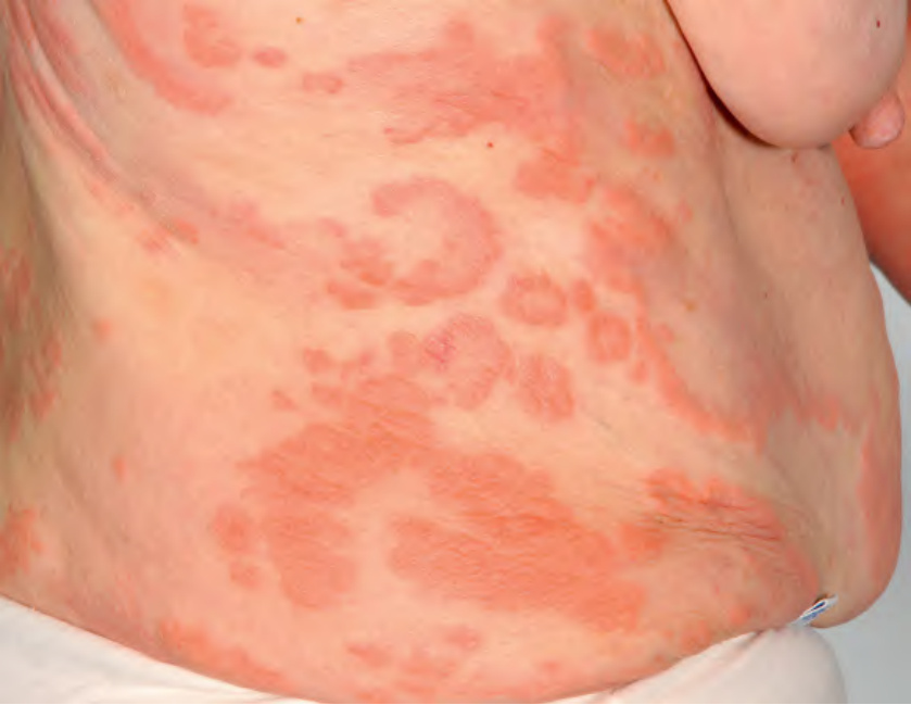
Fig. 30.3 Bullous pemphigoid – urticarial presentation. Multiple, firm annular, arciform and polycyclic urticarial plaques. Note the absence of bullae.
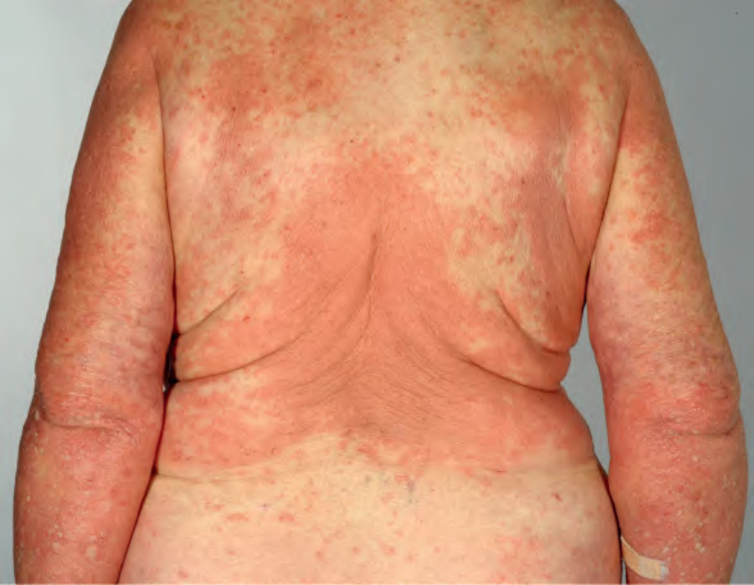
Fig. 30.4 Bullous pemphigoid – eczematous presentation. Large pink eczematous plaques on the trunk and upper extremities.
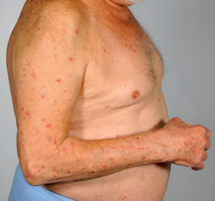
Fig. 30.5 Bullous pemphigoid – nonspecific lesions due to pruritus. Multiple excoriations and nonspecific lesions of prurigo simplex are present. Bullous pemphigoid is in the differential diagnosis of chronic pruritus associated with secondary excoriations.
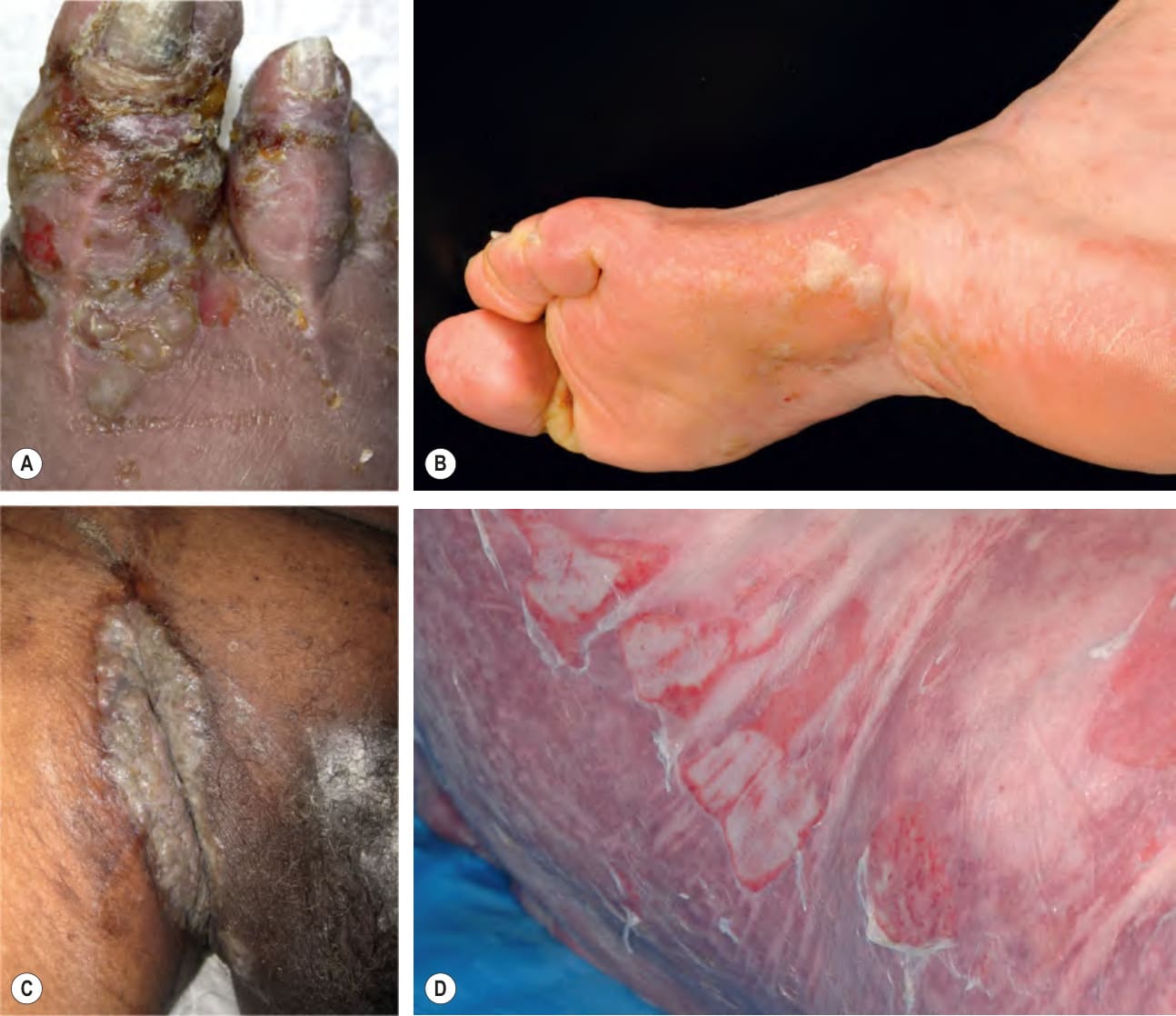
Fig. 30.6 Bullous pemphigoid – uncommon clinical variants.A, B In dyshidrosiform pemphigoid, clusters of vesicles and bullae appear on acral skin and can resemble dyshidrotic eczema or pompholyx. C In pemphigoid vegetans, vegetating plaques can develop in major body folds, including the inguinal crease. D Toxic epidermal necrolysis-like lesions with large erosions.
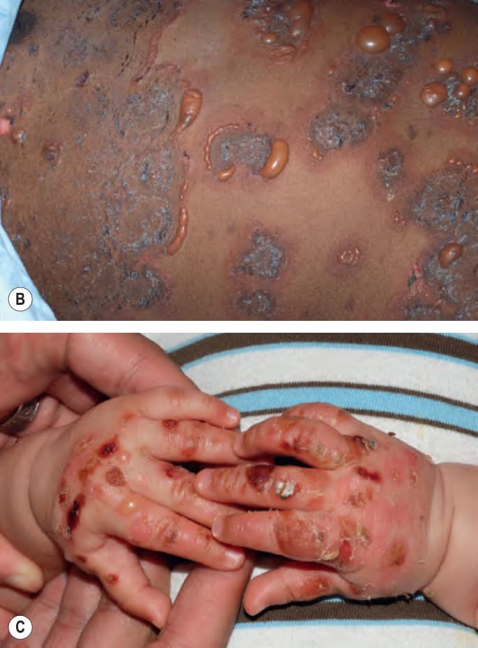
Fig. 30.7 Childhood bullous pemphigoid.A Generalized tense bullae and circular crusted erosions. B Tense vesicles and bullae in an annular or figurate array at the edge of expanding lesions, a presentation that might be diagnosed clinically as linear IgA bullous dermatosis. C A predominance of acral involvement is often seen in infants. B, C, Courtesy Julie V. Schaffer, MD.
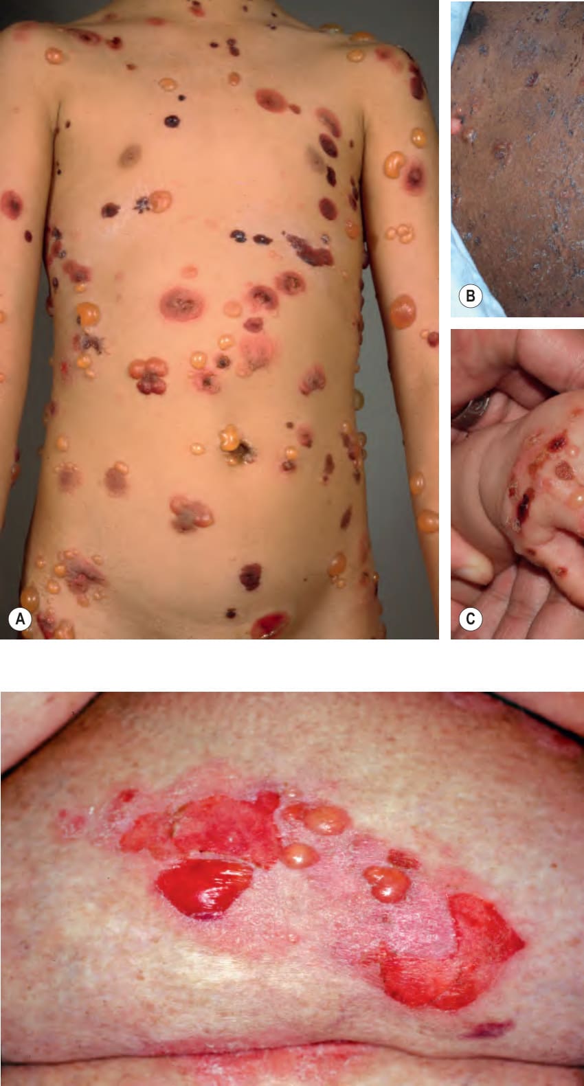
Fig. 30.8 Bullous pemphigoid localized to a psoriatic plaque. No obvious trigger was detected, as the patient was not receiving phototherapy. Courtesy Jean L. Bolognia, MD.
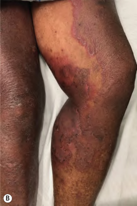
Fig. 30.9 Drug-induced bullous pemphigoid.
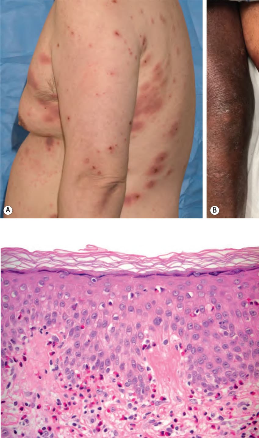
Fig. 30.10 Urticarial phase of bullous pemphigoid – histopathologic features.
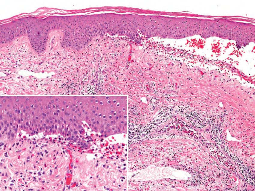
Fig. 30.11 Bullous pemphigoid – histopathologic features. Subepidermal blister which contains fibrin, eosinophils and mononuclear cells (see inset). Courtesy Lorenzo Cerroni, MD.
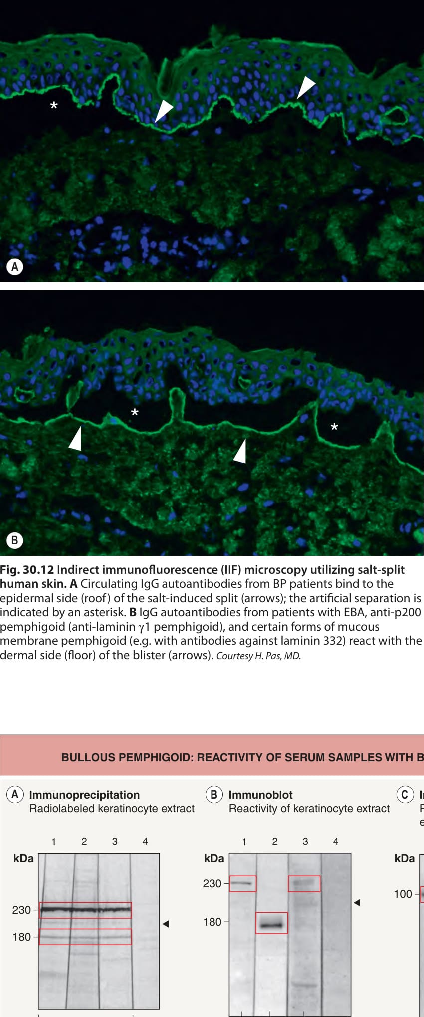
Fig. 30.12 Indirect immunofluorescence (IIF) microscopy utilizing salt-split human skin.A Circulating IgG autoantibodies from BP patients bind to the epidermal side (roof) of the salt-induced split (arrows); the artificial separation is indicated by an asterisk. B IgG autoantibodies from patients with EBA, anti-p200 pemphigoid (anti-laminin γ1 pemphigoid), and certain forms of mucous membrane pemphigoid (e.g. with antibodies against laminin 332) react with the dermal side (floor) of the blister (arrows). Courtesy H. Pas, MD.
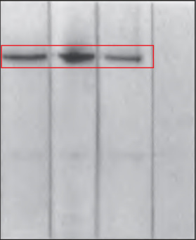
Fig. 30.13 Bullous pemphigoid (BP): reactivity of serum samples with BP180 and BP230.A
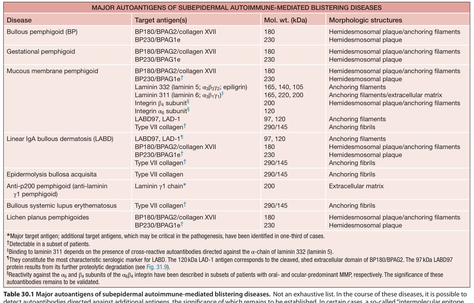
Table 30.1 Major autoantigens of subepidermal autoimmune-mediated blistering diseases.
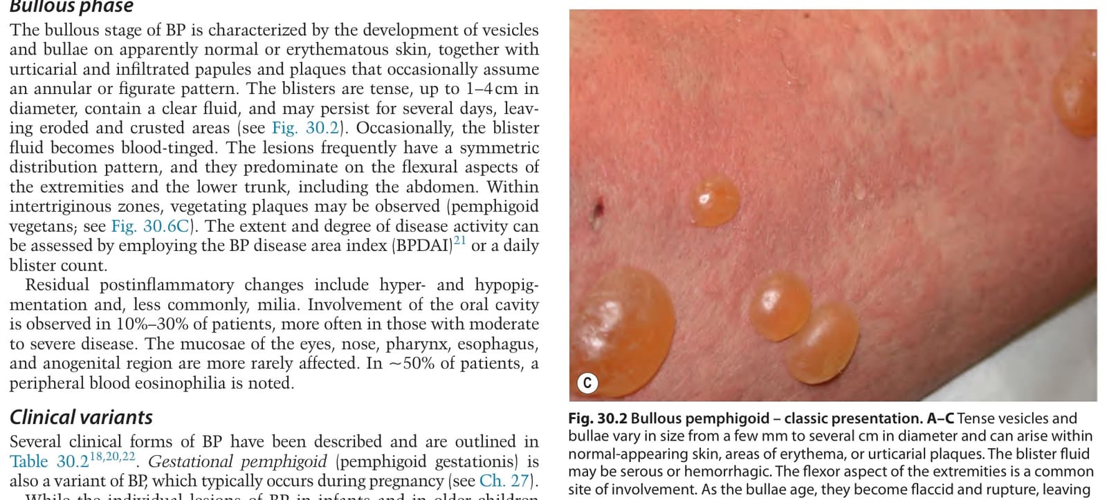
Table 30.2 Unusual clinical variants of bullous pemphigoid.
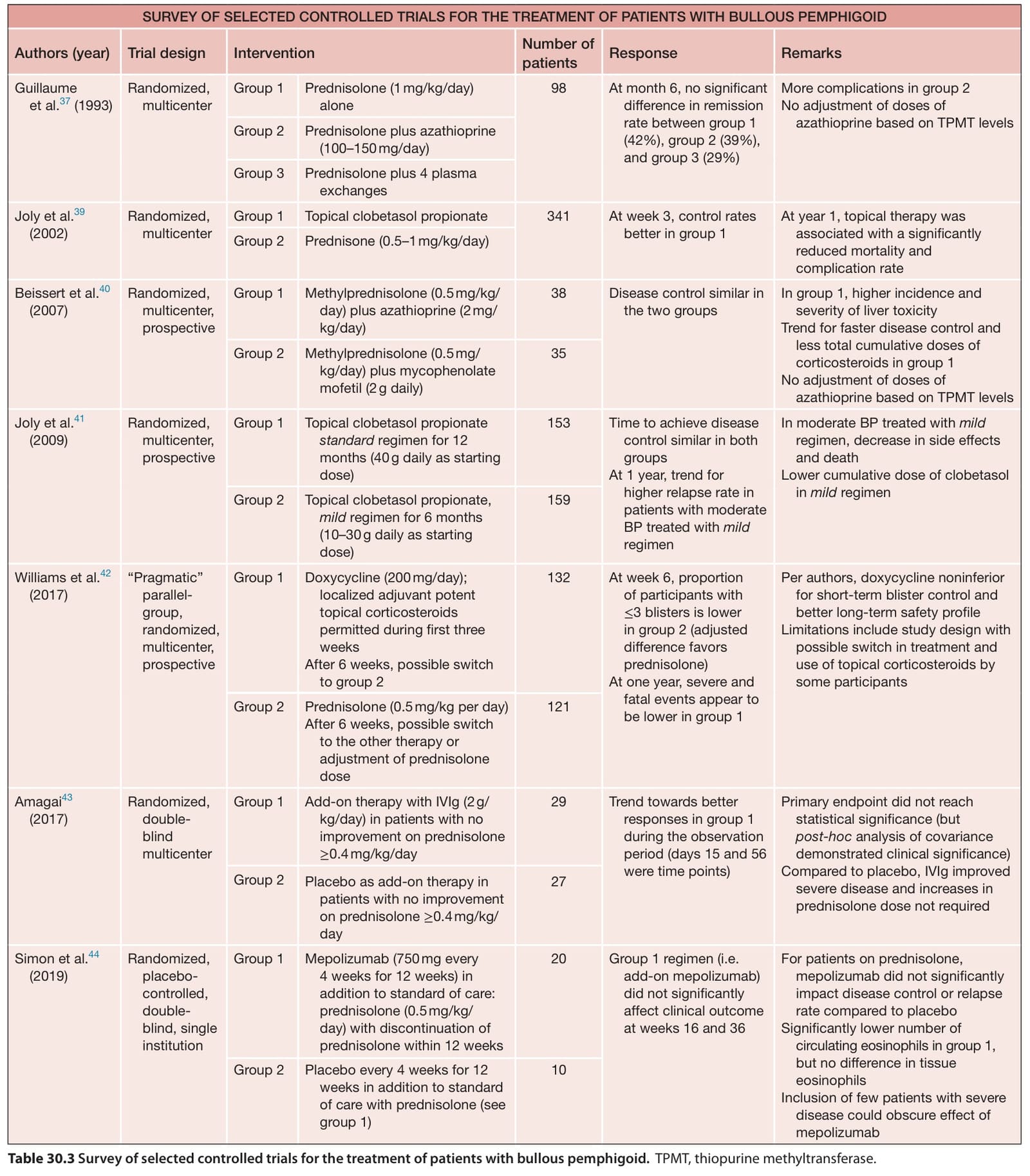
Table 30.3 Survey of selected controlled trials for the treatment of patients with bullous pemphigoid.TPMT, thiopurine methyltransferase.
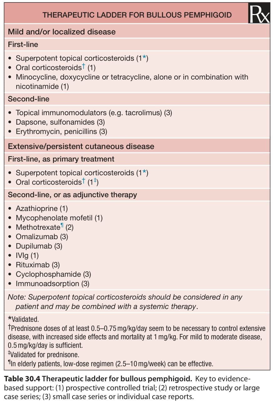
Table 30.4 Therapeutic ladder for bullous pemphigoid.Key to evidencebased support: (1) prospective controlled trial; (2) retrospective study or large case series; (3) small case series or individual case reports.