MUCOUS MEMBRANE PEMPHIGOID
Synonyms (becoming historical): Cicatricial pemphigoid Benign mucous membrane pemphigoid
Subtypes/variants: Anti-laminin 332 (mucous membrane) pemphigoid – anti-epiligrin cicatricial pemphigoid Brunsting–Perry pemphigoid
Key features
Mucous membrane pemphigoid is a chronic, autoimmune, subepithelial blistering disorder characterized by predominant involvement of the external mucosal surfaces and a tendency for scarring
It is associated with tissue-bound and, less often, serum autoantibodies directed against distinct structural components of basement membranes in stratified and some complex epithelia: BP180, BP230, laminin 332 (laminin 5), type VII collagen, and likely integrin β and α subunits
The condition should not be regarded as a clinical entity, but as a
“disease phenotype” shared by a heterogeneous group of diseases with lesions that favor the mucosal surfaces and, less frequently, the skin
As scarring frequently affects the conjunctivae, the disorder can be devastating and ultimately lead to blindness
Diagnosis relies on immunopathologic examinations, especially DIF microscopy, and serological immunoassays
Introduction
Mucous membrane pemphigoid (MMP) is a rare autoimmune subepithelial blistering disorder characterized by predominant involvement of the mucosae, a chronic course, and a tendency towards scarring of the affected areas. The disorder should not be regarded as a clinical entity, but rather as a “disease phenotype” shared by a heterogeneous group of blistering diseases, with predilection for the mucosal surfaces. The majority of patients have linear deposits of immunoglobulins and/or complement components along the epithelial BMZ of the skin and mucosae. Circulating anti-BMZ autoantibodies are detected in the serum of some patients (20%–30% by standard IIF), usually at a low titer or concentration. It is a chronic and progressive disease, which may result in serious mucosal complications. When postinflammatory atrophy or scarring affect the conjunctivae, the disorder can be devastating and ultimately lead to blindness.
History
In 1794, Wichmann first described a chronic blistering disease associated with involvement of the eyes. Civatte separated this disorder from pemphigus on the basis of peculiar histopathologic findings in 1949, and Lever later suggested the name “benign mucous membrane pemphigoid”, a misnomer in view of the potentially severe complications of the disorder. During the latter part of the 20th century, the term “cicatricial pemphigoid” was used primarily. In 2012, based upon an international consensus conference, the term “mucous membrane pemphigoid” was proposed. Demonstration of immunoreactants deposited along the epithelial BMZ by IF microscopy pointed to an autoimmune origin.
Epidemiology
The annual incidence of MMP is ~1 to 2 cases per million population, with a calculated prevalence (in Germany) of 25 cases per million inhabitants. MMP typically occurs in the elderly, with a mean age at the time of diagnosis of between 60 and 80 years. However, isolated cases have been reported in children. Women are affected more frequently than men (estimated ratio 1.5–2:1). Although no geographic or racial predilection has been described, several studies have shown an association with specific immunogenetic HLA class II haplotypes, e.g. HLA- DQw7 (subtype HLA-DQB1*03:01), in both oral and ocular forms of the disease. There is no overall association with malignancy, but in a distinct subtype associated with IgG autoantibodies directed against laminin 332 (also known as anti-epiligrin cicatricial pemphigoid), an increased relative risk for cancer has been suggested. Although this is debated, a search of an associated solid tumor may be recommended in case of anti-laminin 332 reactivity. For example, in a retrospective study of 154 patients, an association of anti-laminin 332 reactivity with malignant neoplasms was noted, with an adjusted odds ratio of 2350a. Recently, MMP has been observed in patients receiving immune checkpoint inhibitors.
Pathogenesis
Mucocutaneous lesions are thought to result from the binding of autoantibodies to the BMZ of stratified epithelia of mucosae and skin. These autoantibodies recognize distinct structural components of the hemidesmosomal adhesion complex. Specifically, they appear to bind predominantly to antigenic sites located extracellularly within the anchoring filament zone rather than within the hemidesmosomal plaque. However, the pathogenicity of various IgG autoantibodies associated with this disease phenotype (see Table 30.1) has not yet been established unequivocally and other mechanisms may also be involved. In a minority of patients with MMP, BP180–NC16A domain-specific T cells are detectable. Increased expression of collagen-binding heat shock protein 47 (HSP47) and TGF-β1 by conjunctival fibroblasts may contribute to the conjunctival scarring seen in patients with ocular disease.
Based upon their autoantibody profile, patients with MMP can be divided into four distinct subgroups. The first subgroup includes those with autoantibodies directed against laminin 332. This subgroup was previously referred to as anti-laminin 332 MMP or anti-epiligrin cicatricial pemphigoid. It is estimated that 18%–30% of patients with MMP belong to this subgroup. Patients with anti-laminin 332 IgG autoantibodies frequently have multiple sites of mucosal involvement, including the pharynx, larynx, and trachea. Their circulating IgG autoantibodies bind to the dermal side of salt-split skin and react with the α chain (G domain) and, less frequently, the β and/or γ chain of laminin 332 (αβγ) (see Fig. 28.5). Occasionally, antibody binding to the α chain of laminin 311 (previously termed laminin 6 [a heterotrimer of αβγ chains]) is also observed because of the presence of cross-reacting autoantibodies. In vitro and in vivo studies have provided convincing evidence that autoantibodies to laminin 332 are pathogenic.
In the second group of patients, involvement is exclusively or pre-dominantly ocular. IgG autoantibodies that bind to the β subunit of αβ integrin, a transmembrane hemidesmosomal component that interacts with laminin 332, have been described anecdotally. Some clinicians refer to this subset as ocular MMP (see Fig. 28.3A).
A third subgroup of patients, who lack a specific clinical phenotype, have tissue-bound and circulating IgG antibodies that react with the same target antigens as in BP, especially BP180. These patients, who have been classified as having anti-BP antigen MMP, represent the largest subgroup. The antigenic regions targeted on BP180 are located within the NC16A domain and on its distal COOH-terminal portion. The latter appears to extend into the lamina densa region of the BMZ (see Fig. 28.3A). There is ample scientific evidence that anti-BP180 IgG antibodies are pathogenic.
The fourth group is rather heterogeneous. It includes patients who have variable involvement of the mucosae without involvement of the skin. While it is yet unclear whether injury occurs as a result of an autoantibody-mediated immune response against proteins of the BMZ, a few patients with an MMP phenotype have shown reactivity against type VII collagen. It has been reported that a small subset of patients with oral MMP have autoantibodies which bind to the α subunit of αβ integrin, but this needs to be validated.
Clinical Features
Mucous membranes
The two most frequently involved sites in patients with the “mucous membrane pemphigoid phenotype” are the oral and conjunctival mucosae. However, the disease may also start in, and affect, any mucosal site, including the nasopharyngeal, anogenital, laryngeal, or esophageal mucosa (in descending order of frequency).
Approximately 90% of patients have oral involvement (often in the absence of skin lesions), and the oral cavity may be the only site of disease activity in up to 30% of patients. Lesions frequently involve the gingiva, buccal mucosa, and palate; the alveolar ridges, tongue, and lips are less commonly affected. A classic oral presentation is desquamative gingivitis accompanied by bleeding erosions and paresthesias (Fig. 30.14A); small intact blisters are rarely observed. Chronic inflammation may lead to periodontal ligament damage and the loss of teeth. In other areas of the mouth, transient vesicles give rise to chronic erosions, especially on the palate (Fig. 30.14B), with variable associated pain. Lesions on the tongue are usually localized to its lateral and ventral surfaces. Adhesions may develop in the area of the uvula and tonsillar fossae as well as between the tongue and the floor of the mouth. White, reticulated striations that resemble oral lichen planus may also be seen.
Conjunctival involvement occurs in ~40% of patients and can result in blindness. The conjunctivae are frequently the only site affected. Although the disease may begin unilaterally, in the majority of patients, lesions appear in both eyes. Ocular involvement starts as a nonspecific chronic conjunctivitis and limbitis, with burning, soreness, foreign-body sensation, decreased vision, and/or mucus production. Periods of exacerbations and subsequent remissions are typical, with eventual progression to a scarring due to subepithelial conjunctival fibrosis (Fig. 30.15).
Conjunctival vesicles or blisters are rare on the tarsal conjunctiva. Chronic inflammation may result in progressive scar tissue formation, leading to a shortened inferior fornix and symblepharon (an adhesion between bulbar and palpebral conjunctival surfaces; Fig. 30.16) as well as ankyloblepharon. Conjunctival fibrosis can also result in trichiasis (inwardly angled eyelashes) and entropion. In uncontrolled disease, trichiasis, entropion, and xerosis (due to scarring of the lacrimal ducts) eventuate in superficial corneal trauma, corneal neovascularization with subsequent corneal ulceration, and blindness.
Nasopharyngeal involvement occurs in a third of patients with MMP and is typically chronic and asymptomatic. It results in crusted intranasal ulcerations, epistaxis, fibrous adhesions between adjacent mucosal surfaces, and airway obstruction. Pharyngeal involvement presents as ulcerations of the posterior or lateral pharynx and dysphagia. Laryngeal involvement is a potentially serious manifestation, presenting as hoarseness, loss of speech, and even life-threatening stenosis, which requires tracheostomy.
Although esophageal disease may be accompanied by dysphagia resulting from erosions of the esophageal mucosa, involvement can be
entirely asymptomatic. Chronic inflammation can lead to strictures and stenosis, with associated dysphagia.
Involvement of genital and anal mucosae is relatively uncommon, but increasingly recognized. Early lesions consist of blisters and chronic erosions. In female patients, progressive disease leads to scarring and narrowing of the introitus. In male patients, adhesions can form between the prepuce and the glans penis. Anal involvement can also lead to scarring and, in severe cases, to stricture formation.
Cutaneous lesions
The skin is involved in 20%–35% of patients. The most frequently involved sites are the scalp, face, neck, and upper trunk. Lesions typically present as erythematous plaques, which become sites for recurrent blister formation and erosions, with subsequent scarring. Their extent and number are generally limited, but an individual patient can have a BP-like clinical presentation.
In the Brunsting–Perry variant, skin lesions are localized predominantly to the head and neck and upper trunk (Fig. 30.17A,B). Mucosal involvement is usually absent or minimal. On the scalp, significant scarring alopecia can result (Fig. 30.17C).
Diagnosis and Differential Diagnosis
The diagnosis of MMP has to be considered in all patients who have blistering or erosive lesions that predominantly involve mucous membranes and whose affected tissues show linear, continuous deposits of immunoreactants along the epithelial BMZ. Unfortunately, there is frequently a significant delay, often more than a year, before the diagnosis is established. As a result, moderate to severe scarring, especially ocular, may have already occurred.
Light microscopy and electron microscopy
The histopathologic features of MMP and BP are often similar. Examination of a fresh vesicle or bulla demonstrates subepithelial or subepidermal blister formation (without acantholysis). A mixed cellular infiltrate of variable intensity is present, with a predominance of neutrophils; the number of eosinophils is increased in older lesions, but they are still fewer than in BP. Older lesions also tend to have scarring in the submucosa or upper dermis. In oral lesions, the inflammatory infiltrate typically contains some plasma cells, a nonspecific finding related to the mouth being a mucosal site. The epithelium of affected conjunctivae is often invaded by inflammatory cells, including mononuclear cells and mast cells, while granulation tissue is found in the submucosa.
In most cases, electron microscopy studies of a fresh blister demonstrate that the dermal–epidermal cleavage occurs within the lamina lucida. In advanced conjunctival lesions with scarring, the lamina densa appears to be discontinuous, focally thickened, or duplicated.
Direct immunofluorescence (DIF) microscopy
By DIF microscopy, 80%–95% of patients with MMP have in vivo bound autoantibodies directed against the BMZ of mucosae and/or skin. The majority of patients exhibit continuous, fine, linear deposits of IgG, IgA, and/or C3 along the epithelial BMZ in perilesional biopsy specimens. It should be noted that DIF microscopy studies of mucosae are more frequently positive (50%–90%) than those of skin (20%–50%). IgG deposits belong primarily to the IgG4 and IgG1 subclasses; deposits of IgM and fibrin are less common. DIF microscopy of salt-split skin is of less diagnostic value. Although a combined pattern with staining of both the epidermal and dermal sides is frequently observed, reactivity with either the epidermal roof or the dermal floor of the separated skin is occasionally noted. If circulating autoantibodies are not detected, analysis of the
Crusted erosion on the lower cheek within an oval area of inflammation and scar. B Mild involvement of the scalp with scarring and atrophy resulting in limited alopecia. C Severe involvement of the scalp with an ulceration within a large area of scarring alopecia. B, Courtesy Karynne O. Duncan, MD; C, Courtesy Michael Gowen, MD.
linear fluorescence pattern of autoantibody deposits at the cutaneous BMZ (n-serrated [pemphigoid group] versus u-serrated [EBA]) or results of the FOAM technique (see above) may prove helpful. Of note, in mucosal specimens, the serration pattern cannot be assessed.
Immunoelectron microscopy
Although electron microscopy studies are not routinely available and are time-consuming, they may be particularly useful in establishing the diagnosis of MMP, especially in patients in whom standard DIF and IIF microscopic findings are negative. Direct immunoelectron microscopy studies in MMP have demonstrated immune deposits distributed either: (1) in the lower lamina lucida and above the lamina densa; or (2) on and around hemidesmosomes, with most of the labeling on the outside of basal keratinocytes below their plasma membranes.
Indirect immunofluorescence (IIF) microscopy
By standard IIF, anti-BMZ autoantibodies can be detected in serum samples from 20%–30% of patients with MMP. Antibodies are mainly of the IgG class, but circulating IgA or IgE may also be detected. Antibody concentrations are usually very low. Some authors have suggested that patients with high serum levels of IgG and IgA anti-BMZ autoantibodies have more severe disease. The sensitivity of IIF can be increased either by the use of normal oral or genital mucosa and conjunctiva as substrates, or by utilizing salt-split skin. Using this latter technique, most patients with MMP have antibodies that bind to the epidermal roof, whereas MMP patients with anti-laminin 332 or anti-type VII collagen reactivity have IgG anti-bodies that bind to the dermal side (see Fig. 30.12). The use of salt-split oral mucosa does not improve the overall sensitivity. Distinguishing MMP with anti-laminin 332 autoantibodies from EBA may be possible by IIF microscopy using “knockout” skin substrates, i.e. substrates deficient in specific basement membrane proteins.
Immunochemical studies, including ELISA
Immunochemical techniques, including ELISA, immunoblots, and immunoprecipitation, have confirmed that the autoantigens targeted in patients with a MMP phenotype are heterogeneous. Serum autoantibodies react with various proteins located within the epithelial BMZ, e.g. BP180, laminin 332, type VII collagen (see Table 30.1). Depending upon the patient population and specific assay, IgG anti-BP180 or anti-laminin 332 antibodies are detected in 30–70% or 15–30% of MMP patients, respectively. These targeted antigens appear to constitute serologic markers for different subgroups of MMP (see above).
Patients may have IgA autoantibodies directed against the ectodomain of BP180. As a result, salivary IgG and IgA autoantibodies directed against BP180–NC16A may serve as diagnostic biomarkers. In some cases, these autoantibodies appear to preferentially bind to antigenic reactive sites present on the proteolytically cleaved and shed portion of the extracellular domain of BP180, i.e. the LABD antigen (LAD-1; see Ch. 31). Lastly, several unidentified target antigens of varying molecular weights have also been reported.
ELISAs that utilize recombinant proteins or affinity-purified native proteins (e.g. BP180, laminin 332, type VII collagen) can be useful in the search for circulating autoantibodies.
Differential diagnosis
Distinction from other subepidermal autoimmune bullous diseases (including BP, EBA, and LABD) may be difficult and often relies on the predominance of mucosal involvement and chronic course. In contrast, pemphigus vulgaris and non-autoimmune blistering diseases are more easily excluded via histology and IF microscopy.
If oral involvement, especially isolated desquamative gingivitis, is the sole manifestation of MMP, it can be difficult to differentiate clinically from pemphigus vulgaris or erosive lichen planus. End-stage, scarring conjunctival lesions may mimic severe chronic infectious conjunctivitis, ocular pseudopemphigoid due to ophthalmologic preparations (including pilocarpine, idoxuridine, guanethidine, β-blockers), or sequelae of Stevens–Johnson syndrome and toxic epidermal necrolysis. While the rare, generalized cutaneous form of MMP can be impossible to distinguish from BP, involvement of multiple mucosal sites favors the former.
Prognosis
MMP is a particularly chronic, potentially devastating, but rarely fatal disease. The most important complication is impairment of vision due to ocular involvement. It can also lead to weight loss as well as respiratory, sexual, or urinary complications. Even with localized involvement, this disease can have a major negative impact on quality of life. Life-threatening complications, due to severe laryngeal, tracheal or esophageal disease, are rare.
Treatment
Treatment can be difficult and at times frustrating, with therapeutic regimens based primarily on clinical experience.
Local therapy is crucial and, for limited involvement, it can be sufficient. Mild to moderate disease activity may be successfully treated with potent topical corticosteroids. Oral lesions can respond to topical corticosteroids (as mouthwashes or topical preparations in a gel or occlusive base supplemented by customized prosthetic devices to provide occlusion) in conjunction with tetracycline mouthwashes and professional dental hygiene. Corticosteroid sprays and inhalers may prove useful in nasal, pharyngeal, or esophageal disease. Recalcitrant lesions of both skin and mucous membranes may improve with intralesional injections of triamcinolone. Esophageal strictures may require dilatations to prevent dysphagia, while severe tracheal involvement may require a tracheostomy to prevent asphyxiation.
Systemic medications include dapsone (50–150 mg/day) as a firstline therapy for controlling oral and cutaneous lesions, and it may also be used for mild ocular disease without rapid worsening. Dapsone can be particularly beneficial in patients with linear deposits of IgA at the BMZ. Cyclophosphamide (1–2 mg/kg/day) is considered the treatment of choice for rapidly progressive or severe ocular disease – alone, in combination with oral corticosteroids, or as pulse therapy. Such regimens are effective in resolving severe conjunctival inflammation and preventing recurrences and secondary scarring. Azathioprine (2 mg/kg/ day) and mycophenolate mofetil (2 g/day) have also been reported to be helpful in reducing the dose of corticosteroids required for control of ocular involvement and cutaneous lesions.
Systemic corticosteroids alone are generally insufficient therapy for patients with significant disease, and they are less effective for mucosal than for cutaneous disease. Patients with significant esophageal or laryngotracheal involvement should be treated aggressively with the combination of prednisone and cyclophosphamide or mycophenolate mofetil to prevent potentially life-threatening complications. Other therapies reported to be of potential value in the treatment of MMP include topical tacrolimus, a combination of tetracycline and niacinamide, minocycline, sulfapyridine, methotrexate, TNF inhibitors, IVIg, topical or systemic cyclosporine, thalidomide, and subconjunctival mitomycin. Case series have also underscored the efficacy of rituximab in controlling progressive ocular involvement and severe extraocular complications. When these manifestations fail to respond to cyclophosphamide plus corticosteroids or there is an intolerance or contraindication to this regimen, rituximab can be considered as second-line therapy.
Surgical therapy may occasionally be necessary for severe scarring involving the eye, larynx, esophagus, or genitalia. It should be performed when the disease is fully controlled by medical therapy. For ocular disease, surgical intervention includes corneal grafts, allograft limbal transplantation, amniotic membrane transplantation, and tarsorrhaphy.
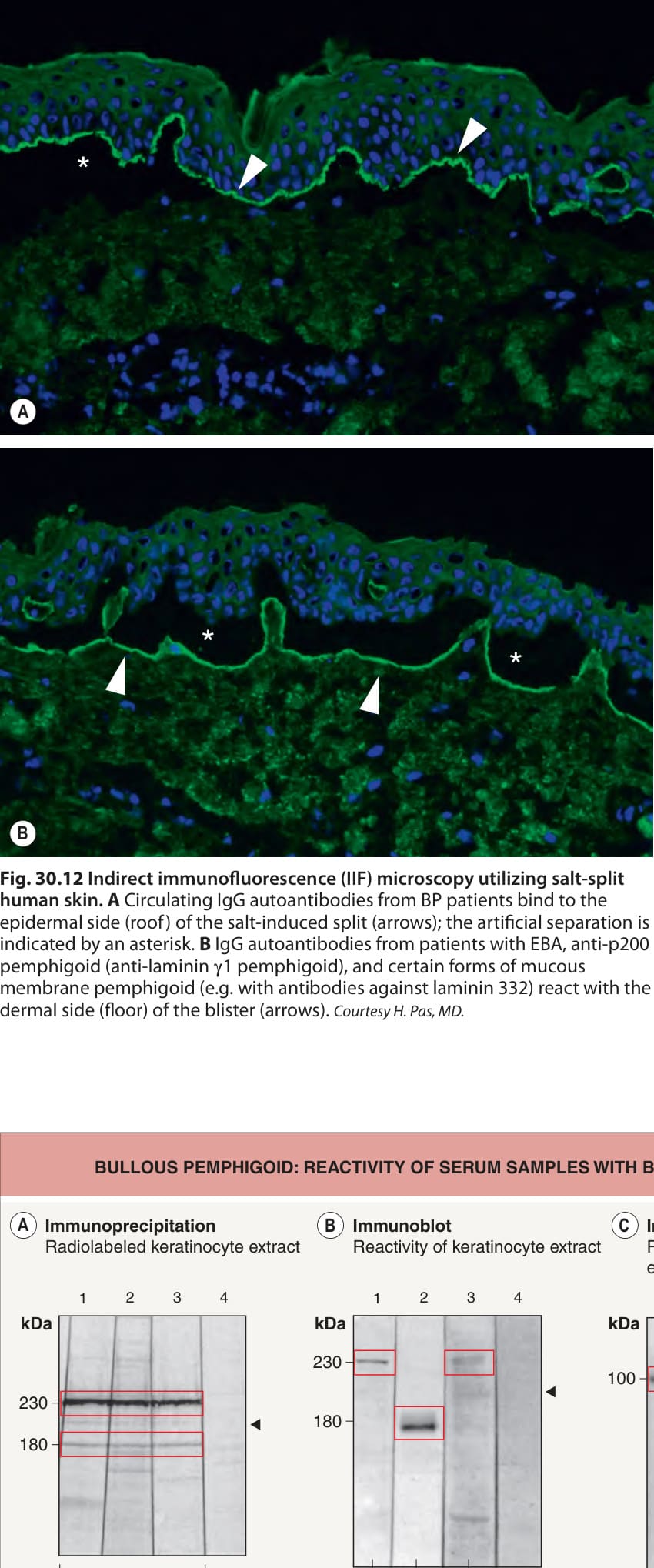
Fig. 30.12 Indirect immunofluorescence (IIF) microscopy utilizing salt-split human skin.A Circulating IgG autoantibodies from BP patients bind to the epidermal side (roof) of the salt-induced split (arrows); the artificial separation is indicated by an asterisk. B IgG autoantibodies from patients with EBA, anti-p200 pemphigoid (anti-laminin γ1 pemphigoid), and certain forms of mucous membrane pemphigoid (e.g. with antibodies against laminin 332) react with the dermal side (floor) of the blister (arrows). Courtesy H. Pas, MD.
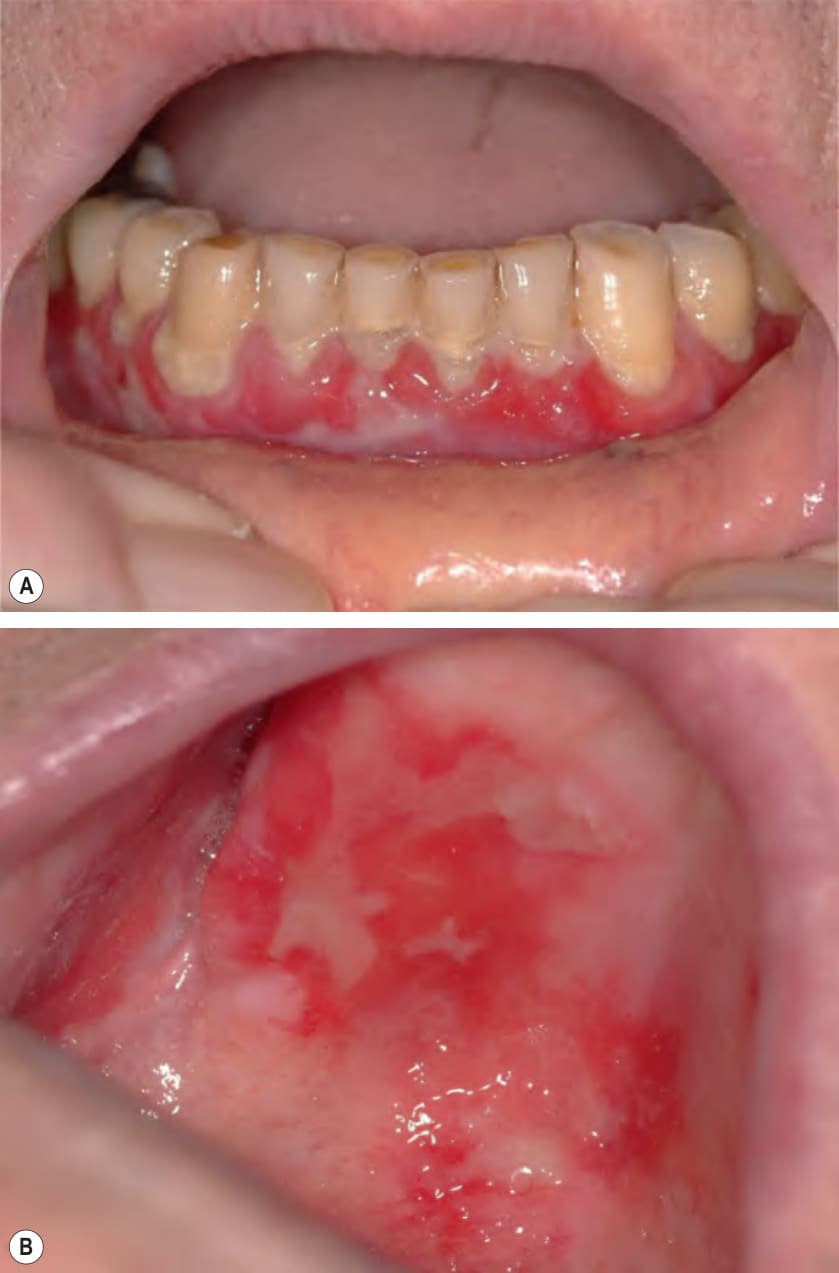
Fig. 30.14 Mucous membrane pemphigoid.A Desquamative gingivitis with erythema and erosions of the gingival margins. Note the sloughing of the mucosa with shaggy margins. B Chronic erosions on the hard palate with irregular borders. Sloughing of mucosa is seen superiorly.
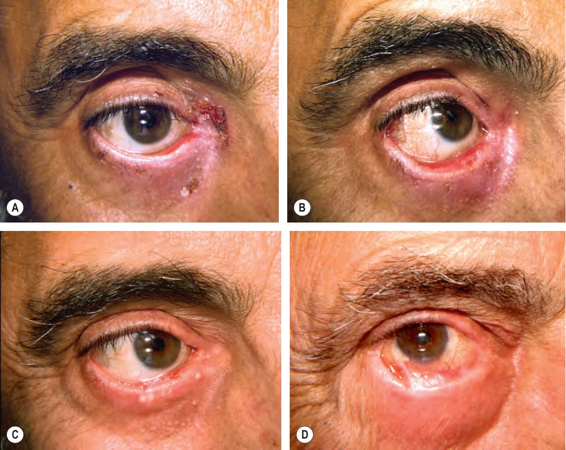
Fig. 30.15 Ocular involvement in mucous membrane pemphigoid.A Erosion and erythema of the lower medial eyelid margin plus scale-crust of the inner canthus and lower eyelid. B Three months later, ectropion and thickening of the lower eyelid in addition to erosions. C Six months later, smaller erosions but scarring and milia formation. D Seven years later, further scarring with significant shortening of the inferior fornix due to symblepharon. Courtesy Louis A. Fragola, Jr, MD.
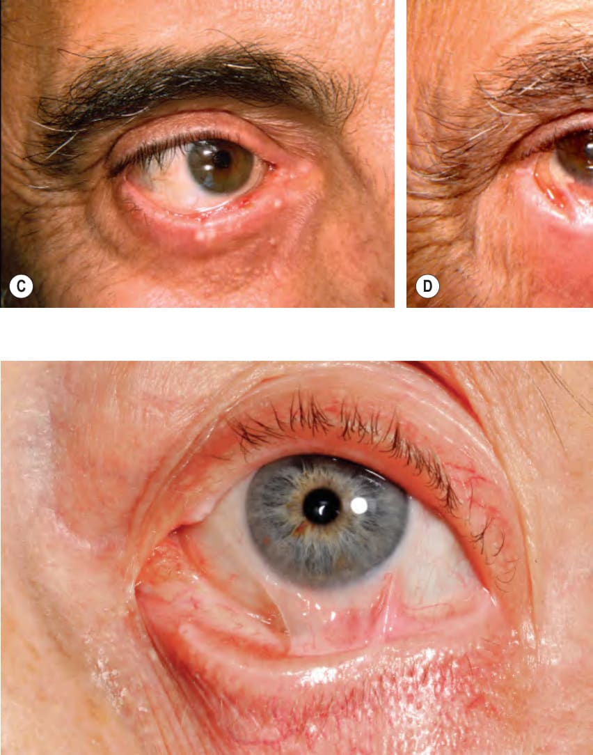
Fig. 30.16 Mucous membrane pemphigoid. Typical ocular involvement as manifested by fibrous tracts, representing partial or incomplete symblepharon.
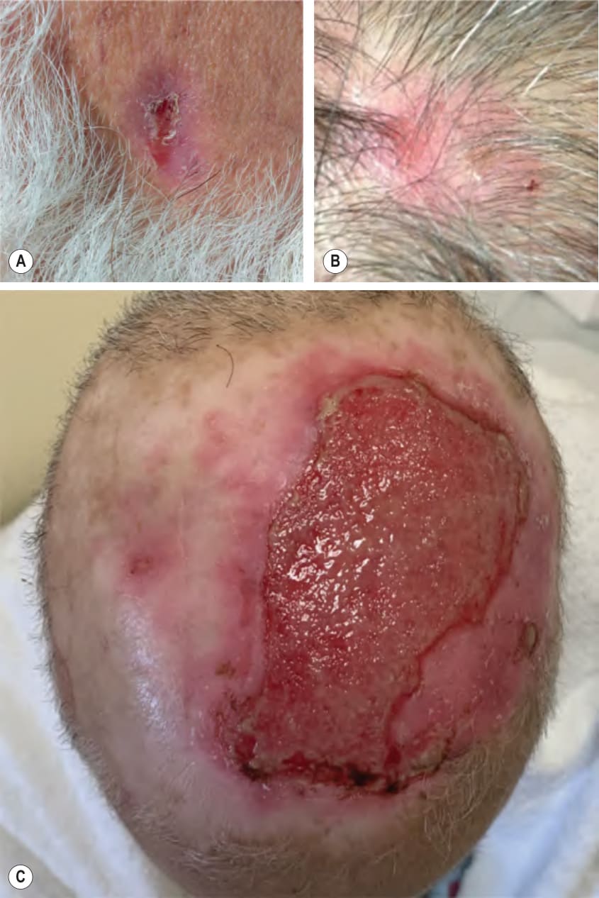
Fig. 30.17 Mucous membrane pemphigoid – Brunsting–Perry variant.A
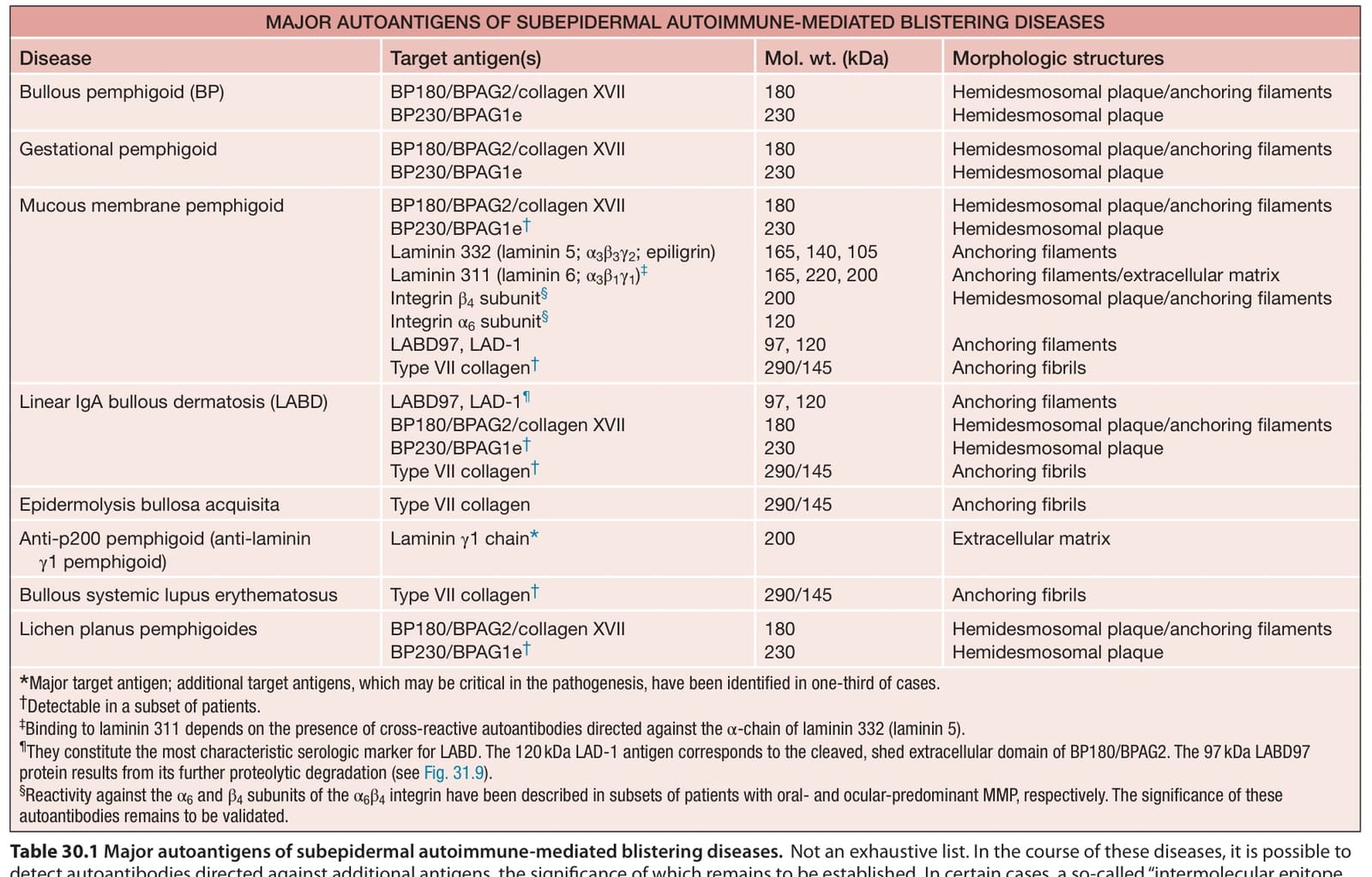
Table 30.1 Major autoantigens of subepidermal autoimmune-mediated blistering diseases.