EPIDERMOLYSIS BULLOSA ACQUISITA
Synonym: Acquired epidermolysis bullosa; dermolytic pemphigoid
Key features
Epidermolysis bullosa acquisita is a rare, acquired, subepidermal bullous disease associated with autoimmunity to type VII collagen, the major component of the anchoring fibrils of the dermal– epidermal BMZ
Patients may present either with a mechanobullous disorder, mimicking dystrophic epidermolysis bullosa, or with clinical features that resemble BP or infrequently MMP, the Brunsting–Perry variant of MMP, or LABD
Diagnosis relies on immunopathologic examinations, especially immunofluorescence microscopy, immunoserologic studies, and occasionally immunoelectron microscopy
The disease is usually chronic and more refractory to treatment than BP
Introduction
Epidermolysis bullosa acquisita (EBA) is a rare, acquired, subepidermal bullous disease associated with autoimmunity to type VII collagen, the major component of the anchoring fibrils of the dermal–epidermal BMZ. Patients may present with either a non-inflammatory variant, the features of which strikingly resemble those observed in patients with dystrophic epidermolysis bullosa (EB), or with an inflammatory variant that mimics BP. Infrequently, the clinical phenotype resembles MMP, the Brunsting–Perry variant of MMP, or LABD.
History
The first cases of an acquired bullous disease with clinical features mimicking hereditary dystrophic epidermolysis bullosa were described by Elliot in 1895. In the early 1970s, before the advent of cutaneous immunopathology, Roenigk and colleagues proposed the initial clinical criteria for EBA. A few years later, IF microscopy studies demonstrated that EBA patients have anti-BMZ antibodies, similar to BP patients. In the early 1980s, immunoelectron microscopy studies separated EBA from BP by demonstrating that IgG deposits in EBA were located in the sublamina densa region of the BMZ. In the 1990s it was shown that EBA autoantibodies bind to type VII collagen, the major component of anchoring fibrils.
Epidemiology
EBA has been reported in both children and adults and may occur at any age. It is one of the rarest subepidermal autoimmune bullous diseases in Western Europe, with an estimated annual incidence of ~0.25 per million people. It may be more common in specific Asian populations (e.g. Koreans) as well as in African-Americans.
Pathogenesis
The pathogenic role of autoantibodies to type VII collagen is supported by several lines of evidence. Patients with EBA have tissuebound and circulating IgG autoantibodies against type VII collagen, the major component of anchoring fibrils (see Fig. 28.3A). In addition, titers of anti-type VII collagen antibodies parallel disease activity in EBA patients, and neonates of mothers with EBA can develop transient blisters due to diaplacental transfer of anti-type VII collagen IgG autoatibodies.
Type VII collagen is located within the lamina densa and sublamina densa region of the BMZ (see Fig. 28.2). It is composed of three identical α-chains, each of which consists of a central 145 kDa collagenous triple helical segment, flanked by a large 145 kDa non-collagenous NH2- terminal domain (NC1) and a short 30 kDa COOH-terminal region (NC2). Patients’ autoantibodies target immunodominant epitopes within the NC1 domain of the protein. However, reactivity with either the central collagenous domain or the NC2 domain is also observed, albeit rarely. It is, as yet, unclear if binding activity to certain antigenic regions of the molecule correlates with a distinct clinical phenotype (see below).
In vitro, EBA autoantibodies, together with the complement system and neutrophil elastase and gelatinase B, are able to mediate leukocyte infiltration (via FcγRIV expression) and dermal–epidermal separation. In mice, studies involving passive transfer of antibodies or active immunization with autologous type VII collagen have shown that autoantibodies to type VII collagen are capable of inducing dermal–epidermal separation, i.e. are pathogenic.
EBA autoantibodies may also directly interfere with the association and assembly of type VII collagen molecules into anchoring fibrils and/ or by critically affecting the interactions between type VII collagen and other matrix proteins such as laminin 332, fibronectin, or type IV collagen.
As in the case of other autoimmune diseases, certain HLA class II alleles appear to be more prevalent in EBA, e.g. DRB115:01 and DR5 in White individuals and African-Americans or DRB113 in Koreans. It is likely that the expression of distinct HLA alleles has a critical impact on the susceptibility to developing EBA. The observation that deficient expression of type VII collagen in patients with various forms of dystrophic EB causes skin fragility and dermal–epidermal separation demonstrates unequivocally the importance of this protein in maintaining the integrity of the epidermal BMZ.
Clinical Features
EBA usually occurs in adult patients, although pediatric cases have been reported. Cutaneous manifestations are heterogeneous and may mimic other bullous diseases.
The classic presentation is that of a non-inflammatory mechanobullous disease characterized by the development of acral blisters that heal with atrophic scarring, milia formation, and dyspigmentation. Cutaneous blisters, which may become hemorrhagic, and evolving erosions appear within non-inflamed skin or on areas of scarring. They are localized to trauma-prone surfaces, especially the elbows, knees, and dorsal aspects of the hands, occiput, back, feet, and toes (Fig. 30.18). As in inherited dystrophic EB, acral involvement in EBA may be mutilating, leading to a “mitten” deformity of the digits, webbing between adjacent fingers or toes (pseudosyndactyly), nail dystrophy, and complete
A Milia and scarring that favor sites of trauma overlying joints, in association with skin fragility. Note the resemblance to dystrophic epidermolysis bullosa. B Postinflammatory hyperpigmentation, mild scarring, and milia in sites of trauma in a patient with systemic lupus erythematosus. Note the ruptured bulla on the left elbow. B, Courtesy Kalman Watsky, MD.
nail loss. Scalp involvement occurs in up to 20% of patients, and, in isolated cases, extensive non-healing erosions with scarring alopecia have been described.
Several inflammatory subtypes of EBA have also been described, which may occur more frequently than the mechanobullous form. The BP-like presentation is the most common inflammatory form of EBA. It is characterized by widely distributed, pruritic vesicles and bullae on an erythematous base. Any cutaneous site may be involved, as well as the oral mucosa. Lesions heal without milia or atrophic scars (Fig. 30.19).
Other inflammatory subtypes of EBA are quite uncommon and include those whose presentation resembles MMP, the Brunsting–Perry variant of MMP with scarring alopecia, and LABD with a polymorphous bullous eruption and a linear band of IgA deposits along the BMZ. Of note, during the course of the disease, the clinical features can convert from one variant to another or, alternatively, a mixture of inflammatory and non-inflammatory features may coexist.
Mucous membrane involvement occurs not only in MMP-like EBA, but is also observed in up to 50% of patients with the mechanobullous and BP-like presentations. Erosions and intact vesicles may be seen in the mouth (Fig. 30.20), larynx and esophagus, and they may lead to dysphagia and laryngeal stenosis. Ocular involvement with severe cicatrizing conjunctivitis and blindness have been reported in the MMP-like phenotype. Lastly, features of childhood EBA have substantial over-lap with those observed in childhood BP and LABD.
In some reports, a number of systemic diseases have been described in association with EBA, particularly Crohn disease and ulcerative colitis (up to 30% of cases) and, more rarely, myeloma, systemic LE, rheumatoid arthritis, thyroiditis and diabetes mellitus.
Widespread erythematous plaques, some of which have erosions and hemorrhagic crusts. Although there is accentuation on the elbows, the differential diagnosis would include bullous pemphigoid. Courtesy Lorenzo Cerroni, MD.
Diagnosis and Differential Diagnosis
Light microscopy and electron microscopy
By light microscopy, biopsy specimens of bullae show a subepidermal cleavage. In the BP-like or MMP-like variants of EBA, there is a variable inflammatory infiltrate within the dermis, composed predominantly of neutrophils admixed with eosinophils and lymphocytes. In the mechanobullous non-inflammatory lesions, there is a minimal-to-absent cellular infiltrate.
Electron microscopy studies of fresh vesicles demonstrate that dermal–epidermal cleavage occurs within the sublamina densa zone. In addition, a reduction in the number of anchoring fibrils may be observed. However, in some cases, separation within the lamina lucida has been noted, most likely reflecting the formation of a subepidermal blister at the locus minoris resistentiae. The level of cleavage is, therefore, not always a reliable criterion for discriminating EBA from other subepidermal bullous disorders.
Direct immunofluorescence (DIF) microscopy
DIF microscopy studies of perilesional skin demonstrate IgG deposits distributed in a continuous, rather broad, linear pattern along the epidermal BMZ. Less commonly, linear deposits of C3, IgA, or IgM are found. In salt-split skin preparations of perilesional skin, the immune deposits are typically located on the dermal side of the cleavage. A u-serrated immune deposition pattern at the epidermal BMZ is very helpful in differentiating EBA from the other subepidermal bullous diseases, i.e. the “pemphigoids”, which have an n-serrated pattern of immunoreactants at the BMZ. Finally, the FOAM technique (see above), employing a confocal laser scanning microscope, localizes the immunoreactants to the level of the BMZ where type VII collagen is expressed.
Indirect immunofluorescence (IIF) microscopy
By standard IIF, circulating anti-BMZ antibodies can be detected in ~50% of patients with EBA. They are primarily of the IgG class, but serum IgA autoantibodies have also been reported. By IIF on saltsplit skin, circulating IgG or IgA antibodies bind to the dermal side of the artificial blister. Although not often available, testing of patients’ sera either against a panel of skin substrate specimens lacking distinct basement membrane molecules (e.g. BP180, type VII collagen, laminin 332) or against HEK cells transfected with the recombinant NC1 domain of type VII collagen can prove helpful in characterizing the antigens targeted by circulating autoantibodies.
Immunoelectron microscopy
Immunoelectron microscopy has traditionally been considered the “gold standard” for diagnosing EBA, especially when circulating autoantibodies are absent and therefore cannot be characterized by IIF microscopy, immunoblot, or ELISA (see below). By immunogold techniques, which allow a more precise localization than peroxidase labeling, EBA autoantibodies specifically bind to the anchoring fibrils and the lamina densa where anchoring fibrils originate and terminate (see Fig. 28.3A).
Other immunochemical studies, including ELISA
In Western immunoblot analyses of dermal extracts, serum autoantibodies from patients with EBA were found to bind to proteins of ~290 kDa and, less frequently, 145 kDa. These proteins have been clearly shown to correspond to type VII collagen and its NH2-terminal NC1 subdomain, respectively. Epitope mapping studies using recombinant forms of type VII collagen have demonstrated that immunodominant epitopes are located within the NC1 domain. ELISAs are now available, utilizing either the recombinant NC1 domain of type VII collagen or a combination of recombinant NC1 and NC2 domains; these assays exhibit high specificity (>96%) and are likely more sensitive than IIF microscopy studies utilizing salt-split skin. Serum concentrations of collagen VII-specific IgG autoantibodies, as determined by ELISA, may correlate with disease severity.
Differential diagnosis
EBA may be confused with inherited dystrophic EB, BP, MMP, LABD, porphyria cutanea tarda, pseudoporphyria, or rare variants of porphyria
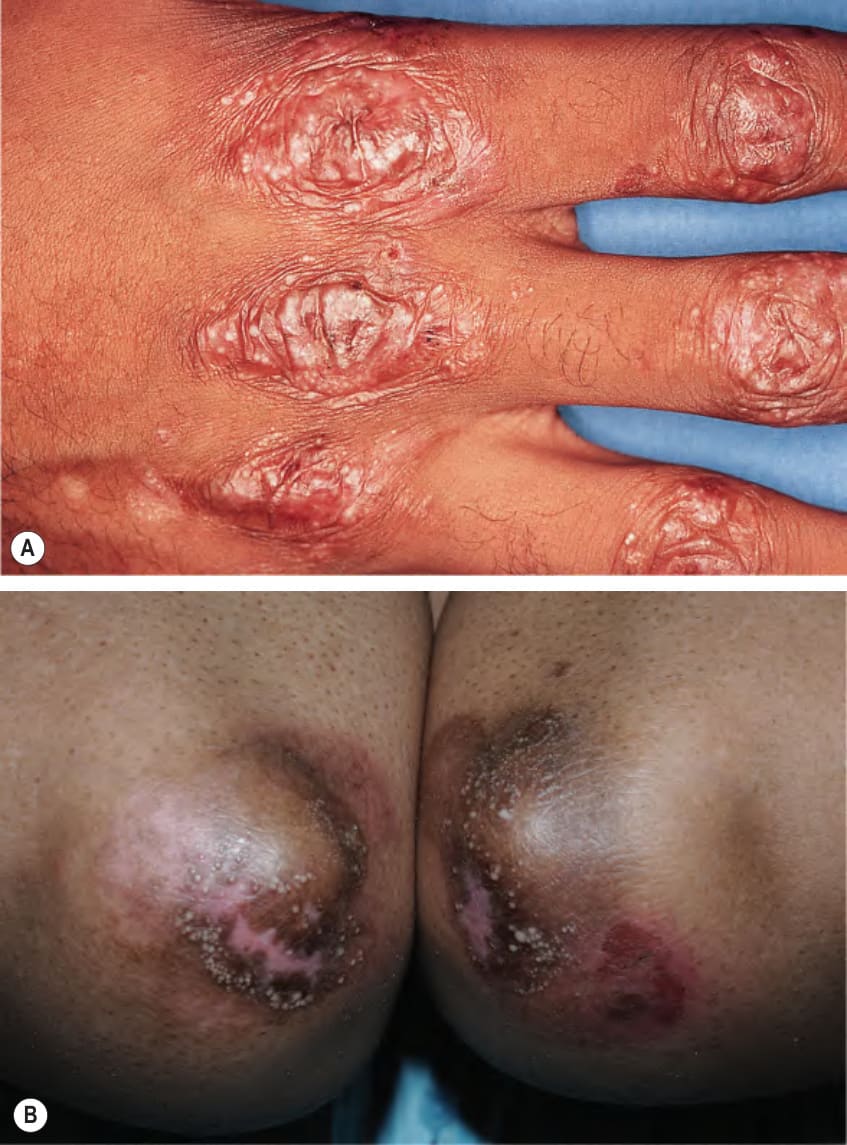
Fig. 30.18 Epidermolysis bullosa acquisita – mechanobullous presentation.
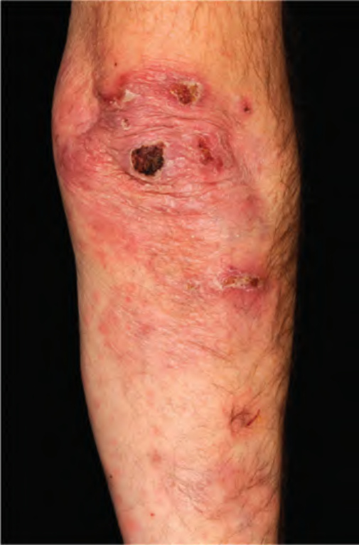
Fig. 30.19 Epidermolysis bullosa aquisita – inflammatory presentation.
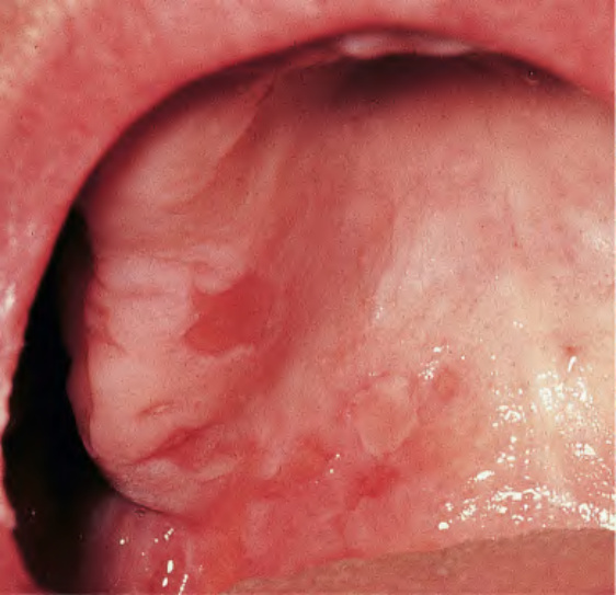
Fig. 30.20 Epidermolysis bullosa acquisita – oral involvement. Multiple erosions of the palate reminiscent of mucous membrane (cicatricial) pemphigoid. Courtesy C. Prost, MD.
(e.g. porphyria variegata). Clinically, the mechanobullous type of EBA most closely resembles mild dominant dystrophic EB, but the lack of a family history, late onset, and the positive DIF findings generally allow for distinction. However, some patients with inherited EB do develop autoantibodies against type VII collagen. The involvement of the hands may mimic porphyria cutanea tarda, but the latter can easily be excluded by porphyrin studies. Occasionally, patients with EBA on incriminating medications are misdiagnosed as having pseudoporphyria. The inflammatory type of EBA is clinically indistinguishable from BP, anti-p200 (anti-laminin γ1) pemphigoid, or MMP, including the Brunsting–Perry variant. These conditions may be distinguished by u-serrated versus n-serrated DIF pattern analysis, FOAM technique, ELISA, IIF with salt-split skin as a substrate, Western immunoblot analysis of both epidermal and dermal extracts, and direct immunoelectron microscopy studies.
Bullous systemic LE usually presents as a transient, widespread, inflammatory subepidermal blistering condition (see Ch. 41), and, less frequently, as a mechanobullous eruption with milia formation and scarring. In a number of these cases, circulating autoantibodies show binding to type VII collagen and the possibility of systemic LE-associated EBA could be considered. In addition to differences in histology, adult EBA exhibits a poor response to treatment, while a significant improvement following initiation of dapsone or azathioprine therapy is generally seen in patients with bullous systemic LE.
Treatment of EBA is difficult and often unsatisfactory. Because of the rarity of the condition, most data on treatment are based on anecdotal reports. Systemic corticosteroids and immunosuppressive agents, such as azathioprine, methotrexate, mycophenolate mofetil, cyclophosphamide or, more recently, rituximab, are sometimes helpful in controlling the BP-like variant of EBA, which may eventually go into clinical remission. Colchicine, dapsone, and cyclosporine have also been reported to have some benefit. In patients with severe EBA unresponsive to conventional immunosuppressive therapy, IVIg ± rituximab may prove effective. A combination of dapsone plus prednisolone has been recommended by some authors for the treatment of childhood EBA.