PATHOGENESIS
EB is caused by mutations within genes that encode structural proteins that reside within the epidermis (EB simplex), dermal–epidermal junction (junctional EB), or uppermost papillary dermis (dystrophic EB). The site within which each of these proteins resides determines the ultrastructural location where the blisters arise (Fig. 32.1, see Table 32.1).
EB Simplex
The most common forms of EB simplex (EBS) are transmitted in an autosomal dominant manner and are associated with blistering at the level of the basal keratinocyte. EBS most often results from a dominant-negative mutation (see Ch. 54) within the keratin 5 (KRT5) or 14 (KRT14) genes, expression of which is primarily within the basal layer of the epidermis. The clinical severity and other phenotypic features of EBS are closely associated with the genotype. For example, mutations in the helix initiation and termination motifs of KRT5 and KRT14 lead to the severe subtype of EBS (severe EBS, previously known as EBS-Dowling–Meara), whereas the specific phenotype of EBS with mottled pigmentation almost always arises from a particular missense mutation in the V1 domain of KRT5.
Dominant stabilizing mutations in the kelch-like protein 24 ubiquitin ligase gene (KLHL24) lead to EBS via increased ubiquitination and degradation of keratin 14. An autosomal recessive form of EBS due to mutations in the gene encoding plectin (PLEC) is associated with muscular dystrophy, which is not surprising considering that plectin is expressed in skeletal muscle as well as in the hemidesmosomes of basilar keratinocytes. Other EBS patients with plectin or rarely αβ integrin deficiencies present with pyloric atresia, while autosomal recessive variants of EBS caused by mutations in bullous pemphigoid antigen 1/dystonin (DYS) or exophilin 5 (EXPH5) genes have also been reported.
Junctional EB
Junctional EB (JEB) is almost always transmitted in an autosomal recessive manner (see Table 32.1). The severe subtype of JEB (severe JEB,
previously known as JEB-Herlitz) typically results from homozygous or compound heterozygous truncating mutations within a gene encoding one of the three subunits of laminin 332, a key component of the lamina lucida of the dermal–epidermal junction (see Ch. 28). In laryngo-onycho-cutaneous syndrome, the underlying mutations affect only the A isoform of the laminin α3 subunit. The milder intermediate form of JEB (intermediate JEB, previously known as JEB-non-Herlitz and generalized atrophic benign EB) results from mutations within the genes for either a subunit of laminin 332 or, less frequently, type XVII collagen/bullous pemphigoid antigen 2. JEB with pyloric atresia, which is much more common than EBS with pyloric atresia, is also caused by mutations within either of the two genes that encode the subunits of αβ integrin. A form of JEB associated with respiratory and renal involvement results from mutations in the integrin α chain.
Dystrophic EB
Dystrophic EB (DEB) is transmitted in either an autosomal dominant or autosomal recessive manner and is caused by mutations in the type VII collagen gene (COL7A1). Dominant DEB (DDEB) results from dominant-negative mutations, typically a missense mutation that leads to substitution of another amino acid for a glycine within the triplehelical domain of this collagen. Although the resultant protein is structurally abnormal, immunohistochemical staining for type VII collagen along the dermal–epidermal junction is usually indistinguishable from that of normal skin.
Recessive DEB (RDEB) is due to compound heterozygous or, less frequently, homozygous mutations within the type VII collagen gene. Premature stop codons, which result in truncated proteins, are
(EB).A Ultrastructural sites of blister formation in three major subtypes of EB simplex (EBS). The ultrastructure of normal intact skin is presented for comparison. In all three major subtypes of EBS – localized, intermediate, and severe – blisters arise within the lowermost intracytoplasmic portion of the basilar keratinocyte. In severe EBS, keratin filaments tend to coalesce into larger electron-dense clumps, especially in lesional skin. Keratin filaments are absent or reduced in the rare autosomal recessive form of EBS, and in EBS with muscular dystrophy there is lack of integration of keratin filaments into the underlying hemidesmosome. B Ultrastructural sites of blister formation in two major subtypes of junctional EB (JEB). All forms of JEB are characterized by blister formation within the lamina lucida of the dermal–epidermal junction. As such, the lamina densa remains firmly attached to the dermis which forms the base of the blister cavity. Sub-basal dense plates and anchoring filaments are absent in generalized severe JEB and often attenuated in other subtypes of JEB. Hemidesmosomes are absent or extremely sparse and rudimentary in severe JEB, whereas they are normal or reduced in number and size in intermediate JEB. C Ultrastructural sites of blister formation in three major subtypes of dystrophic EB (DEB). Blister formation occurs beneath the lamina densa in all forms of dystrophic EB. Anchoring fibrils appear normal in size and structure in dominant DEB (DDEB) skin, although they may be somewhat reduced in number. Consistent with the presence of sublamina densa cleavage, these anchoring fibrils remain attached to the blister roof, which is composed of intact epidermis and an attached basement membrane that includes the lamina densa. In contrast, anchoring fibrils are usually completely absent in lesional skin from patients with severe recessive DEB (RDEB). In intermediate RDEB, anchoring fibrils are reduced in number and rudimentary in appearance.
characteristic of the severe subtype (severe RDEB, previously known as RDEB-Hallopeau–Siemens). Consistent with the severity of these mutations, anchoring fibrils are undetectable or extremely sparse and poorly formed in skin biopsy specimens, and immunohistochemical staining with antibodies against the major epitopes of the type VII collagen molecule is absent or barely detectable. Milder forms of RDEB are associated with less severe biallelic mutations in the type VII collagen gene.
A single nucleotide polymorphism in the matrix metalloproteinase 1 gene promoter has been identified as a disease modifier in RDEB. It is likely that additional modifying genes account for some of the inter-and intra-familial phenotypic variability observed in this and other forms of EB. There is also evidence for secondary effects from loss of protein expression in EB. For example, loss of collagen VII in RDEB fibroblasts leads to alterations in dermal matrix proteins, metalloproteinases, and transforming growth factor-β (TGF-β), which are thought to further influence keratinocyte adhesion, dermal–epidermal integrity, and dermal fibrosis.
Severe RDEB and (to a lesser extent) other forms of DEB, JEB, and Kindler EB are associated with an increased risk for the development of cutaneous SCCs. The pathogenesis of these tumors is multifactorial, and the cutaneous microenvironment plays a critical role in the initiation of carcinogenesis, tumor progression, and metastatic potential. Increased dermal stiffness, bacterial colonization, chronic inflammation, and defects in the innate immune system contribute to the early development and aggressive nature of these tumors.
In self-improving DEB (previously known as bullous dermolysis of the newborn), inheritance is usually dominant and blistering is typically confined to the first 1–2 years of life. Clinical expression coincides with a time during which type VII collagen is present primarily within basal keratinocytes in these patients’ skin, rather than along the dermal–epidermal junction (see Fig. 32.1D). This suggests that there may be a temporary disruption in the transport of this protein from the keratinocyte cytoplasm to the underlying ECM.
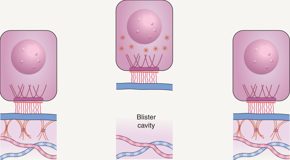
Fig. 32.1 Blister formation in epidermolysis bullosa
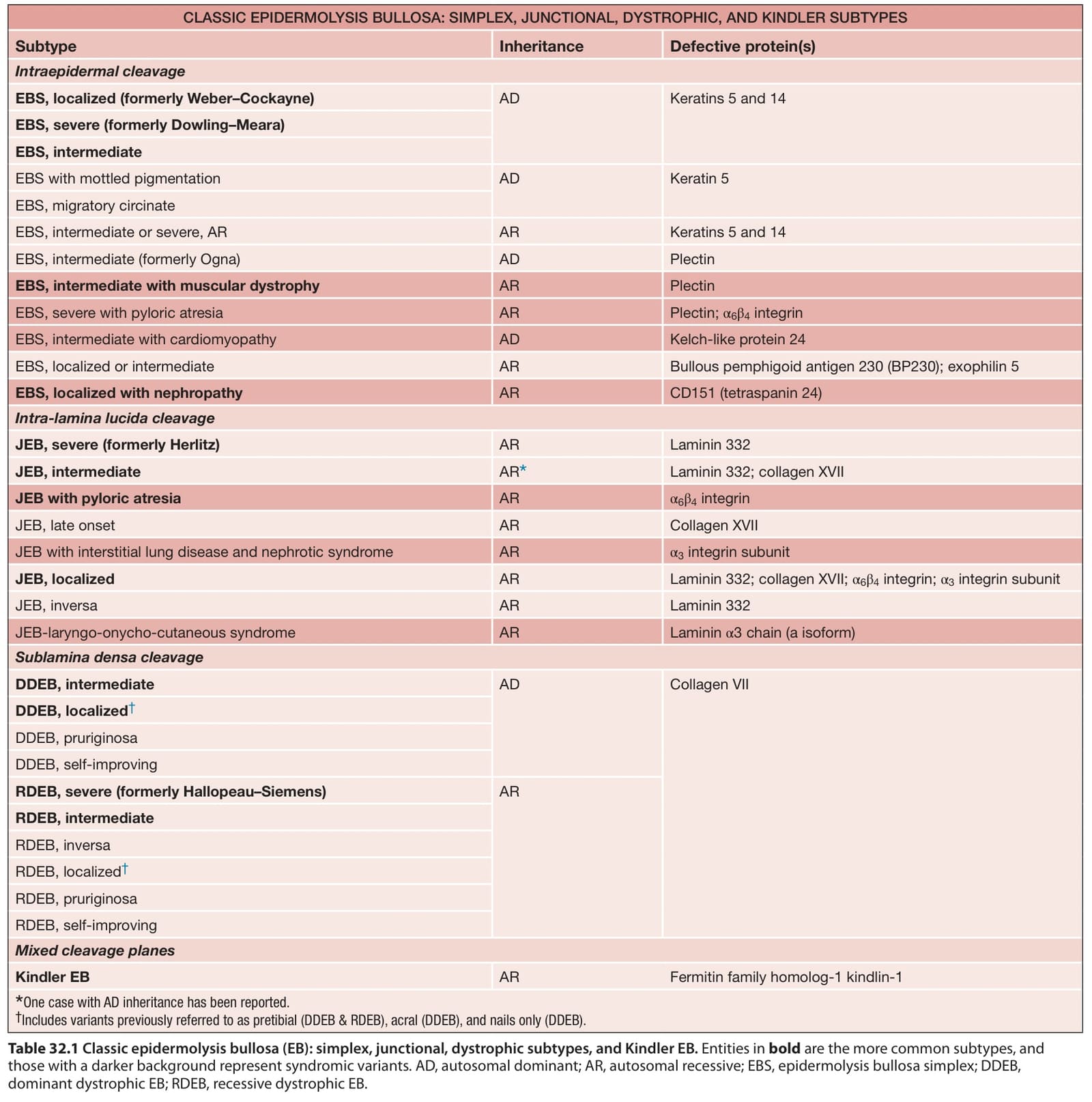
Table 32.1 Classic epidermolysis bullosa (EB): simplex, junctional, dystrophic subtypes, and Kindler EB. Entities in bold are the more common subtypes, and those with a darker background represent syndromic variants. AD, autosomal dominant; AR, autosomal recessive; EBS, epidermolysis bullosa simplex; DDEB, dominant dystrophic EB; RDEB, recessive dystrophic EB.