CLINICAL FEATURES
Cutaneous Findings
All forms of inherited EB are characterized by mechanically fragile skin, erosions, and (with rare exceptions) macroscopic blisters (Figs. 32.2 & 32.3). Scarring is almost always atrophic and can occur in any subtype of EB (Fig. 32.4), including localized EBS. However, scarring is most frequent in the subtypes that are characterized clinically by generalized disease activity and ultrastructurally by disruption of the basement
membrane, particularly the lamina densa (Fig. 32.5). Whereas scarring is estimated to occur in only 15% of patients with localized EBS, it is present in essentially every patient with RDEB. Other cutaneous findings that have similar variations in frequency across the major types and subtypes of EB include dystrophic or absent nails (see Fig. 32.4), milia, and scarring alopecia of the scalp.
Some cutaneous findings have diagnostic implications (Fig. 32.6). Reticulated hyperpigmented macules distinguish a rare subtype of EBS termed EBS with mottled pigmentation. Grouped (“herpetiform”) blisters, often in an arcuate (Fig. 32.7) or polycyclic array, are highly characteristic of severe EBS, which is also associated with the gradual development of a diffuse palmoplantar keratoderma (Fig. 32.8). Migratory circinate erythema with vesiculation of the advancing edge has been described in patients with EBS caused by a frameshift mutation that leads to an elongated keratin 5 protein. Excessive or exuberant granulation tissue, usually in a symmetric distribution involving the periorificial areas, skin folds, upper back, nape of the neck, and periungual areas, is typical of severe JEB (Fig. 32.9). Extremely pruritic papules that can coalesce into a linear arrangement on the lower extremities, are characteristic of DEB pruriginosa (Fig. 32.10).
Distribution of skin disease activity is also useful in the subclassification of EB, although the pattern of involvement tends to be less distinctive in infants than in adults. Patients with the “inversa” subtypes of JEB and RDEB experience severe disease activity primarily in intertriginous areas such as the axillae and inguinal creases. In contrast, patients with localized JEB have involvement mainly in acral sites. Individuals with localized DEB may have lesions almost exclusively on the shins (previously referred to as pretibial DEB). A rare
subtype previously known as RDEB centripetalis initially features acral blistering, followed by slow progression of disease activity toward the trunk over the years.
Extracutaneous Findings
The molecular defects that affect the skin in patients with EB may also lead to manifestations in other tissues with an epithelial lining or surface, including the eye, oral cavity, and gastrointestinal, genitourinary, and respiratory tracts. Major extracutaneous complications of EB are summarized in Table 32.2. Although exceptions exist, extracutaneous involvement occurs most frequently in RDEB and JEB, and it can result in blisters, erosions, ulcers, and scarring. Rare subtypes of JEB and EBS present at birth with pyloric atresia as well as skin fragility and blistering. In other forms of EB, extracutaneous disease may become apparent as early as the first few months of life. Repeated blistering of the external eye can result in neovascularization and blindness. Chronic involvement of the esophagus leads to scarring, stricture formation and, rarely, even complete obstruction. Involvement of the small intestine presents with chronic malabsorption, whereas disease activity within the large intestine tends to produce constipation and painful anal fissures or strictures. Recurrent genitourinary tract blistering may result in urethral or ureterovesical
strictures; if persistent, the latter may eventuate in ureteric reflux and hydronephrosis. Tracheolaryngeal blistering and associated soft tissue edema, seen most often in infants and young children with severe JEB, may lead to potentially fatal acute airway obstruction. JEB with respiratory and renal involvement is a rare subtype associated with severe interstitial lung disease.
Dental enamel hypoplasia, which occurs exclusively in JEB, is associated with pitting of the surfaces of primary and permanent
EBS, EB simplex; GERD, gastroesophageal reflux disease; intermed, intermediate; sev, severe; ILD, interstitial lung disease; inv, inversa; JEB, junctional EB; MD, muscular dystrophy; PA, pyloric atresia; RDEB, recessive dystrophic EB.
teeth. If untreated, affected individuals lose teeth during childhood due to excessive caries. Severe caries and tooth loss also occur in severe RDEB, likely resulting from impaired clearance of food from the mouth and poor dental hygiene in the setting of intraoral injury and scarring, ankyloglossia, and microstomia.
Pseudosyndactyly (“mitten” deformities) of the hands and feet primarily affects patients with RDEB, especially severe RDEB, although it occasionally occurs to a lesser degree in DDEB and JEB (Fig. 32.11B & C). Initially presenting as proximal web formation between adjacent digits, the digits may eventually become totally fused and
encased by scar tissue. Lack of mobility leads to progressive bone resorption and muscular atrophy, and hand function is severely compromised.
Osteoporosis, detectable by dual-emission X-ray absorptiometry (DEXA) scanning, is common in severe RDEB and severe JEB. Radiographs may demonstrate vertebral crush fractures in severe cases. EBS due to plectin deficiency is associated with mild to severe muscular
dystrophy. Although the muscle disease presents during infancy in some patients, weakness often develops insidiously during later childhood or even early adulthood in those who are less severely affected.
Chronic renal failure occasionally develops in patients with severe forms of EB, most notably severe RDEB, which is associated with a ~10% risk of death from renal failure by age 35 years. Renal disease may result from untreated outflow obstruction, glomerulonephritis, secondary systemic amyloidosis, or IgA nephropathy. Nephrotic syndrome associated with altered expression of laminin isoforms in renal basement membranes has been reported in an infant with generalized JEB and it occurs congenitally as a result of integrin α mutations in JEB with respiratory and renal involvement. A small subset of patients with severe forms of EB, especially RDEB, develop potentially fatal dilated cardiomyopathy. Although as yet unproven, selenium or carnitine deficiency may be a contributing factor.
Although it was a common occurrence several decades ago, potentially lethal bacterial sepsis is now relatively rare in inherited EB, presumably due to improved wound care and the availability and judicious use of broad-spectrum antibiotics. When sepsis does occur in EB, it tends to affect infants with severe disease. In contrast, failure to thrive is still common among infants with severe JEB and may lead to death.
Cutaneous Malignancies
A major complication, usually of RDEB, is the development of multiple cutaneous SCCs. These tumors most often arise in chronic non-healing wounds or hyperkeratotic lesions (Fig. 32.12). Histologically, they are usually well differentiated. However, the borders of the lesions are often indistinct and they are difficult to completely excise, with a tendency to recur locally. In addition, SCCs in EB patients frequently metastasize and are strikingly unresponsive to chemotherapy or radiotherapy. Indeed, they represent the leading cause of death in EB at or after mid-adolescence, with death from an SCC of cutaneous origin occurring in most patients within 5 years of the diagnosis of their first SCC. These tumors occur primarily in RDEB, especially when severe, although SCCs can also develop in adults with other forms of DEB, JEB, and Kindler EB. The cumulative risk of at least one SCC in patients with severe RDEB is 7.5% by age 20 years but rises to 68%, 80%, and 90% by ages 35, 45, and 55 years, respectively. In contrast, the risk of SCC in other forms of RDEB is <25% by age 45 years.
Melanoma occurs in a small number of children with severe RDEB, with a cumulative risk of 2.5% by the age of 12 years. Although this is a relatively infrequent complication, careful surveillance during childhood is indicated. Children with EB, in particular those with inter-mediate JEB, can develop large, irregularly shaped, darkly pigmented melanocytic nevi (“EB nevi”) (Fig. 32.13) that clinically resemble melanoma, although they are histologically and biologically benign. A large size from the onset rather than a relatively slow horizontal expansion is a clue to the diagnosis of these EB-associated nevi.
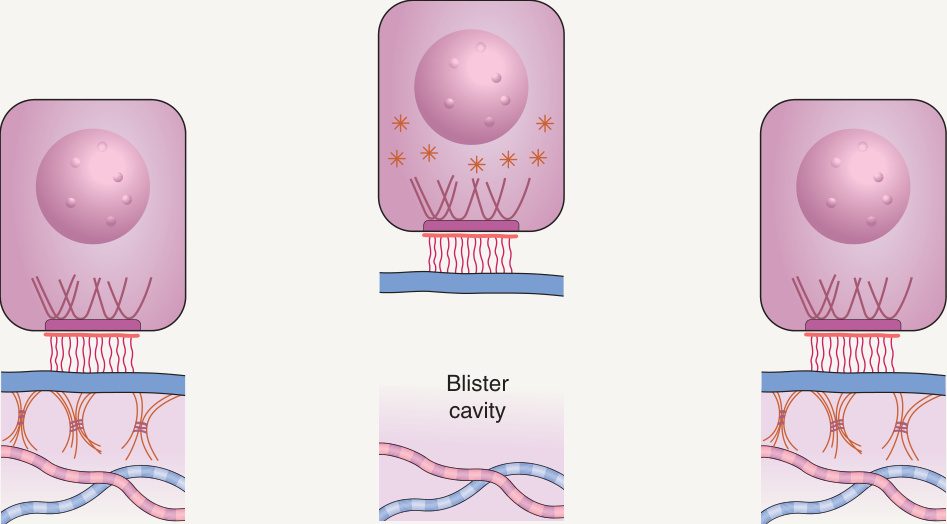
Fig. 32.1 Blister formation in epidermolysis bullosa
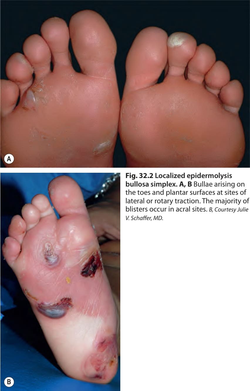
Fig. 32.2 Localized epidermolysis bullosa simplex.A, B Bullae arising on the toes and plantar surfaces at sites of lateral or rotary traction. The majority of blisters occur in acral sites. B, Courtesy Julie V. Schaffer, MD.
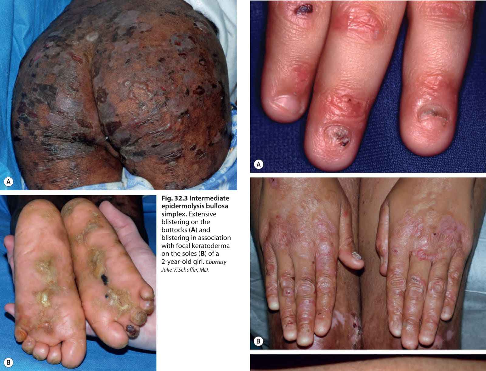
Fig. 32.3 Intermediate epidermolysis bullosa simplex. Extensive blistering on the buttocks (A) and blistering in association with focal keratoderma on the soles (B) of a 2-year-old girl. Courtesy Julie V. Schaffer, MD.
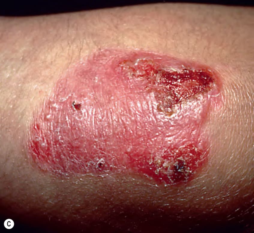
Fig. 32.4 Dominant dystrophic epidermolysis bullosa.A, B Erosions, scarring and milia on the fingers. Note the partial (A) to almost complete (B) loss of the nails. C A discrete area of scarring on the elbow with blisters, crusting, and milia. B, Courtesy Julie V. Schaffer, MD.
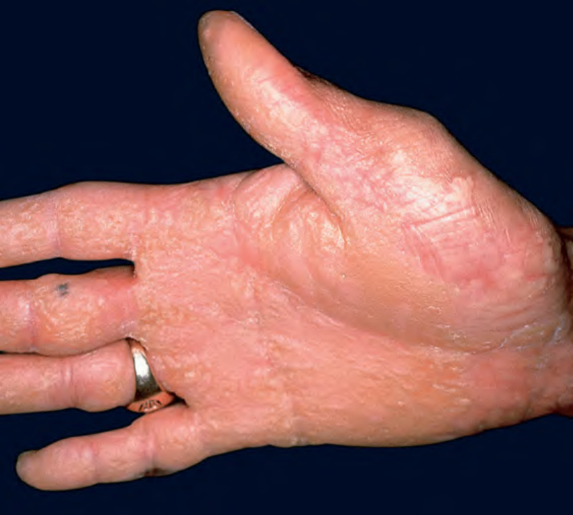
Fig. 32.5 Frequency of selected cutaneous findings within each major subtype of inherited epidermolysis bullosa (EB). Note the increasing frequency from localized EB simplex (EBS) to recessive dystrophic EB (RDEB). JEB, junctional EB; DDEB, dominant dystrophic EB.
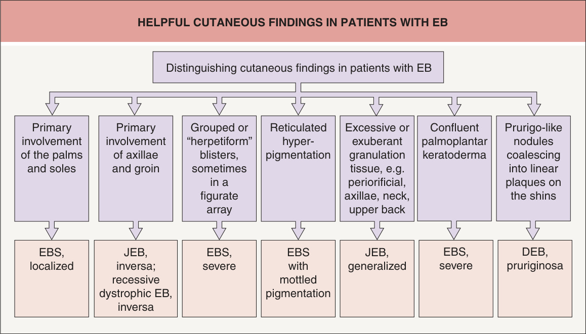
Fig. 32.6 Helpful cutaneous findings in patients with epidermolysis bullosa (EB). DEB, dystrophic EB; EBS, EB simplex; JEB, junctional EB.
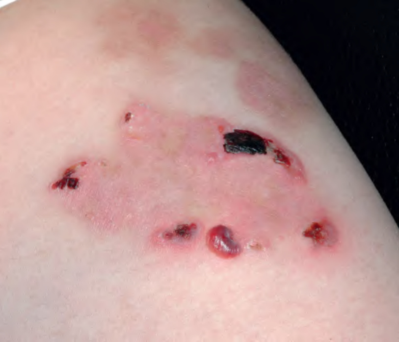
Fig. 32.7 Severe epidermolysis bullosa simplex. Clustered vesicles in an arcuate array on the thigh of this child. Courtesy Julie V. Schaffer, MD.
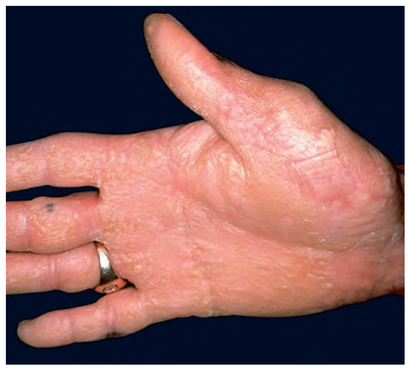
Fig. 32.8 Severe epidermolysis bullosa simplex. Diffuse palmar keratoderma in an adult.
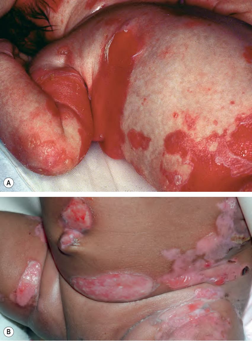
Fig. 32.9 Severe junctional epidermolysis bullosa.A Blisters on the elbow and large areas of denuded skin; note the bright red color in the axilla and groin. B Blisters and large erosions on the abdomen of an infant. B, Courtesy Julie V. Schaffer, MD.
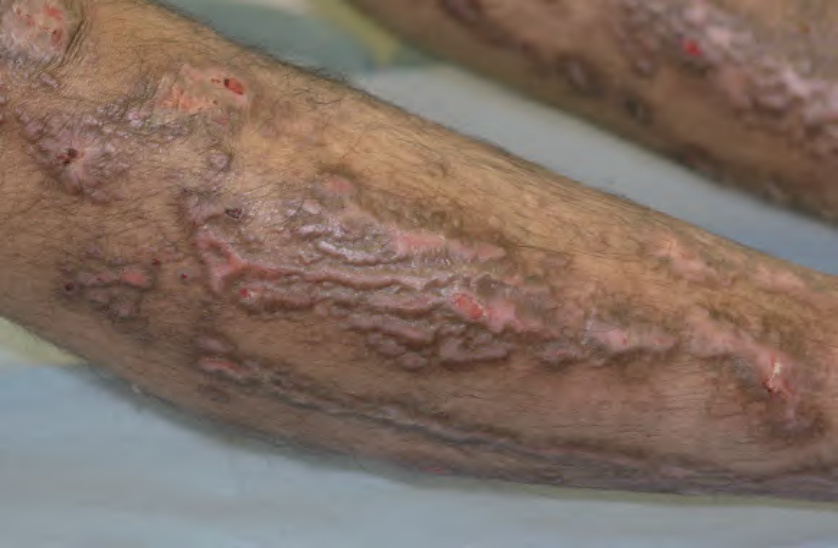
Fig. 32.10 Dystrophic epidermolysis bullosa pruriginosa. Prurigo-like papulonodules coalescing into linear plaques on the shin as a consequence of chronic scratching.
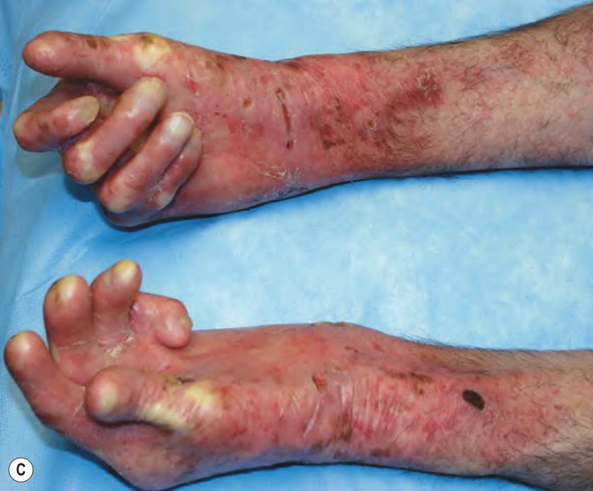
Fig. 32.11 Severe recessive dystrophic epidermolysis bullosa.A Area on the neck with chronic and recurrent wounds in a 7-year-old child. B Early proximal interdigital web formation together with atrophic scarring and loss of nails in a 5-year-old girl. C Partial “mitten” deformities of the hands in an older child. A, B, Courtesy Julie V. Schaffer, MD.
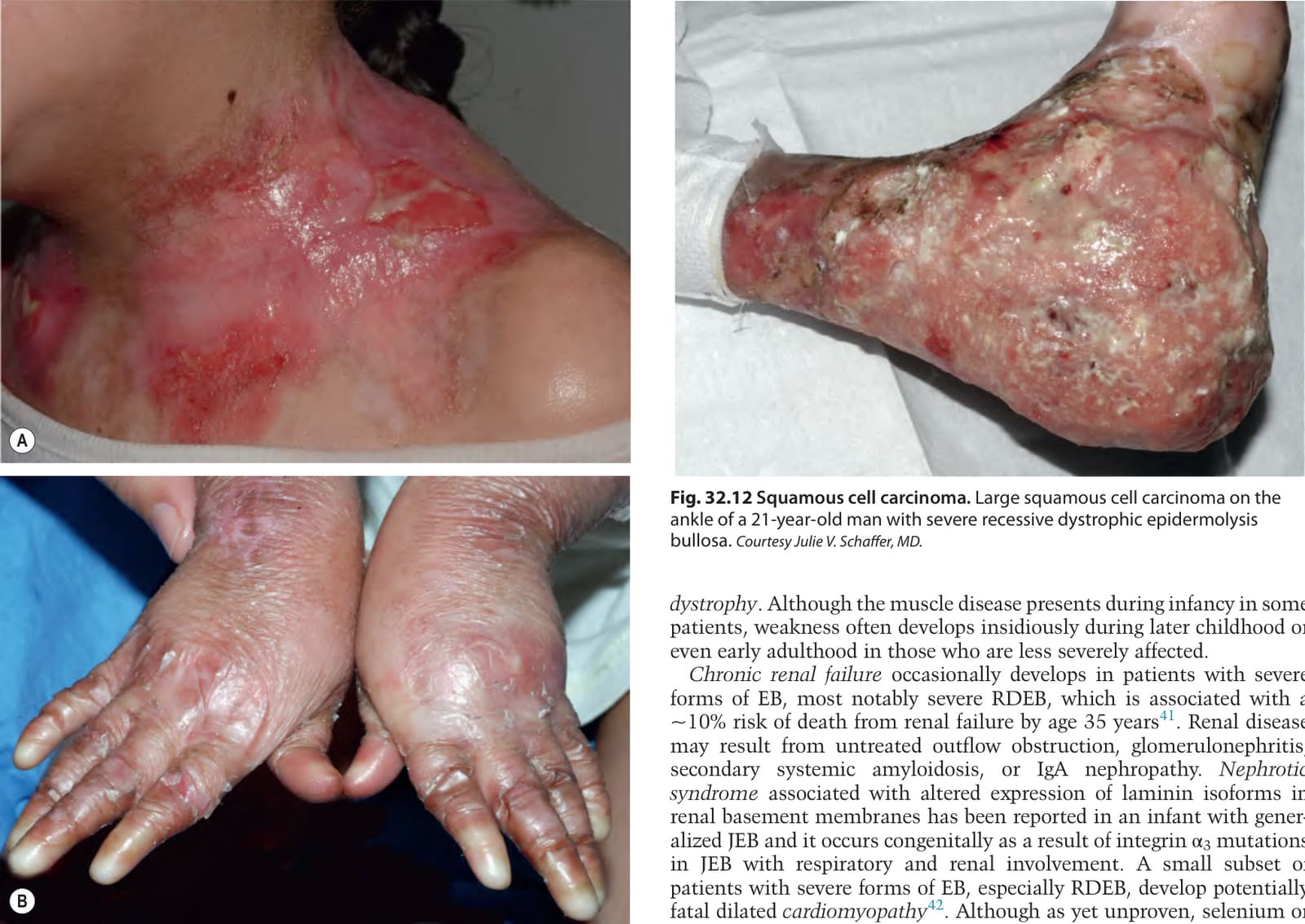
Fig. 32.12 Squamous cell carcinoma. Large squamous cell carcinoma on the ankle of a 21-year-old man with severe recessive dystrophic epidermolysis bullosa. Courtesy Julie V. Schaffer, MD.
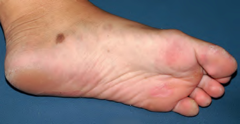
Fig. 32.13 Large acquired melanocytic nevus at a site of blistering in a teenage girl with junctional epidermolysis bullosa.Courtesy Julie V. Schaffer, MD.
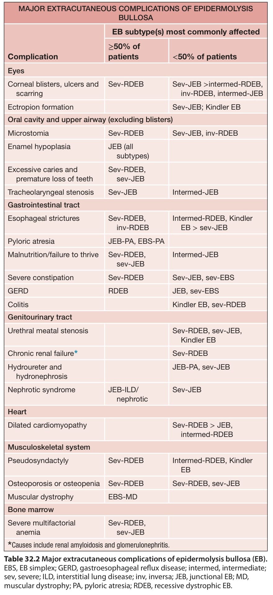
Table 32.2 Major extracutaneous complications of epidermolysis bullosa (EB).