APPROACH TO DIAGNOSIS
Patients with EB are classified on the basis of the clinical phenotype, mode of inheritance, ultrastructural level of blister formation, antigenic profile in the skin, and underlying genetic defect. Although skin
findings play an important role in determination of the EB phenotype, different subtypes of EB have considerable overlap in their cutaneous manifestations. In addition, the severity, distribution, and type of skin findings in an individual patient may change over time, such as evolution from generalized to localized blistering (or the opposite) or delayed development of sequelae such as exuberant granulation tissue, scarring, and nail dystrophy.
Standard light microscopy has no role in the diagnosis of classic forms of inherited EB, since it may be difficult to distinguish between even lower intraepidermal and subepidermal blister formation, let alone between intra-lamina lucida (i.e. JEB) and sublamina densa (i.e. DEB) types, without more sophisticated staining techniques. The diagnostic tests employed for EB include immunofluorescence antigen mapping (IFM), transmission electron microscopy (TEM), and increasingly genetic analysis. TEM can define the ultrastructural level of blister formation and provide a quantitative and qualitative assessment of specific structures, such as basilar tonofilaments, hemidesmosomes, sub-basal dense plates, anchoring filaments, and anchoring fibrils; however, currently it is routinely performed in only a few reference laboratories. A diagnostic algorithm is presented in Fig. 32.14.
Genetic analysis has become the first-line method for EB diagnosis and classification, with increasing availability and decreasing cost of massively parallel (next-generation) sequencing panels that include all known EB genes. Molecular diagnosis allows more accurate genetic counseling and is required for DNA-based prenatal/preimplantation testing.
Differential Diagnosis
Although it is difficult to determine the subtype of inherited EB on the basis of clinical findings alone, the diagnosis of EB itself is usually straightforward, especially beyond early childhood. There is a limited differential diagnosis for a chronic mechanobullous disease with early onset and/or a positive family history. Other genetic diseases that may present with blistering early in life include acantholytic erosive disorder and other skin fragility syndromes due to defects in desmosomal proteins as well as peeling skin syndromes and epidermolytic ichthyosis (see Ch. 57) (Fig. 32.15A,B). Of note, the former disorders were previously categorized as forms of EBS. Genodermatoses that can be associated, at least transiently, with skin fragility or blisters and therefore potentially misdiagnosed as EB are outlined in Table 32.3. Less often, EB may be confused with acrodermatitis enteropathica, incontinentia pigmenti stage 1, pachyonychia congenita, or early lipoid proteinosis.
The differential diagnosis of blisters and erosions in neonates or young infants may also include infectious diseases (e.g. herpes simplex viral infection, staphylococcal scalded skin syndrome, bullous impetigo), sucking blisters, mastocytosis, autoimmune blistering diseases, and congenital erosive and vesicular dermatosis (see Ch. 34). Diagnostic considerations in patients with acral blistering, erosions, and ulcerations with progression to digital resorption may include congenital erythropoietic porphyria (see Table 32.3 and Ch. 49) and SAVI (STING-associated vasculopathy with onset in infancy [Fig. 32.15C]; see Ch. 45).
Congenital localized absence of skin (CLAS), also referred to as aplasia cutis congenita, can occur in any form of EB (see Ch. 64). In patients with EB, CLAS is distinguished by the associated skin fragility and blistering as well as its frequent location on the lower extremities (referred to as Bart syndrome), although involvement may be more widespread, especially in forms of EBS or JEB associated with pyloric atresia.
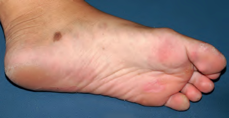
Fig. 32.13 Large acquired melanocytic nevus at a site of blistering in a teenage girl with junctional epidermolysis bullosa.Courtesy Julie V. Schaffer, MD.
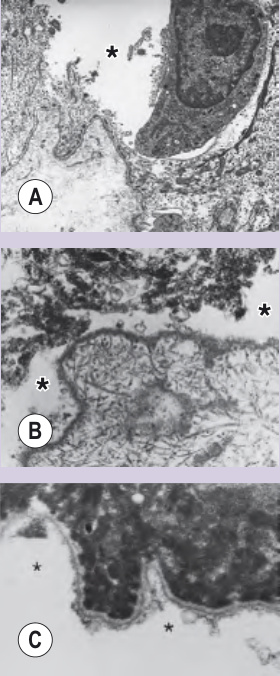
Fig. 32.14 Approach to the laboratory diagnosis of epidermolysis bullosa (EB).A Electron microscopy (EM) of an induced blister in localized EBS demonstrates skin cleavage (asterisk) within the inferior-most portion of the basilar keratinocyte. B EM of a spontaneous blister from a patient with the severe JEB reveals skin cleavage (asterisks) within the lamina lucida. Hemidesmosomes, sub-basal dense plates, and anchoring filaments are all absent. In contrast, anchoring fibrils are still present in normal amounts within the underlying dermis. C EM of the roof of a spontaneous blister from a patient with severe RDEB reveals cleavage (asterisks) beneath the level of the lamina densa. Anchoring fibrils are absent along the epidermal roof of the blister. BM, basement membrane; DDEB, dominant dystrophic EB; EBS, EB simplex; JEB, junctional EB; RDEB, recessive dystrophic EB.
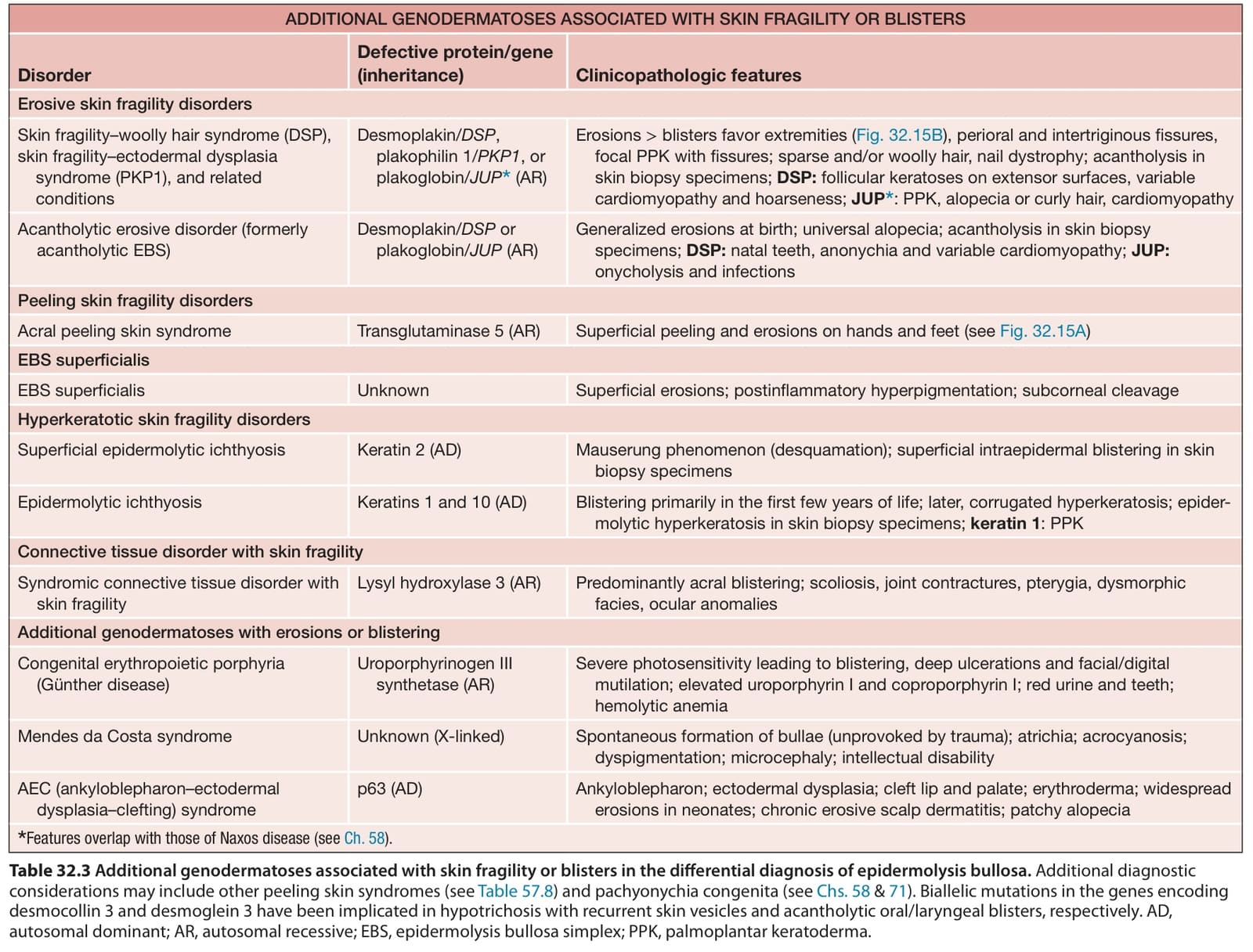
Table 32.3 Additional genodermatoses associated with skin fragility or blisters in the differential diagnosis of epidermolysis bullosa. Additional diagnostic considerations may include other peeling skin syndromes (see Table 57.8) and pachyonychia congenita (see Chs. 58 & 71). Biallelic mutations in the genes encoding desmocollin 3 and desmoglein 3 have been implicated in hypotrichosis with recurrent skin vesicles and acantholytic oral/laryngeal blisters, respectively. AD, autosomal dominant; AR, autosomal recessive; EBS, epidermolysis bullosa simplex; PPK, palmoplantar keratoderma.