診斷方法(APPROACH TO DIAGNOSIS)
EB 病人依臨床表現型、遺傳模式、水疱形成的超微結構層次、皮膚中的抗原輪廓,以及潛在的基因缺陷來分類。雖然皮膚
表現在判定 EB 表現型上扮演重要角色,不同 EB 亞型在皮膚表現上有相當多的重疊。此外,個別病人皮膚表現的嚴重度、分布與型態可能隨時間改變,例如由全身性水疱形成演變為局部性(或反之),或延遲出現後遺症,如旺盛的肉芽組織、疤痕與指甲失養。
標準光學顯微鏡在典型遺傳性 EB 的診斷中毫無角色,因為若無更精密的染色技術,即使要區分較低位的表皮內水疱與表皮下水疱形成都可能有困難,更遑論區分透明板內(即 JEB)與緻密板下(即 DEB)型。用於 EB 的診斷檢驗包括免疫螢光抗原定位(immunofluorescence antigen mapping, IFM)、穿透式電子顯微鏡(transmission electron microscopy, TEM),以及日益普及的基因分析。TEM 可界定水疱形成的超微結構層次,並對特定結構(如基底 tonofilaments、半橋粒、基底下緻密斑、錨定絲與錨定纖維)提供量性與質性評估;然而目前僅有少數參考實驗室常規執行此項檢查。診斷演算法呈現於圖 32.14。
基因分析已成為 EB 診斷與分類的第一線方法,因為涵蓋所有已知 EB 基因的大規模平行(次世代)定序套組其可得性日增且成本下降。分子診斷可提供更準確的遺傳諮詢,且為以 DNA 為基礎的產前/著床前檢測所必需。
鑑別診斷(Differential Diagnosis)
雖然單憑臨床表現要判定遺傳性 EB 的亞型有其困難,EB 本身的診斷通常相當直接,特別是在幼兒期之後。對於發病早期且/或有陽性家族史的慢性機械性水疱病,其鑑別診斷有限。其他可能在生命早期以水疱形成表現的遺傳性疾病,包括 acantholytic erosive disorder 與其他因橋粒蛋白缺陷所致的皮膚脆弱症候群,以及 peeling skin syndromes 與 epidermolytic ichthyosis(見第 57 章)(圖 32.15A、B)。值得注意的是,前述疾病先前曾被歸類為 EBS 的型別。可能至少暫時性地與皮膚脆弱或水疱相關、因而可能被誤診為 EB 的遺傳性皮膚病,概述於表 32.3。較少見的情況下,EB 可能與 acrodermatitis enteropathica、incontinentia pigmenti stage 1、pachyonychia congenita 或早期的 lipoid proteinosis 混淆。
新生兒或幼小嬰兒水疱與糜爛的鑑別診斷,也可能包括感染性疾病(例如單純疱疹病毒感染、staphylococcal scalded skin syndrome、bullous impetigo)、吸吮性水疱(sucking blisters)、肥大細胞增生症、自體免疫水疱病,以及 congenital erosive and vesicular dermatosis(見第 34 章)。對於出現肢端水疱、糜爛與潰瘍並進展至指(趾)吸收的病人,診斷考量可能包括 congenital erythropoietic porphyria(見表 32.3 與第 49 章)與 SAVI(STING-associated vasculopathy with onset in infancy[圖 32.15C];見第 45 章)。
先天性局部皮膚缺失(congenital localized absence of skin, CLAS),亦稱為 aplasia cutis congenita,可發生於任何型別的 EB(見第 64 章)。在 EB 病人中,CLAS 的區辨特點在於伴隨的皮膚脆弱與水疱形成,以及其常位於下肢(稱為 Bart syndrome),儘管侵犯範圍可能更為廣泛,特別是在與幽門閉鎖相關的 EBS 或 JEB 型別中。
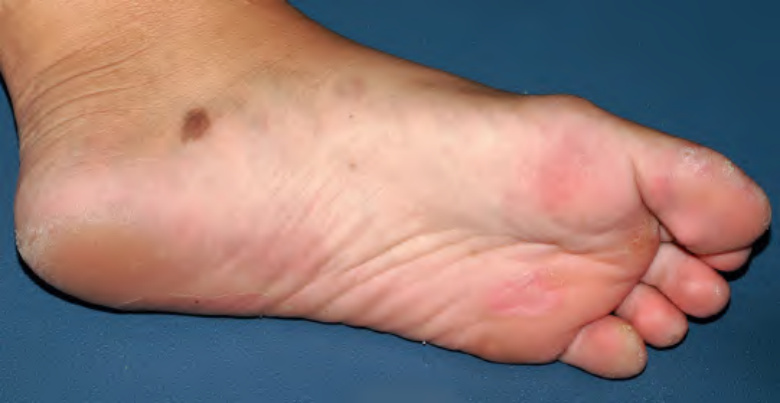
圖 32-13:一名罹患 junctional epidermolysis bullosa 的青少女,於水疱形成部位出現大型後天性黑色素細胞痣。
Fig. 32.13 Large acquired melanocytic nevus at a site of blistering in a teenage girl with junctional epidermolysis bullosa.Courtesy Julie V. Schaffer, MD.
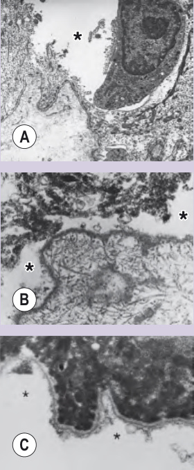
圖 32-14:水疱性表皮鬆解症(EB)實驗室診斷的方法。A localized EBS 誘發水疱的電子顯微鏡(EM)顯示皮膚裂隙(星號)位於基底 keratinocyte 的最下方部分。B 一名 severe JEB 病人自發性水疱的 EM 顯示皮膚裂隙(星號)位於透明板內。半橋粒、基底下緻密斑與錨定絲皆缺失。相對地,錨定纖維在下方真皮內仍以正常量存在。C 一名 severe RDEB 病人自發性水疱頂部的 EM 顯示裂隙(星號)位於緻密板層次以下。錨定纖維沿著水疱的表皮頂部缺失。
Fig. 32.14 Approach to the laboratory diagnosis of epidermolysis bullosa (EB).A Electron microscopy (EM) of an induced blister in localized EBS demonstrates skin cleavage (asterisk) within the inferior-most portion of the basilar keratinocyte. B EM of a spontaneous blister from a patient with the severe JEB reveals skin cleavage (asterisks) within the lamina lucida. Hemidesmosomes, sub-basal dense plates, and anchoring filaments are all absent. In contrast, anchoring fibrils are still present in normal amounts within the underlying dermis. C EM of the roof of a spontaneous blister from a patient with severe RDEB reveals cleavage (asterisks) beneath the level of the lamina densa. Anchoring fibrils are absent along the epidermal roof of the blister. BM, basement membrane; DDEB, dominant dystrophic EB; EBS, EB simplex; JEB, junctional EB; RDEB, recessive dystrophic EB.
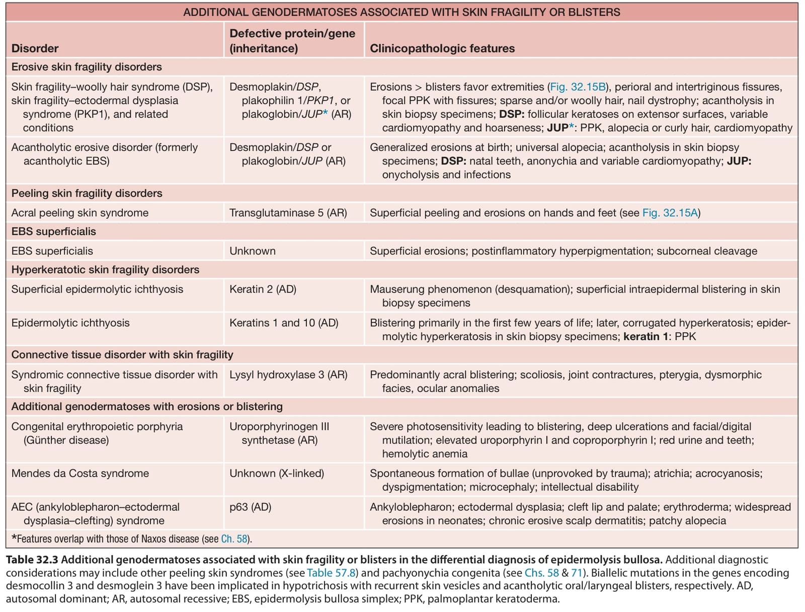
表 32-3:水疱性表皮鬆解症鑑別診斷中,其他與皮膚脆弱或水疱相關的遺傳性皮膚病。其他診斷考量可能包括其他 peeling skin syndromes(見表 57.8)與 pachyonychia congenita(見第 58 與 71 章)。編碼 desmocollin 3 與 desmoglein 3 之基因的雙等位基因突變,分別被認為與 hypotrichosis with recurrent skin vesicles 及 acantholytic oral/laryngeal blisters 有關。
Table 32.3 Additional genodermatoses associated with skin fragility or blisters in the differential diagnosis of epidermolysis bullosa. Additional diagnostic considerations may include other peeling skin syndromes (see Table 57.8) and pachyonychia congenita (see Chs. 58 & 71). Biallelic mutations in the genes encoding desmocollin 3 and desmoglein 3 have been implicated in hypotrichosis with recurrent skin vesicles and acantholytic oral/laryngeal blisters, respectively. AD, autosomal dominant; AR, autosomal recessive; EBS, epidermolysis bullosa simplex; PPK, palmoplantar keratoderma.