TREATMENT
Day-to-day management of EB revolves around the prevention of mechanical trauma, wound care, and avoidance of infection. Protective bandaging, padding over bony prominences, and soft/loosefitting clothing can be helpful. Bathing or soaking with ~0.005% sodium hypochlorite (0.5 cup household bleach [6%–8.25% sodium hypochlorite] in a full standard bathtub) or 0.25% acetic acid (1 part white vinegar [5% acetic acid] to 19 parts water) may help to reduce bacterial colonization. Antibiotics should be used judiciously, with avoidance of chronic treatment with topical mupirocin or oral antibiotics. Problematic plantar hyperhidrosis in EBS patients may be reduced by the topical application of aluminum chloride hexahydrate, although its effect on blistering is less certain, and injection of botulinum toxin A can decrease plantar blistering and associated pain in some individuals with EBS.
A number of dressings are available for cutaneous wounds in EB patients (Table 32.4; see Ch. 145). As a general rule, only those that are non-adhesive or “low-tack” should be applied to EB skin. Soft silicone dressings, some of which incorporate an absorbent foam backing, are widely used. Silver-impregnated dressings may be helpful for heavily colonized or infected wounds, although long-term application should be avoided to prevent excessive systemic silver absorption and cutaneous argyria. Less expensive Vaseline®-impregnated gauze is another suitable dressing for non-infected wounds. Topical therapies under investigation include diacerein 1% cream, which downregulates production of interleukin-1β and keratin 14, thereby stabilizing the keratinocyte intermediate filament network and reducing blister formation in patients with EBS.
Systemic drugs that may be of benefit include tetracycline or erythromycin for EBS and dupilumab, thalidomide, or cyclosporine for symptomatic relief in DEB pruriginosa. There are few reports on
the use of systemic retinoids in any form of EB, although these drugs appear to be tolerated, at least in low dosage, in patients with RDEB. Whether long-term treatment with low-dose systemic retinoids would
help to prevent the development of SCCs in severe RDEB patients remains to be determined. The angiotensin receptor blocker losartan interferes with TGF-β signaling, which is upregulated in the skin of RDEB patients and may contribute to the risk of developing SCC. Losartan was shown to reduce cutaneous fibrosis and digital fusion in a mouse model of EB and is currently under investigation in RDEB patients.
Although not yet in mainstream clinical practice, translational therapeutic approaches for EB utilizing cell-, protein- and gene-based methods have been a focus of attention in recent years. In clinical trials investigating the value of allogeneic bone marrow-derived stem cell transplantation in patients with RDEB, significant clinical responses were observed, with production of collagen VII at the dermal–epidermal junction and improved wound healing. However, there was also substantial associated morbidity and mortality, although ameliorated to some extent by using reduced-intensity conditioning regimens. An alternative approach has been to use intradermal or intravenous injections of allogeneic mesenchymal stem cells in patients with severe RDEB, which has led to improved wound healing as well as reduced inflammation and itch. Injections of allogeneic fibroblasts into RDEB wounds also improved wound healing in a small clinical trial. In addition, intradermal and intravenous delivery of recombinant collagen VII has been explored in animal models of RDEB but not yet in human studies.
Gene therapy for EB is currently under investigation in several clinical trials. It was first successfully performed in a man with intermediate JEB, with ex vivo gene correction of autologous keratinocytes to restore laminin 332. A skin equivalent was generated and grafted onto the patient’s legs, with amelioration of blistering that persisted for years post-procedure. Similar approaches utilizing ex vivo-gene correction have subsequently been employed in a child with severe JEB, with graft coverage of ~80% of his body surface area, and patients with RDEB. In vivo topical gene therapy with beremagene geperpavec (B-VEC), a non-replicating COL7A1-containing herpes simplex virus type 1 vector, was recently shown in a randomized placebo-controlled study to induce collagen VII expression and promote healing when applied to wounds in patients with RDEB. Gene editing using technologies such as CRISPR/Cas9 and base editing to restore expression of functional proteins in EB has also been explored. Another promising method is the use of drugs such as aminoglycoside antibiotics (e.g. topical or intravenous gentamicin) to induce read-through of nonsense mutations and improve wound healing in RDEB and JEB patients.
Revertant mosaicism in the skin of EB patients represents “natural gene therapy” that can restore wild-type function to a clone of cells, providing a model for potential gene manipulation strategies and possibly explaining the tendency for improvement with age in some subtypes of EB. Mitotic gene conversion, true back mutations, and second-site mutations that prevent a premature termination codon or restore the reading frame have been shown to reverse disease-causing type XVII collagen mutations in individuals with intermediate JEB, leading to decreased blistering in discrete areas of skin. Second-site nonsense mutations have also been shown to abrogate blistering by silencing a dominant-negative keratin 14 allele in those with severe EBS, and revertant mosaicism has also been observed in RDEB patients. Furthermore, patients with intermediate JEB have had successful local treatment with autologous grafts harvested from areas of revertant skin.
Strategies for the medical and surgical management of long-term complications of EB are outlined in Table 32.5. Multidisciplinary clinics can provide care and support for the wide range of needs that patients with EB and their families have. Families can also obtain helpful information from several support group websites, in particular www.debra.org and www.debra-international.org.
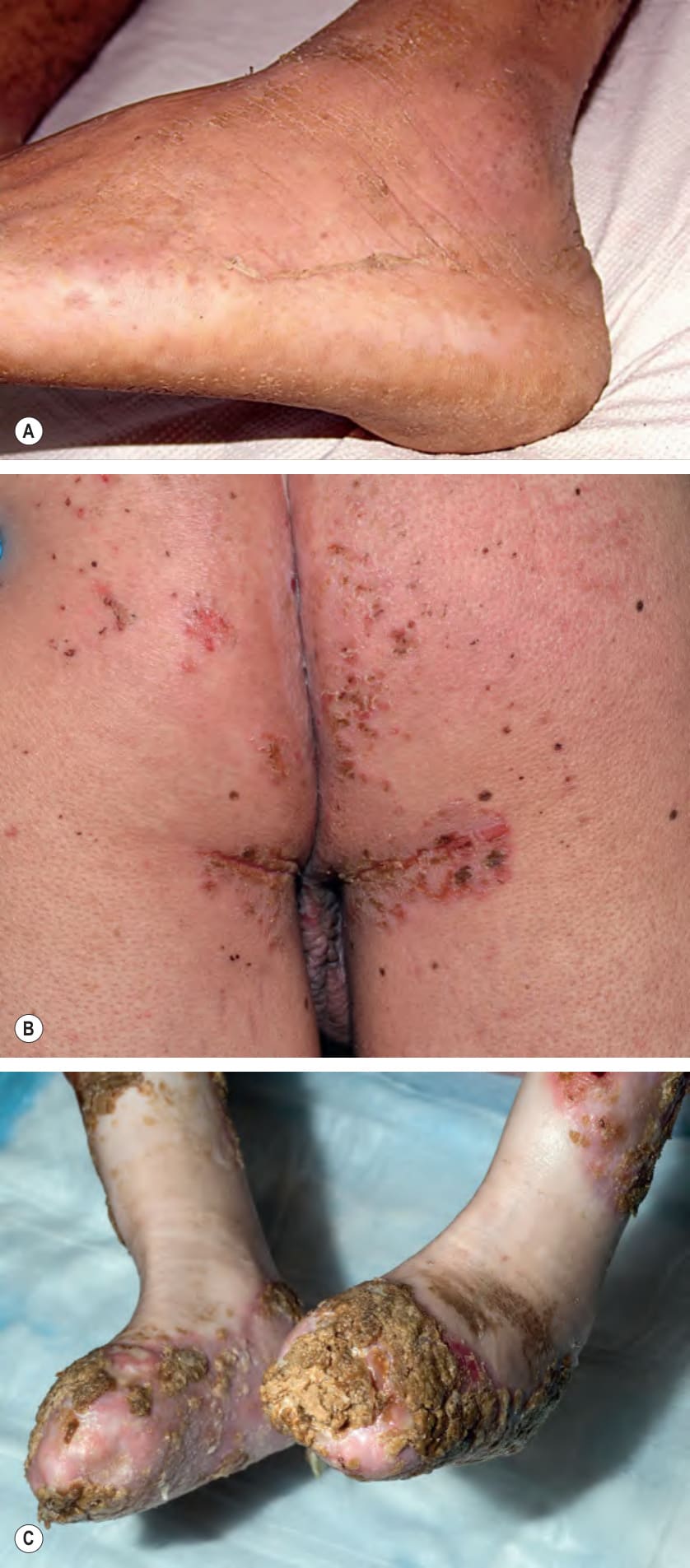
Fig. 32.15 Differential diagnosis of epidermolysis bullosa.A Acral peeling skin syndrome presenting with superficial peeling of the skin on the ankle and dorsal aspect of the foot. B Skin fragility–ectodermal dysplasia syndrome due to plakophilin 1 deficiency. Note the scattered erosions and crusts on the buttocks. C SAVI (STING-associated vasculopathy with onset in infancy) represents a vasculopathic genetic disorder that can result in digital resorption resembling that in recessive dystrophic epidermolysis bullosa. A, Courtesy E. Sprecher, MD, PhD; B, Courtesy Antonio Torrelo, MD; C, Courtesy Edward Cowen, MD.
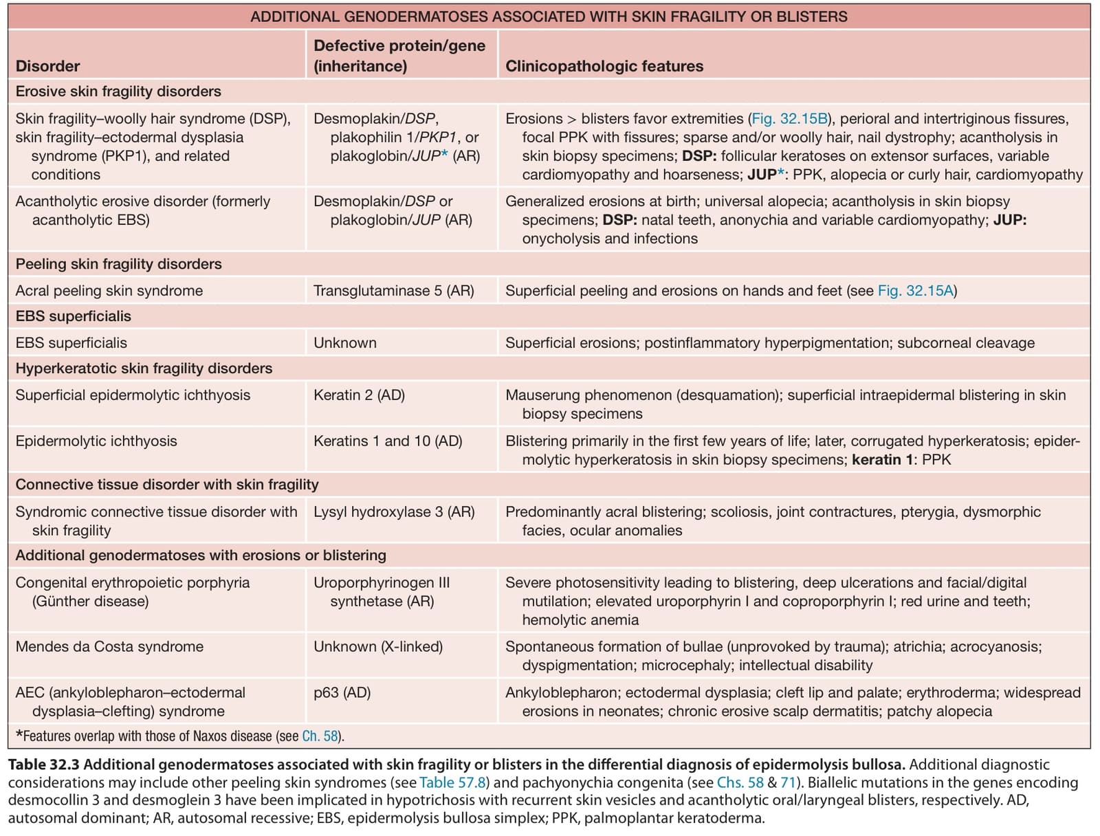
Table 32.3 Additional genodermatoses associated with skin fragility or blisters in the differential diagnosis of epidermolysis bullosa. Additional diagnostic considerations may include other peeling skin syndromes (see Table 57.8) and pachyonychia congenita (see Chs. 58 & 71). Biallelic mutations in the genes encoding desmocollin 3 and desmoglein 3 have been implicated in hypotrichosis with recurrent skin vesicles and acantholytic oral/laryngeal blisters, respectively. AD, autosomal dominant; AR, autosomal recessive; EBS, epidermolysis bullosa simplex; PPK, palmoplantar keratoderma.
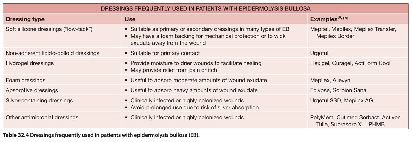
Table 32.4 Dressings frequently used in patients with epidermolysis bullosa (EB).
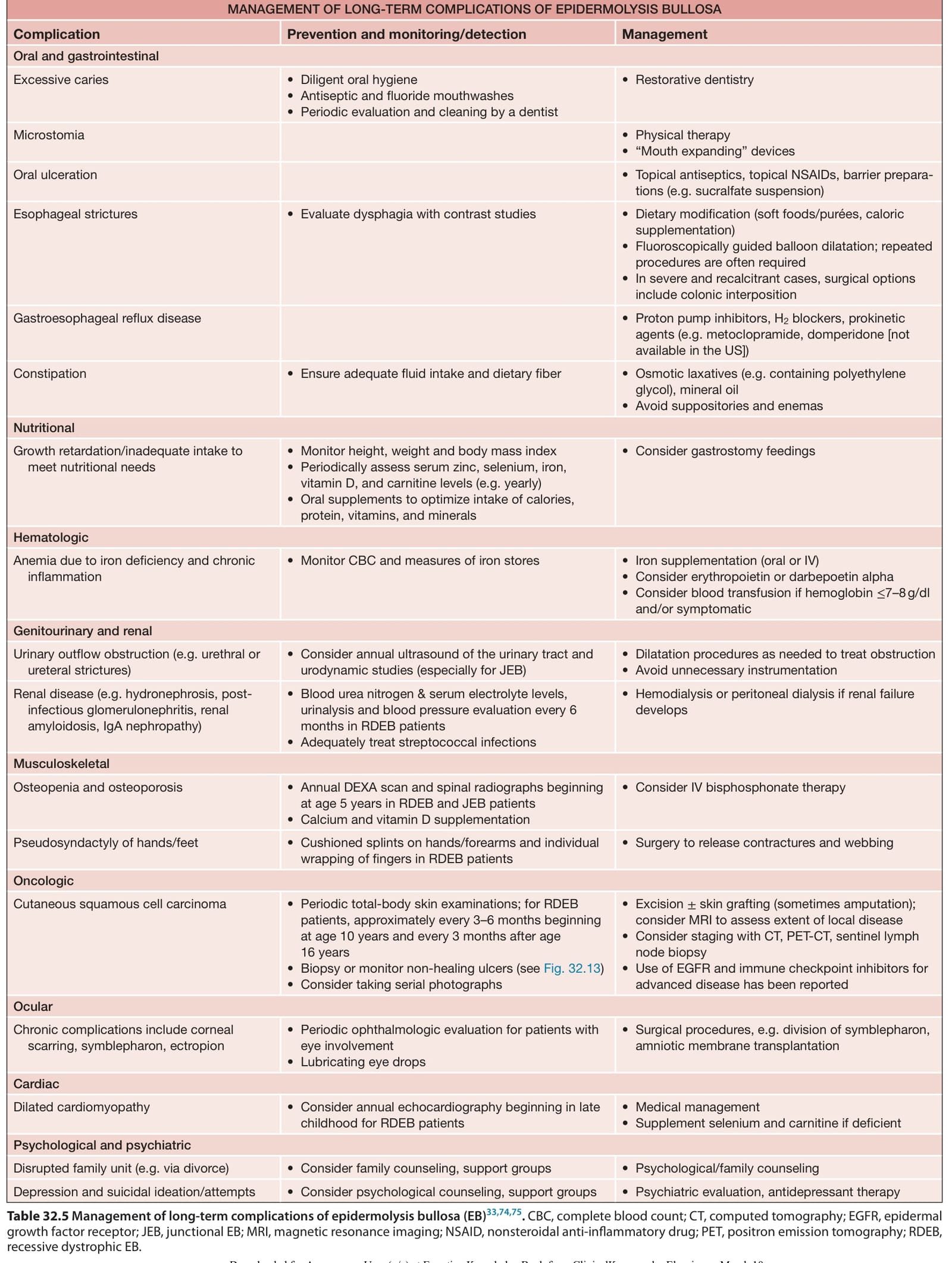
Table 32.5 Management of long-term complications of epidermolysis bullosa (EB). CBC, complete blood count; CT, computed tomography; EGFR, epidermal growth factor receptor; JEB, junctional EB; MRI, magnetic resonance imaging; NSAID, nonsteroidal anti-inflammatory drug; PET, positron emission tomography; RDEB, recessive dystrophic EB.