KINDLER EPIDERMOLYSIS BULLOSA
Synonyms: Kindler syndrome Kindler–Weary syndrome Bullous acrokeratotic poikiloderma of Kindler and Weary Hereditary acrokeratotic poikiloderma
Introduction
Kindler EB is a rare autosomal recessive genodermatosis that features skin fragility and photosensitivity during early life as well as progressive poikiloderma, cutaneous atrophy, and mucosal inflammation.
History
In 1954, Theresa Kindler described a patient with acral blistering and photosensitivity during infancy, followed later in life by progressive poikiloderma and atrophy. Twenty years later, Weary reported ten members of a single family with similar findings in addition to widespread atopic dermatitis during early childhood and keratotic papules over the joints of the arms and legs that appeared in childhood and persisted indefinitely. Oral involvement and other mucosal manifestations were subsequently emphasized.
Pathogenesis
Kindler EB is characterized ultrastructurally by basement membrane reduplication and mixed planes of cleavage that can be within basal keratinocytes, through the lamina lucida, and/or below the lamina densa. It results from mutations in the FERM domain-containing kindlin-1 gene (FERMT1) which encodes kindlin-1, a component of focal adhesions that connect actin filaments in basal keratinocytes to the underlying ECM (see Ch. 28). Through integrin-mediated signaling, kindlin-1 affects the shape, polarity, adhesion, proliferation, and motility of keratinocytes. There is also evidence that kindlin-1 has a role in regulating cutaneous epithelial stem cell homeostasis, with loss of kindlin-1 leading to increased risk of skin cancer as well as cutaneous atrophy due to stem cell exhaustion and premature senescence of keratinocytes.
Clinical Features
Erosions are occasionally present at birth, most often on the forearms and shins, and blistering during infancy is most prominent on the hands and feet. In contrast to other forms of EB, skin fragility tends to decrease considerably during childhood. Photosensitivity manifesting as an increased susceptibility to sunburn also improves over time. Reticulated hyperpigmentation and telangiectasias begin to develop in sun-exposed areas during childhood, and after puberty these findings spread to sun-protected sites. A characteristic feature in adult patients with Kindler EB is poikiloderma (Fig. 32.16A), which persists throughout life. Tissue paper-like atrophy is frequently seen, particularly on the dorsal surfaces of the hands and feet (Fig. 32.16 B,C), and mild webbing of the digits (see Fig. 32.16C) and palmoplantar hyperkeratosis may also be evident. Eczematous dermatitis, typically beginning during infancy and resolving by early childhood, occurs in some patients.
Patients with Kindler EB often have erosive gingivitis and poor dentition. Mucosal involvement may result in intraoral and corneal scarring, ectropion, colitis, and strictures of the esophagus, urethra, vagina, and anus. Patients have an increased risk of SCC of the lip and oral mucosa as well as acral skin.
Pathology
In older patients, skin biopsy specimens show the typical changes of poikiloderma, including epidermal atrophy, vacuolization of the basal cell layer, variable epidermal melanin content, dermal melanophages, and capillary dilation. Immunostaining of skin biopsy specimens with anti-kindlin-1 antibodies often demonstrates a marked reduction or absence of this protein. Ultrastructural findings are noted above.
Differential Diagnosis
The predominantly acral distribution of the blisters and associated atrophy and photosensitivity distinguish Kindler EB from other forms of EB, although a form of JEB characterized by progressive cutaneous atrophy similar to that in Kindler EB has been described. As poikiloderma becomes apparent during early childhood, the differential diagnosis may include Rothmund–Thomson syndrome and other entities outlined in Table 63.9.
Treatment
Management is akin to other forms of EB, with the addition of sun protection and assiduous oral hygiene.
Additional figures available in our eBook (see inside front cover for access code).
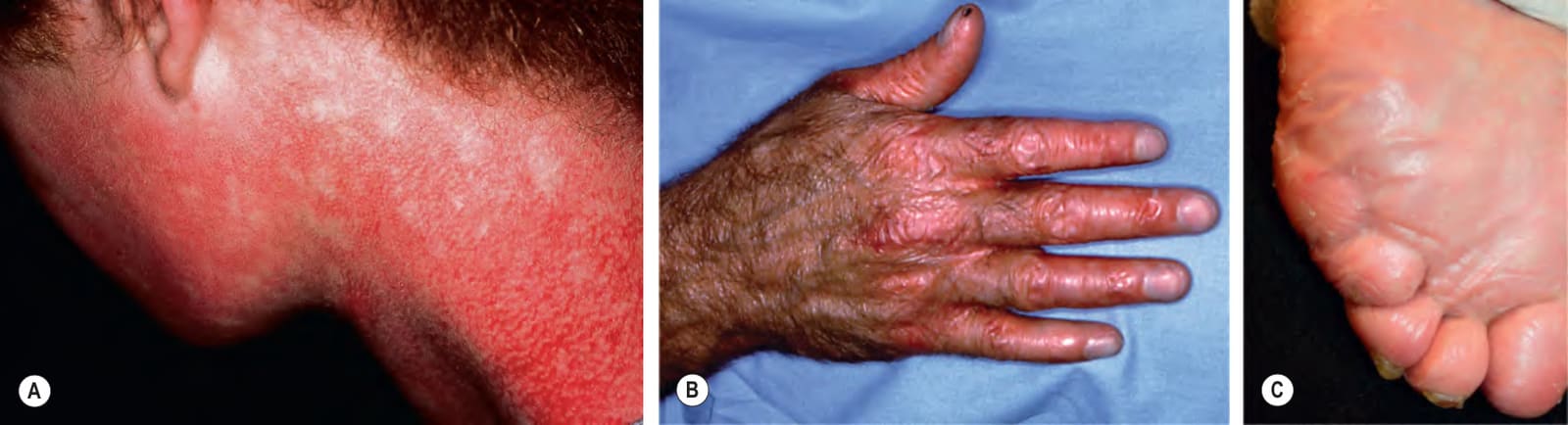
Fig. 32.16 Kindler epidermolysis bullosa.A Poikiloderma of the face and neck with “skip” areas. B Erythema and atrophy on the dorsal hand. C Progressive syndactyly of fourth and fifth toes in an adult. Note the near absence of dermatoglyphics. This patient also had ectropion and urethral strictures. C, Courtesy Jean L. Bolognia MD.
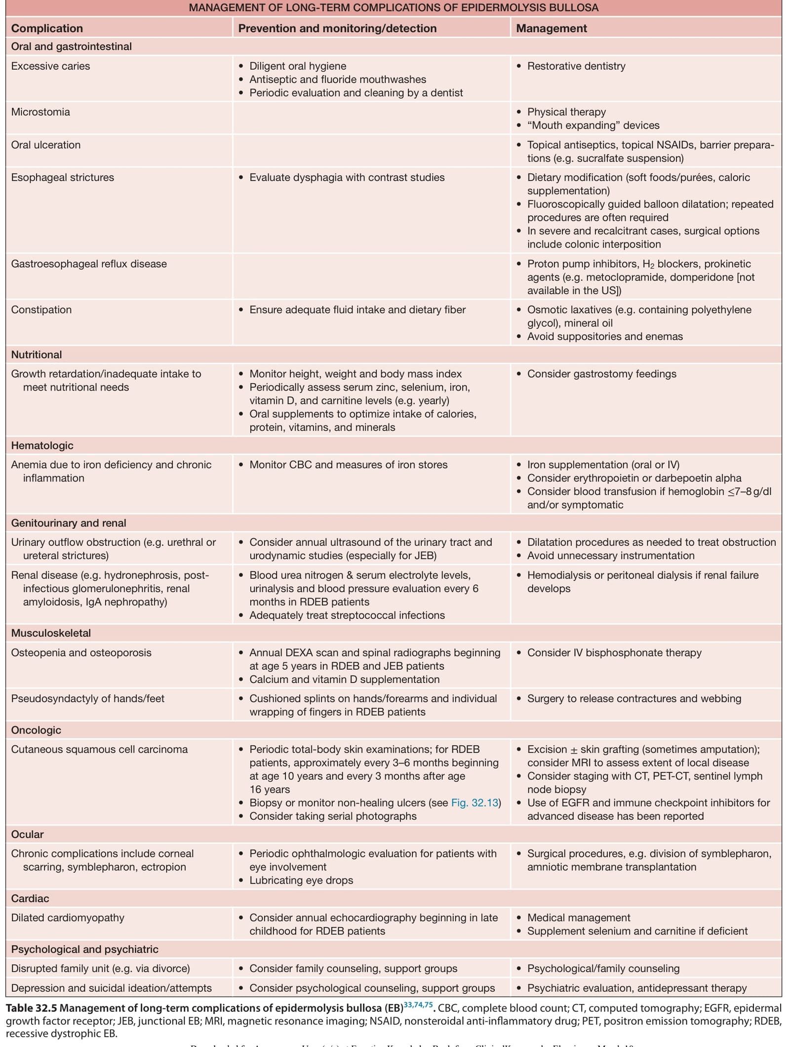
Table 32.5 Management of long-term complications of epidermolysis bullosa (EB). CBC, complete blood count; CT, computed tomography; EGFR, epidermal growth factor receptor; JEB, junctional EB; MRI, magnetic resonance imaging; NSAID, nonsteroidal anti-inflammatory drug; PET, positron emission tomography; RDEB, recessive dystrophic EB.