BULLOUS INSECT BITE REACTIONS
setting of hematologic malignancies): Eosinophilic dermatosis of hematologic malignancy Eosinophilic dermatosis associated with hematological disorders
Key features
Bullous insect bite reactions are fairly common, particularly in children, although patients often do not recall being bitten
Severe insect bite reactions as well as insect bite-like reactions can occur in patients with hematologic malignancies, in particular chronic lymphocytic leukemia
Introduction
Although most insect bite reactions present as pruritic erythematous papules (see Ch. 85), vesiculobullous reactions are not uncommon.
History
An exaggerated reaction to insect bites in patients with chronic lymphocytic leukemia (CLL) was first described in 1965 by Robert Weed, and it has been increasingly recognized over the past two decades.
Epidemiology
There seems to be no difference in the incidence of insect bites based on sex, race, or age. Certain individuals may be bitten more frequently because of their body temperature, odor, use of perfumes, or carbon dioxide excretion. Children tend to have more severe reactions than adults because the latter tend to become desensitized by repeated exposures over time.
Patients with CLL, as well as other hematologic malignancies (e.g. non-Hodgkin lymphomas, acute leukemias, multiple myeloma), can develop both exaggerated reactions to insect bites and insect bite-like reactions. The latter term is used to describe lesions that resemble insect bites clinically and histologically, but for which a clear link to insect bites cannot be established. These exaggerated reactions can precede the diagnosis of the hematologic malignancy; in a large retrospective study of patients with CLL, the eruption preceded the diagnosis in 20% of patients. Of note, no relationship to disease activity has been observed. More recently, these reactions have been referred to as “eosinophilic dermatosis associated with hematological disorders” or “eosinophilic dermatosis of hematologic malignancy”; the term “eosinophilic dermatosis of myeloproliferative disease” is inaccurate, as CLL is not a myeloproliferative disorder (see Ch. 121).
Pathogenesis
Most lesions are the result of an individual’s immune response to insect antigens introduced by the bite. Both humoral (IgE-mediated) and cellmediated immunity (delayed hypersensitivity) are involved. Initial bites typically do not produce significant lesions, but sensitization occurs over a few weeks. Subsequent bites then induce a stronger reaction and even bullae formation. With time, repeated bites can result in desensitization. In the case of bullous bed bug reactions, cutaneous vasculitis may be playing a role.
In patients with NK/T cell lymphomas, severe hypersensitivity to mosquito bites has been well documented and a possible role for the EBV-induced clonal proliferation of NK cells has been raised. For patients with ALK-positive anaplastic large cell lymphoma, the possibility has been raised that insect bite-associated antigens lead to an influx of T lymphocytes, some of which bear t(2;5). The release of cytokines then results in activation of these cells, expression of NPM-ALK (nucleophosmin–anaplastic lymphoma kinase) fusion protein, and subsequent uncontrolled proliferation. In patients with B cell malignancies, an increased production of IL-4 and IL-5 may explain associated tissue eosinophilia.
Clinical Features
Insect bites usually appear as intensely pruritic erythematous papules or nodules. They are usually grouped, and a linear arrangement is commonly seen. However, vesicular and bullous bite reactions are not uncommon, and blisters can develop centrally within papules or as bland vesiculobullae (Fig. 33.7). Flea bites are the most likely to cause
blisters, especially on the legs, and bedbug bites can also be bullous. Some insects (e.g. fire ants) can produce pustular lesions. In patients with hematologic malignancies, the exaggerated reactions consist of persistent papulonodules, vesicles, bullae, and even necrotic lesions (Fig. 33.8).
Pathology
The typical pattern is that of a superficial and deep perivascular and periadnexal lymphocytic infiltrate with abundant eosinophils. A wedgeshaped pattern is characteristic. During the initial phase, intraepidermal spongiotic vesicles may be present and they can be associated with eosinophilic spongiosis. Pronounced dermal edema leads to the formation of subepidermal blisters. Sometimes, epidermal necrosis develops. Flame figures (extruded eosinophilic granules aggregated onto collagen fibers that are classically associated with eosinophilic cellulitis) can be observed, particularly in the exaggerated reactions seen in patients with hematologic malignancies.
Differential Diagnosis
The presence of intense pruritus and lesion distribution usually suggests a bullous bite reaction. However, these reactions can mimic bullous pemphigoid, linear IgA bullous dermatosis, bullous erythema multiforme, bullous impetigo, allergic contact dermatitis, or even viral infections.
Treatment
Most patients with bullous insect bites can be managed with drainage of blisters plus topical corticosteroids and systemic antihistamines to relieve pruritus. In the most severe reactions, a short course of systemic corticosteroids may be necessary. Systemic corticosteroids can lead to improvement of eosinophilic dermatosis of hematologic malignancy, but lesions tend to recur when the dose is reduced. Dupilumab may represent a therapeutic option in these patients. Protective clothing and insect repellants are important preventative measures (see Ch. 85).
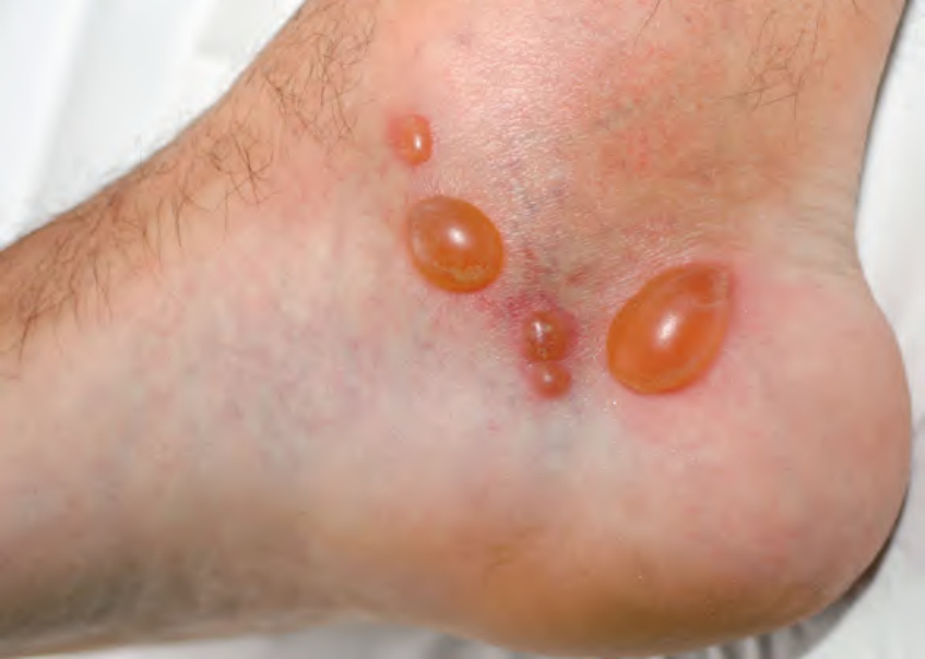
Fig. 33.7 Bullous insect bite reactions. The serous blister fluid is sterile and the ankle is a common site. Courtesy Luis Requena, MD.
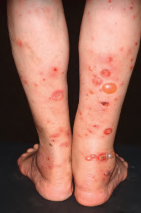
Fig. 33.8 Exaggerated insect bite-like reactions in a patient with B cell chronic lymphocytic leukemia. Both bullae and erosions at sites of ruptured bullae are present. Reprinted with permission from Cerroni L. Skin Lymphomas: The Illustrated Guide, 5ed. Oxford: Wiley- Blackwell, 2020.
Table 33.1 Bullous drug eruptions. Occasionally, eczematous drug reactions (e.g. secondary to warfarin, calcium channel blockers, angiotensin converting enzyme [ACE] inhibitors, angiotensin II receptor blockers) and systemic contact dermatitis can be papulovesicular; patients with drug reaction with eosinophilia and systemic symptoms (DRESS)/drug-induced hypersensitivity syndrome (DIHS) and drug-induced Sweet syndrome can also develop vesicles. Coma blisters and bullous small vessel vasculitis are discussed in the text. EGFR, epidermal growth factor receptor; NSAIDs, nonsteroidal anti-inflammatory drugs.