PATHOGENESIS OF ACNE
The development of acne involves the complex interplay of four main factors: (1) follicular hyperkeratinization; (2) hormonal influences on sebum production and composition; (3) inflammation; and (4) C. acnes residing in the sebaceous follicle (Fig. 36.1). Appreciating the anatomy and physiology of the pilosebaceous unit is essential to understanding the pathogenesis of acne and designing effective treatment regimens. Genetics and exogenous factors such as diet, medications, and environmental exposures can also play a role in acne pathogenesis. The structure and function of sebaceous glands are reviewed in detail in Chapter 35, with summaries of their roles in acne in Table 35.3 and Fig. 35.4.
Follicular Hyperkeratinization
The microcomedo is thought to be the precursor of all clinically apparent acne lesions. It forms in the upper portion of the follicle within the lower portion of the infundibulum, the infrainfundibulum. Corneocytes, which are normally shed into the hair follicle lumen and extruded, accumulate due to increases in both follicular keratinocyte proliferation and corneocyte cohesiveness, leading to the development of a hyperkeratotic plug that acts as a bottleneck. In acne patients, non-invasive high-resolution imaging of facial skin has confirmed that follicular dilation and accumulation of a keratotic plug precede the appearance of an inflammatory papule. The exact inciting event for microcomedo formation is unknown, but data support a putative role for interleukin-1α (IL-1α) and increases in skin pH.
Hormonal Influences on Sebum Production and Composition
The sebaceous gland expresses multiple receptors including androgen, estrogen, progesterone, retinoid, and peroxisome proliferator-activated receptors (PPARs; see Table 35.3). Sebum production is controlled primarily by androgens, with additional influences from other hormones and neuropeptides. Androgens are produced both outside the pilosebaceous unit, mainly by the gonads and adrenal glands, and locally within the sebaceous gland via the action of androgen-metabolizing
enzymes such as 3β-hydroxysteroid dehydrogenase (HSD), 17β-HSD and 5α-reductase (see Fig. 70.16). Androgen receptors, found in the cells of the basal layer of the sebaceous gland and the outer root sheath of the hair follicle, are responsive to testosterone and 5α-dihydrotestosterone (DHT), the most potent androgens. DHT has a 5- to 10-fold greater affinity than testosterone for the androgen receptor and is the principal androgen mediating sebum production.
The impact of androgens on sebaceous gland activity begins during the neonatal period. From birth until approximately 6–12 months of age, infant boys have elevated levels of luteinizing hormone (LH), which stimulates testicular production of testosterone. In addition, both male and female infants exhibit increased levels of dehydroepiandrosterone (DHEA) and DHEA sulfate (DHEAS) secondary to a large, androgen-producing “fetal zone” in the adrenal gland that involutes during the first year of life. Sebaceous gland activity in infants is not the result of persistent maternal hormonal stimulation, as was previously hypothesized. Both testicular and adrenal androgen production decrease substantially by 1 year of age and remain at a stable nadir until adrenarche.
With the onset of adrenarche, typically at 7–8 years of age, circulating levels of DHEAS begin to rise due to its production by the adrenal gland. This hormone serves as a precursor for the synthesis of more potent androgens within the sebaceous gland (see Fig. 70.16). The rise in DHEAS serum levels in prepubescent children is associated with increased sebum production and often the initial development of comedonal acne. Although the overall composition of sebum is similar in adolescents irrespective of the presence or absence of acne, the sebum in those with acne tends to have higher levels of squalene monounsaturated fatty acids and less linoleic acid. In infants with facial acne, the skin surface lipid composition has also been shown to differ from those without acne.
The physiologic role of estrogens and progesterone in modulating sebum production is not fully understood. Estrogens, when administered systemically in sufficient amounts, decrease sebum production, although the dose needed is greater than the dose required to suppress ovulation and increases the risk of thromboembolic events. However, acne often responds to treatment with lower-dose oral contraceptives containing 20–50 mcg of ethinyl estradiol or its esters because suppression of ovulation itself inhibits ovarian androgen production. Postulated mechanisms for estrogen-mediated downregulation of sebogenesis include direct opposition of androgens within the sebaceous gland, a negative feedback loop that decreases androgen production via inhibition of pituitary gonadotropin release, and regulation of genes that affect sebaceous gland activity. Although the role of progesterone in human sebum production remains to be fully determined, it can increase sebum production in both male and female rats, likely via modulation of the androgen receptor.
Inflammation in Acne
Although tremendous progress has been made in our understanding of acne as an inflammatory process, several questions remain regarding inflammation during acne lesion development. It is clear that when a follicle involved with acne ruptures, it exudes keratin, sebum, C. acnes, and cellular debris into the surrounding dermis, thereby intensifying inflammation. However, inflammation is also seen early in acne lesion formation. For example, in acne-prone sites, the number of CD4+ T cells and levels of IL-1 are increased perifollicularly prior to hyperkeratinization. In addition, insulin-like growth factor-1 has been found to increase the expression of inflammatory markers and sebum production in cultured sebocytes. TREM2 macrophages induced by lipids such as squalene have been shown to promote inflammation in acne lesions and block oxidative killing of C. acnes23a.
The type of inflammatory response determines the clinical lesion observed. If neutrophils predominate (typical of early lesions), suppuration occurs and a pustule is formed. Neutrophils also promote the inflammatory response by releasing lysosomal enzymes and generating reactive oxygen species; levels of the latter in the skin and plasma may correlate with acne severity. An influx of lymphocytes (predominately T helper cells), foreign body-type giant cells, and neutrophils results in inflamed papules, nodules, and cysts. The type of inflammatory response also plays a role in the development of scarring. Early, nonspecific inflammation results in less scarring than does a delayed, specific inflammatory response.
Cutibacterium acnes colonization and activities
C. acnes (previously known as Propionibacterium acnes) is a Gram-positive, anaerobic/microaerophilic rod that is found on the skin surface and within the sebaceous follicle, often together with smaller numbers of C. granulosum. In adults, C. acnes is the predominant organism in the microbiome of facial and other sebaceous skin. C. acnes produces porphyrins (primarily coproporphyrin III) that fluoresce with Wood’s lamp illumination.
C. acnes is considered a commensal organism of the skin rather than a pathogen per se. Although studies have documented increased levels of C. acnes on the facial skin of acne patients, the C. acnes density does not correlate with clinical severity. In a comparison of the microbiome of facial skin in adults with and without acne, C. acnes ribotypes 4 and 5 were more frequently found in acne patients, suggesting that these strains are either more capable of inducing acne or better suited to survive in an acne environment. In addition, acne-associated C. acnes phylotypes (IA-2, IB-1, IC) stimulated peripheral blood monocytes to produce the proinflammatory cytokines IL-17 and IFN-γ, whereas phylotypes found in healthy skin (II [RT6], III) preferentially induced monocytes to secrete the anti-inflammatory cytokine IL-10. However, another study found that while isolated peripheral blood monocytes from patients with acne exhibited a more robust release of inflammatory cytokines in response to C. acnes stimulation than did monocytes from individuals without acne, no differences were observed when C. acnes strains isolated from acne lesions were compared to strains from the normal skin of unaffected adults.
The pathogenicity of C. acnes includes the direct release of lipases, chemotactic factors, and enzymes that contribute to comedo rupture, as well as stimulation of inflammatory cells and keratinocytes to produce proinflammatory mediators and reactive oxygen species. Interactions between the skin’s innate immune system and C. acnes play an important role in acne pathogenesis. One mechanism is via Toll-like receptors (TLRs), a class of transmembrane receptors that mediates the recognition of microbial pathogens by immune cells (monocytes, macrophages, and neutrophils) as well as by keratinocytes (see Ch. 4). TLR2, which recognizes lipoproteins and peptidoglycans as well as CAMP factor 1 produced by inflammatory strains of C. acnes, is found on the surface of macrophages surrounding acne follicles. C. acnes has also been shown to increase expression of TLR2 and TLR4 by keratinocytes. Through activation of the TLR2 pathway, C. acnes stimulates the release of proinflammatory mediators such as IL-1α, IL-8, IL-12, tumor necrosis factor (TNF), and matrix metalloproteinases. IL-8 leads to neutrophil recruitment, the release of lysosomal enzymes, and subsequent disruption of the follicular epithelium, while IL-12 promotes Th1 responses (see Ch. 4).
C. acnes has been shown to activate NLRP3 inflammasomes in neutrophils and monocytes, resulting in the release of proinflammatory IL-1β. As noted above, C. acnes also stimulates Th17 responses within acne lesions. Lastly, C. acnes can induce monocytes to differentiate into two distinct innate immune cell subsets: (1) CD209+ macrophages, which more effectively phagocytose and kill C. acnes and whose development is promoted by tretinoin; and (2) CD1b+ dendritic cells that activate T cells and release proinflammatory cytokines.
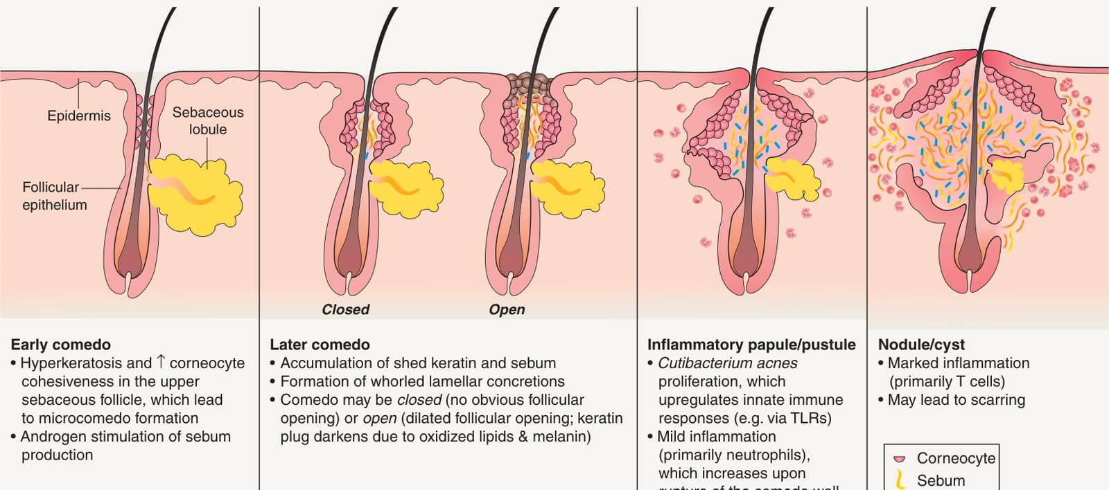
Fig. 36.1 Pathogenesis of acne.