CLINICAL FEATURES
Acne is typically found in sites with well-developed sebaceous glands, most often the face and upper trunk. Despite evidence that inflammation is present in even the earliest comedones, acne lesions are divided into non-inflammatory and inflammatory groups based upon their clinical appearance. Non-inflammatory acne is characterized by open and closed comedones (Fig. 36.2). The histologic features of these follicular-based lesions are reflected in their clinical appearance. Closed comedones (whiteheads) are generally small (~1 mm), skin-colored papules with no apparent follicular opening or associated erythema. These lesions may be subtle and better appreciated upon palpation, stretching, or sidelighting of the skin. In contrast, open comedones (blackheads) have a conspicuous dilated follicular opening that is filled with an inspissated core of shed keratin (see Fig. 36.4). Melanin deposition and lipid oxidation within the debris may be responsible for the black color. Larger macrocomedones are deep-seated and may be cystic.
Inflammatory acne is characterized by papules, pustules, and nodules of varying severity (Figs. 36.3–36.6). Erythematous papules typically range from 1 to 5 mm in diameter. Pustules tend to be approximately equal in size and are filled with white purulent material and normal microbiota, including C. acnes. As the severity of lesions progresses, nodules form and become markedly inflamed, indurated, and tender. The pseudocysts of acne are deeper and filled with a combination of pus and serosanguineous fluid. In patients with severe nodulocystic acne, these lesions frequently coalesce to form large, complex, inflamed plaques that can include sinus tracts.
Early treatment of acne is essential for the prevention of lasting cosmetic disfigurement due to scarring. Erythema and
postinflammatory hyperpigmentation (Fig. 36.7) often persist after resolution of inflammatory acne lesions. Although the pigmentary changes usually fade over many months if the acne is brought under control, occasionally they can be permanent. Unfortunately, pitted scars (Fig. 36.8A) or hypertrophic scars (most commonly on the trunk; Fig. 36.8B,C) are often sequelae of nodulocystic acne. Skin-colored, dome-shaped papules on the nose and chin with histopathologic features indistinguishable from angiofibromas represent an additional form of acne scarring that has a predilection for male patients with darkly pigmented skin (Fig. 36.9).
Acne Variants
Post-adolescent acne in women
Inflammatory acne beyond 25 years of age is most common in women and may be associated with a high level of psychological stress. The majority of affected women present with findings similar to those of adolescent acne, with a mixture of inflammatory and comedonal lesions involving various facial sites and sometimes the trunk. Although the mandibular area is involved in ~80% of women with acne, a distinct smaller subset has inflammatory papules, pustules, and nodules exclusively in this location. Half of women report persistence of their acne since its onset, often during adolescence, while one-quarter describe periods of remission followed by recurrences. Premenstrual flares are common, but only ~20% of women with acne have irregular menses. Up to 30% of those in the latter group have other signs of hyperandrogenism, such as hirsutism and androgenetic alopecia (see below). A predominantly comedonal, adult-onset form of acne that is associated with smoking has also been described.
Acne fulminans
Acne fulminans is the most severe form of acne and is characterized by the abrupt development of nodular and suppurative acne lesions in association with systemic manifestations. This uncommon variant primarily affects boys 13–16 years of age. Patients typically have mild to moderate acne prior to the onset of acne fulminans, when numerous microcomedones suddenly erupt and become markedly inflamed. There is rapid coalescence into painful, oozing, friable plaques with hemorrhagic crusts (Fig. 36.10). The face, neck, trunk, and arms are all affected. Lesions tend to ulcerate and can lead to significant scarring.
Osteolytic bone lesions may accompany the cutaneous findings; the clavicle and sternum are most commonly affected, followed by the ankles, humerus, and iliosacral joints. Systemic manifestations include fever, arthralgias, myalgias, hepatosplenomegaly, and severe malaise. Erythema nodosum may also arise in association with acne fulminans. Laboratory abnormalities vary and include an elevated ESR, proteinuria, leukocytosis, and anemia. Laboratory studies are not required to establish the diagnosis, but their evolution may parallel the clinical course and response to therapy. The related synovitis, acne, pustulosis, hyperostosis, and osteitis (SAPHO) syndrome, which can accompany acne fulminans, is discussed in detail in Chapter 26. Acne fulminans has also been associated with late-onset congenital adrenal hyperplasia and anabolic steroid use, including therapeutic testosterone.
Recommended treatment of acne fulminans includes prednisone 0.5–1 mg/kg/day as monotherapy for at least 2–4 weeks, followed by initiation of low-dose isotretinoin (e.g. 0.1 mg/kg/day) after the acute inflammation subsides; after at least 4 weeks of this combination, the isotretinoin dose can be slowly increased and the prednisone tapered over a period of 1–2 months. Paradoxically, an acne fulminans-like flare occasionally develops during the first few weeks of isotretinoin therapy
Multiple coalescing papules, papulopustules, and small nodules are present on the cheeks (A,B) and temple (A). Early scarring is evident laterally (A). B, Courtesy Kalman Watsky, MD.
for acne; this may be avoided by starting with a low dose of isotretinoin and concomitant administration of oral corticosteroids at the first sign of a flare or possibly preemptively in high-risk patients (see Ch. 126). Additional treatment options for acne fulminans include topical or intra-lesional corticosteroids, oral antibiotics (generally of limited efficacy), TNF inhibitors, IL-1 antagonists, and immunosuppressive agents (e.g. azathioprine, cyclosporine). Dapsone may be particularly beneficial in the treatment of acne fulminans associated with erythema nodosum.
Acne conglobata and associated conditions
Acne conglobata (see Fig. 36.6B) is a severe form of nodulocystic acne that may have an eruptive onset but without systemic manifestations.
A Numerous nodules. B Acne conglobata with coalescence of purulent nodules on the forehead, with associated eyelid edema. This form is best treated with prednisone and a low dose of isotretinoin initially. B, Courtesy Julie V. Schaffer, MD.
This recalcitrant acne variant is part of the follicular occlusion tetrad, along with dissecting cellulitis of the scalp, hidradenitis suppurativa, and pilonidal sinus (see Ch. 38).
The association of sterile pyogenic arthritis, pyoderma gangrenosum, and acne conglobata can occur in the context of an autosomal dominant autoinflammatory disorder referred to as PAPA syndrome, which can be caused by mutations in PSTPIP1 which encodes proline–serine– threonine phosphatase interacting protein 1. Dysfunction of PSTPIP1, which has a role in actin reorganization and interacts with the pyrin protein (defective in familial Mediterranean fever), compromises the physiologic signaling required for maintenance of a proper inflammatory response. PSTPIP1 mutations have also been identified in some patients with pyogenic arthritis, pyoderma gangrenosum, acne, and suppurative hidradenitis (PAPASH) and pyoderma gangrenosum, acne, and suppurative hidradenitis (PASH) syndromes. Of note, a possible association between nodulocystic acne and inflammatory bowel disease may potentially confound links that have been observed between the latter disorder and acne therapies such as tetracyclines and isotretinoin.
Solid facial edema
An unusual and disfiguring complication of acne vulgaris is solid facial edema (Morbihan disease). Clinically, there is a distortion of the midline face and cheeks due to soft tissue swelling (Fig. 36.11). The woody induration may be accompanied by erythema. Impaired lymphatic drainage and fibrosis (potentially induced by mast cells) in the setting of chronic inflammation are thought to be involved in the pathogenesis of solid facial edema, and a report of its occurrence in identical twins with acne suggests that genetic factors may also have a role. Similar changes have been described in patients with rosacea (see Table 36.2). Although fluctuations in severity are common, solid facial edema does not usually resolve spontaneously. Treatment with isotretinoin (0.2–1 mg/kg/day) for 4–6 months has been reported to lead to improvement, although a more extended course of 9–24 months is often required. Combination of isotretinoin with ketotifen 1–2 mg/day (not available in the US) or prednisone 10–30 mg/day may have additional benefit.
Neonatal cephalic pustulosis (neonatal acne)
Neonatal cephalic pustulosis occurs in more than 20% of healthy newborns (see Ch. 34). Lesions usually appear at about 2 weeks of age and generally resolve within the first 3 months of life. Small papulopustules, but not comedones, arise primarily on the cheeks, forehead, eyelids and chin, although the neck and upper trunk can also be involved (see Fig. 34.6).
An inflammatory response to Malassezia spp. (e.g. sympodialis, furfur) has been proposed as the etiology by some investigators. Additional support for this view comes from the clinical response to treatment with topical imidazoles (e.g. ketoconazole 2% cream). Active sebaceous glands in neonates and their relatively high sebum excretion rate (see Pathogenesis) are also thought to play a role. The substantial decline in sebum production after the first few months of life helps to explain the limited duration of neonatal cephalic pustulosis. Given the
transient and benign nature of this eruption, parental reassurance alone is usually adequate. However, as noted previously, therapy with topical imidazoles can be effective.
Infantile acne
Infantile acne typically has its onset at 2–12 months of age. In contrast to neonatal cephalic pustulosis, comedo formation is prominent and pitted scarring may develop in up to half of patients (Fig. 36.12). Deep, suppurative nodules are occasionally seen. Its pathogenesis reflects the androgen production intrinsic to this stage of development (see Pathogenesis), including elevated levels of LH that stimulate testicular production of testosterone in boys during the first 6–12 months of life and elevated levels of DHEA produced by the infantile adrenal gland in both boys and girls. These androgen levels normally decrease substantially by 12 months of age and remain at nadir levels until adrenarche. Patients with infantile acne should be assessed for signs of hyperandrogenism, precocious puberty, or abnormal growth; if these findings are present, endocrinologic evaluation including a hand/wrist X-ray to determine bone age and laboratory testing of hormone levels should be performed.
Infantile acne usually resolves within 6–18 months and remains quiescent until around puberty, with an increased risk of severe acne during adolescence having been observed. Topical retinoids (e.g. tretinoin, adapalene) and benzoyl peroxide are first-line treatments for infantile acne. Oral antibiotics (e.g. erythromycin, azithromycin) can be helpful for patients with a more severe inflammatory component, and isotretinoin is occasionally required for recalcitrant or nodulocystic presentations.
Mid-childhood acne
Acne presenting between 1 and 6 or 7 years of age is categorized as “mid-childhood acne”. Because this is an uncommon time for acne development due to quiescent androgen production, the possibility of an underlying hyperandrogenic condition such as premature adrenarche, congenital adrenal hyperplasia, or an androgen-secreting tumor should be considered. In addition to a thorough history and physical examination to assess for signs of hyperandrogenism (see below) and precocious puberty, the child’s growth curve should be carefully reviewed and a hand/wrist bone age X-ray performed if there are signs of accelerated growth. A complete endocrine evaluation is required if any abnormalities are present. Treatment strategies are the same as for infantile acne.
Preadolescent acne
It is common for acne to begin to develop with the onset of adrenarche, typically between 7 or 8 to 11 years of age, which is often prior to other signs of pubertal maturation. Preadolescent acne tends to be primarily comedonal and favors the forehead and central face (“T-zone”). Polycystic ovary syndrome (PCOS) and other endocrinologic abnormalities should be considered when the acne is unusually severe or accompanied by
signs of hyperandrogenism. Treatment is similar to that for adolescent acne, although tetracycline courses of >21 days should be avoided in children <8 years of age.
Acne excoriée
Acne excoriée occurs more often in young women, in whom it may be referred to as acne excoriée des jeunes filles. Typical comedones and inflammatory papules are systematically excoriated in a ritualistic manner, leaving crusted erosions that may scar (see Ch. 7). Linear erosions suggest self-manipulation, and an underlying psychiatric component should be considered. Individuals with an anxiety, obsessive– compulsive, or body dysmorphic disorder are particularly at risk, and antidepressants or psychotherapy may be helpful in these patients.
Acne associated with endocrinologic abnormalities
Although most patients with acne do not have overt endocrinologic abnormalities, hyperandrogenism should be suspected in women and late adolescent girls with irregular menstrual periods. Acne in such patients is often severe or more difficult to treat, and the onset can be fairly abrupt. Other signs and symptoms of hyperandrogenism in women and children include hirsutism (see Ch. 70), coarsening of the voice, a muscular habitus, androgenetic alopecia, clitoromegaly with variable posterior labial fusion, and increased libido. Insulin resistance and acanthosis nigricans can occur in association with hyperandrogenism in the HAIR-AN syndrome. These patients are at increased risk for accelerated cardiovascular disease and diabetes mellitus, and they should be followed by appropriate medical specialists.
The evaluation of patients suspected of having hyperandrogenism includes a thorough history and physical examination; the age of the patient and pubertal status are also important parameters. Prepubertal children, adolescent girls, and women with signs of hyperandrogenism should undergo appropriate evaluation, and laboratory studies should not be performed while the patient is taking oral contraceptives. Initial tests typically include serum levels of total and free testosterone, DHEAS, and 17-hydroxyprogesterone. Patients with clinical findings suggestive of hypercortisolism should also be initially assessed with either a late-night salivary cortisol level, 24-hour urine cortisol level, or low-dose dexamethasone suppression test. In prepubertal children, growth charts should be assessed and X-rays of the hand and wrist performed to evaluate bone age (see Mid-childhood acne).
Understanding pathways of hormone production is essential in the evaluation of hyperandrogenic states (see Fig. 70.18). For example, an elevated serum DHEAS or 17-hydroxyprogesterone level indicates an adrenal source of excess androgen production. The degree to which levels of these hormones are increased is then useful in discerning an etiology. DHEAS values in the range of 4000–8000 ng/ml or 17-hydroxyprogesterone levels >3 ng/ml may be indicative of congenital adrenal hyperplasia. Defects in adrenal enzymes, most commonly 21-hydroxylase and less often 11-hydroxylase, lead to this condition. Patients with severe deficiencies of these enzymes become symptomatic during infancy, whereas those with partial deficiencies present in adolescence. If the serum DHEAS is >8000 ng/ml, with or without an elevated testosterone level, an adrenal tumor should be suspected.
If the testosterone levels (total and free) are elevated and the DHEAS level is relatively normal, an ovarian source is likely. Polycystic ovary syndrome (PCOS) is the most common condition associated with an elevated serum testosterone, with levels typically ranging from 100 to 200 ng/dl. An increased LH/FSH ratio (>2–3) is also commonly observed. Symptoms of PCOS include irregular menstrual periods, hirsutism, obesity, insulin resistance, and reduced fertility (see Fig. 70.14). When levels of serum testosterone exceed 200 ng/dl, an ovarian tumor should be considered.
Acne associated with genetic syndromes
Apert syndrome (acrocephalosyndactyly type I) is an autosomal dominant disorder that features disfiguring synostoses of the bones of the hands and feet, vertebral bodies, and cranium. Affected individuals have an increased incidence of severe, early-onset acne that tends to be nodulocystic and have a more widespread distribution than in classic acne, often involving the entire extensor aspects of the arms, buttocks, and thighs. Acne in patients with Apert syndrome is typically resistant to therapy, although isotretinoin has been reported to be beneficial. Other cutaneous findings in this disorder can include marked seborrhea, nail anomalies (e.g. a single nail for the second through fourth digits), and diffuse pigmentary dilution of the hair and skin. Of note, Apert syndrome results from activating mutations in FGFR2, which encodes fibroblast growth factor receptor 2; mosaicism for the same FGFR2 mutations has been found to underlie the Munro acne nevus (see Ch. 62). FGFR2 signaling has been shown to have effects on follicular keratinocyte proliferation, sebaceous lipogenesis, and inflammatory cytokine production.
Frank–ter Haar syndrome (Borrone dermato-cardio-skeletal syndrome), an autosomal recessive disorder caused by SH3PXD2B mutations that affect epidermal growth factor signaling, is characterized by dysmorphic facies, thick skin, severe acne, vertebral abnormalities, and mitral valve prolapse. Early-onset, severe acne and hidradenitis suppurativa have been reported in patients with Patau syndrome (trisomy 13). PAPA syndrome and related conditions are discussed in the section on acne conglobata.
Acneiform Eruptions
Drug-induced acne
Acne or acneiform eruptions (e.g. folliculitis) can be seen as a side effect of a number of medications (Table 36.1). An abrupt, monomorphous eruption of inflammatory papules and pustules is often observed in drug-induced acne (Fig. 36.13), in contrast to the heterogeneous morphology of lesions seen in acne vulgaris. When a history of prescription medication use is not elicited, a comprehensive review of all over-the-counter medications and supplements, as well as recent medical procedures, may reveal the responsible agent (see Table 36.1). Bodybuilders and athletes should be questioned about the use of anabolic steroids as well as protein supplements. It is important to
Abrupt eruption of monomorphous follicular papules and pustules on the chest.
note that gender-affirming hormonal therapy (e.g. testosterone) can exacerbate acne in transgender patients. In one study, a third of transgender patients receiving masculinizing hormonal therapy had acne.
High-dose intravenous or oral corticosteroids commonly induce characteristic acneiform eruptions with a concentration of lesions on the chest and back (see Fig. 36.13). Steroid-induced acne (and rosacea) can also result from the inappropriate use of topical corticosteroids on the face. Inflamed papules and pustules develop on a background of erythema that favors the distribution of corticosteroid application. Lesions eventually resolve following discontinuation of the corticosteroid, although “steroid dependency” can lead to prolonged and severe flares post-withdrawal (see Ch. 37).
Epidermal growth factor receptor (EGFR) inhibitors, kinase inhibitors (e.g. MEK inhibitors), and immune checkpoint inhibitors (e.g. anti-PD-1, -PD-L1, -CTLA-4) can induce acneiform papulopustular eruptions (Fig. 36.14; see Tables 21.16 & 21.18). Janus kinase (JAK) inhibitors (e.g. upadacitinib, tofacitinib) have also been associated with exacerbation of acne. Eruptions of comedo-like and cystic lesions similar to chloracne have been described in patients receiving the BRAF inhibitor vemurafenib.
Occupational acne, acne cosmetica, and pomade acne
Exposure to insoluble, follicle-occluding substances in the workplace is responsible for occupational acne (see Ch. 16). Offending agents include cutting oils, petroleum-based products, chlorinated aromatic hydrocarbons, and coal tar derivatives. Comedones dominate the clinical picture, with varying numbers of papules, pustules, and cystic lesions distributed in exposed as well as typically covered areas. Primarily comedonal facial acne, with a predominance of closed comedones, can also develop in sites chronically exposed to follicle-occluding cosmetics (acne cosmetica) or hair products. The latter, referred to as pomade acne, favors the forehead and temples. Face mask-induced acneiform eruptions (“maskne”) became more common during the SARS-CoV-2 pandemic.
Chloracne (metabolizing acquired dioxin-induced skin
Chloracne results from systemic exposure to halogenated aromatic hydrocarbons, which accelerate keratinization and convert sebocytes to keratinocytic differentiation via activation of the aryl hydrocarbon receptor and induction of cytochrome P450 1A1 expression. Chloracne typically develops several weeks after exposure, which can occur via percutaneous absorption, inhalation, or ingestion. The following agents, found in electrical conductors and insulators, insecticides, fungicides, herbicides and wood preservatives, have all been implicated: polychlorinated naphthalenes, biphenyls, dibenzofurans and dibenzodioxins; polybrominated naphthalenes and biphenyls; tetrachloroazobenzene; and tetrachloroazoxybenzene.
Comedo-like lesions and yellowish cysts with relatively little associated inflammation most commonly affect the malar and retroauricular areas of the head and neck (see Fig. 16.13), as well as the axillae and scrotum. The extremities, buttocks, and trunk are variably involved. Cystic lesions can heal with significant scarring, and the condition may persist for several years following cessation of exposure. Additional findings may include hypertrichosis and grayish discoloration of the skin.
Initial management is aimed at removal of the source of exposure. Topical or oral retinoids may be beneficial, but chloracne is often recalcitrant to therapy.
Acne mechanica
Acne mechanica is due to repeated mechanical and frictional obstruction of the pilosebaceous outlet. Comedo formation is the result. Mechanical factors include rubbing by helmets, chin straps, suspenders, and collars as well as friction between the inner thighs. Orthopedic causes include acne mechanica in the axillae due to the use of crutches and on amputee stumps due to friction from prostheses. A classic example of acne mechanica is fiddler’s neck, where repetitive trauma from violin placement on the lateral neck results in a well-defined, lichenified, hyperpigmented plaque interspersed with comedones. Linear and geometrically distributed areas of involvement suggest acne mechanica. Treatment is aimed at eliminating the inciting forces.
Tropical acne
Tropical acne is a follicular acneiform eruption that results from exposure to heat and humidity. This can occur in tropical climates or secondary to scorching occupational environments, as in furnace workers. Historically, tropical acne caused significant morbidity among military troops. Markedly inflamed nodulocystic acne involving the trunk and buttocks is typically seen, and secondary staphylococcal infection is a frequent complication. Treatment is often of limited efficacy until the patient returns to a more moderate climate.
Radiation acne
Radiation acne is characterized by comedo-like papules occurring at sites of previous exposure to therapeutic ionizing radiation. The lesions begin to appear as the acute phase of radiation dermatitis starts to resolve. The ionizing rays induce epithelial metaplasia within the follicle, creating adherent hyperkeratotic plugs that are resistant to expression.
“Pseudoacne” of the nasal crease
The transverse nasal crease is a horizontal anatomical demarcation line found in the lower third of the nose, which corresponds to the separation point between the alar cartilage and the triangular cartilage. Milia, cysts, and comedones can line up along this fold (Fig. 36.15A). These acne-like lesions are not hormonally responsive and arise during early childhood prior to the onset of puberty. Treatment consists of mechanical expression or topical therapy with a retinoid or benzoyl peroxide as needed.
Idiopathic aseptic facial granuloma (idiopathic facial
This painless nodule with an acneiform appearance typically develops on the cheeks of young children (mean age, ~3.5 years), although older children are occasionally affected. Multiple lesions are uncommon. Histopathologic evaluation reveals a dermal lymphohistiocytic infiltrate with foreign body-type giant cells. In general, cultures are negative and the lesions do not respond to antibiotic therapy. Eventually, after an average of one year, the lesions resolve spontaneously. It has been suggested that this entity represents a form of childhood rosacea as >40% of patients have at least two other clinical signs of rosacea including recurrent chalazions, facial flushing, telangiectasias, or papulopustules.
Childhood flexural comedones
This entity is characterized by discrete, double-orifice comedones localized to the axillae and, less commonly, the groin. The majority of patients have a single lesion and the average age at diagnosis is 6 years, with boys and girls equally affected. Occasionally, the condition is familial. There is no association with hidradenitis suppurativa, acne vulgaris, or precocious puberty. In addition to hidradenitis suppurativa, the differential diagnosis includes comedones associated with resolving molluscum contagiosum or pseudoxanthoma elasticum.
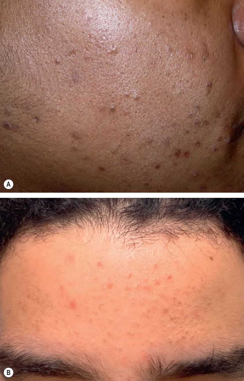
Fig. 36.2 Comedonal acne vulgaris. On the cheek (A) and forehead (B), there are open and closed comedones as well as postinflammatory hyperpigmentation (A) and inflammatory papules (B). B, Courtesy Kalman Watsky, MD.
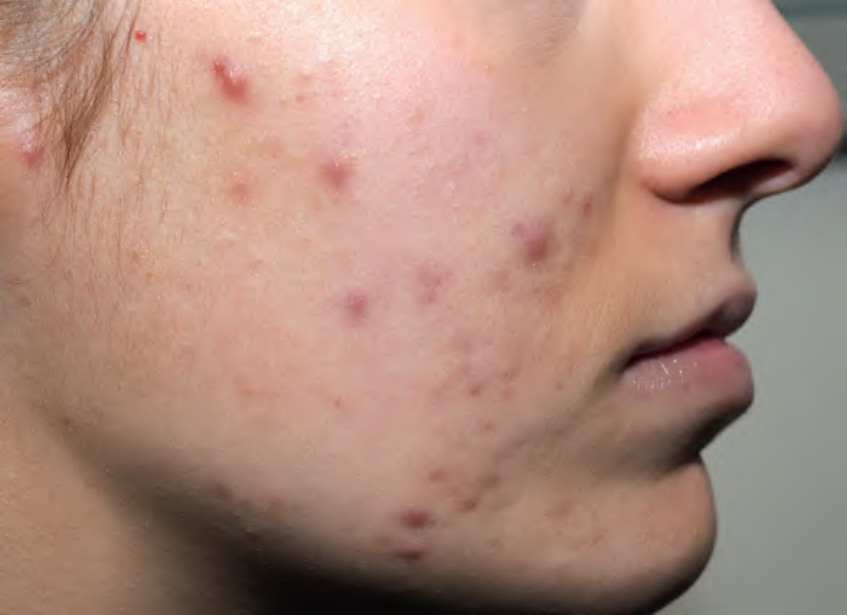
Fig. 36.3 Moderate acne vulgaris. Scattered papulopustules and papulonodules are present on the cheek. Courtesy Kalman Watsky, MD.
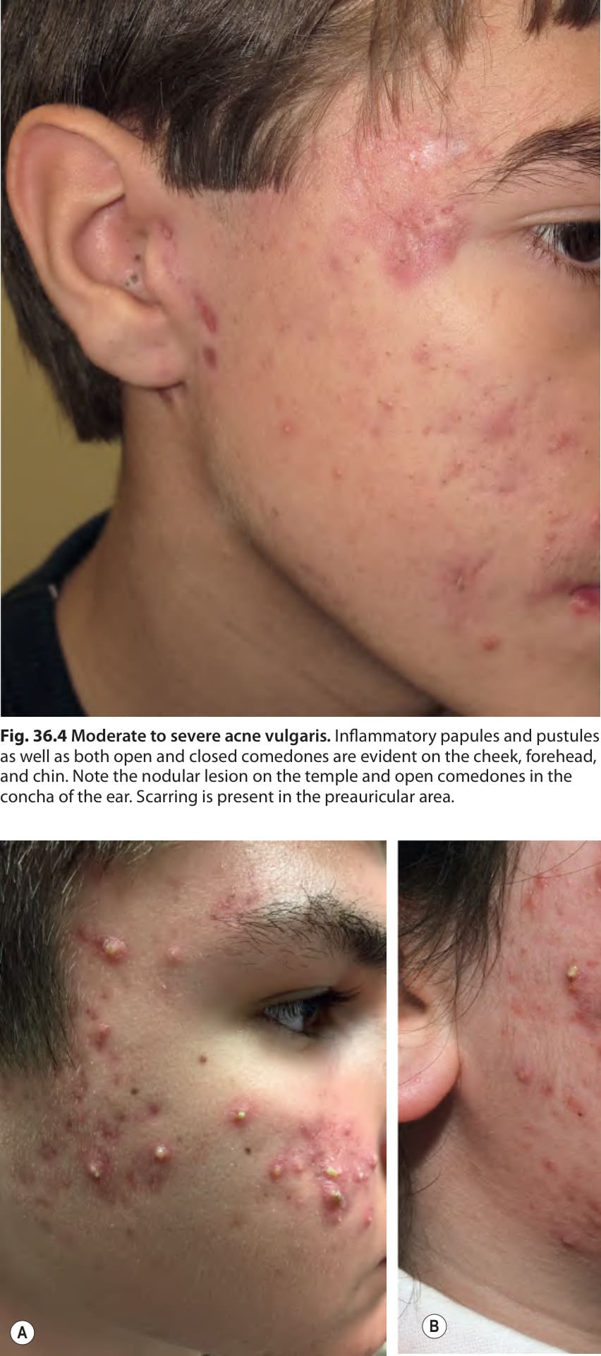
Fig. 36.4 Moderate to severe acne vulgaris. Inflammatory papules and pustules as well as both open and closed comedones are evident on the cheek, forehead, and chin. Note the nodular lesion on the temple and open comedones in the concha of the ear. Scarring is present in the preauricular area.
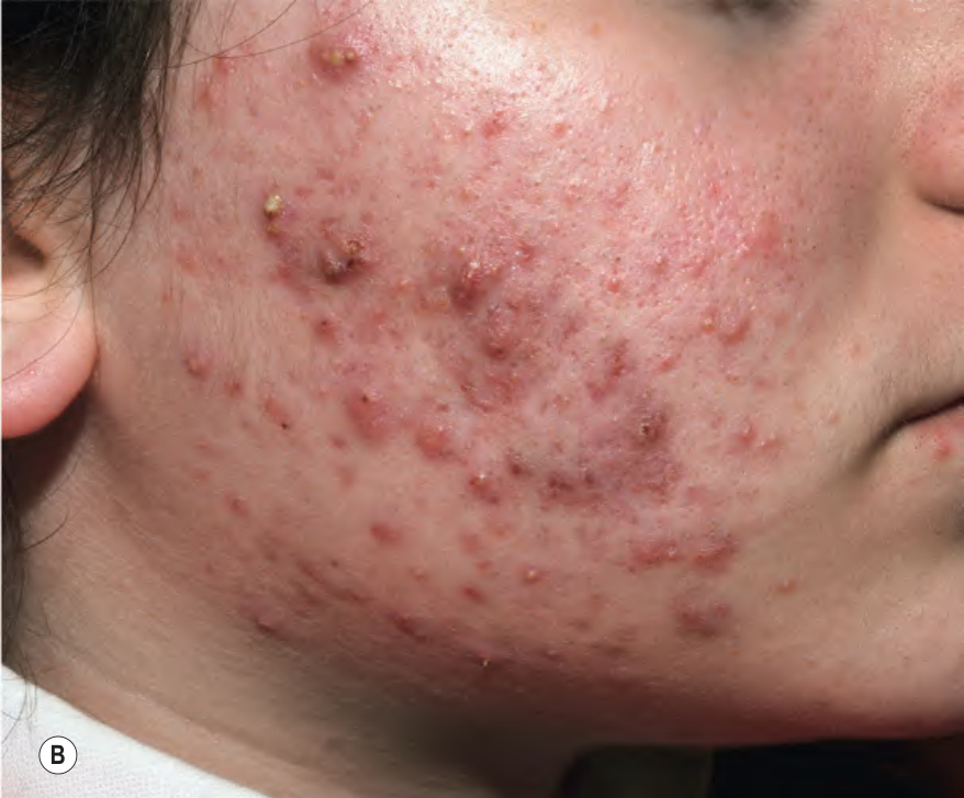
Fig. 36.5 Moderate to severe acne vulgaris.
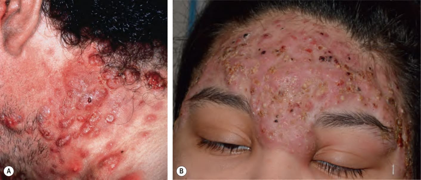
Fig. 36.6 Severe to very severe nodular acne.
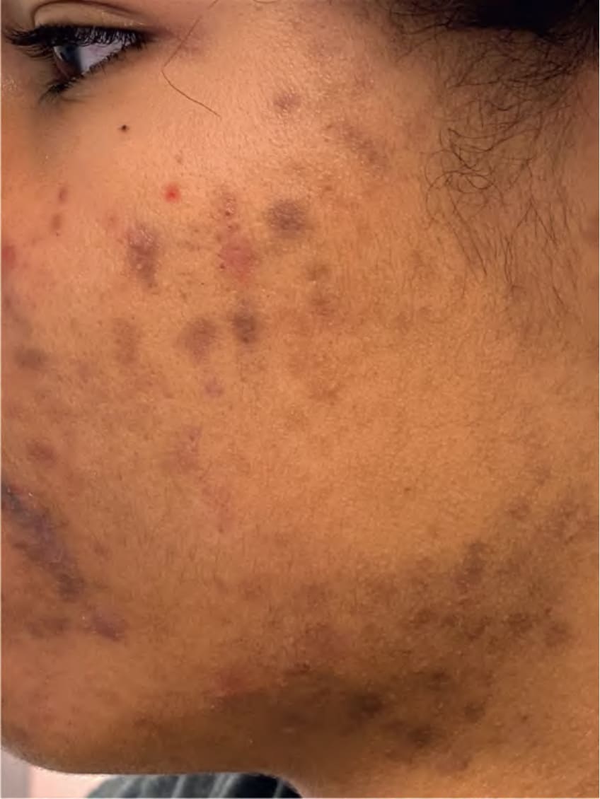
Fig. 36.7 Postinflammatory hyperpigmentation secondary to acne. Such pigmentary changes are common in patients with darker skin tones.
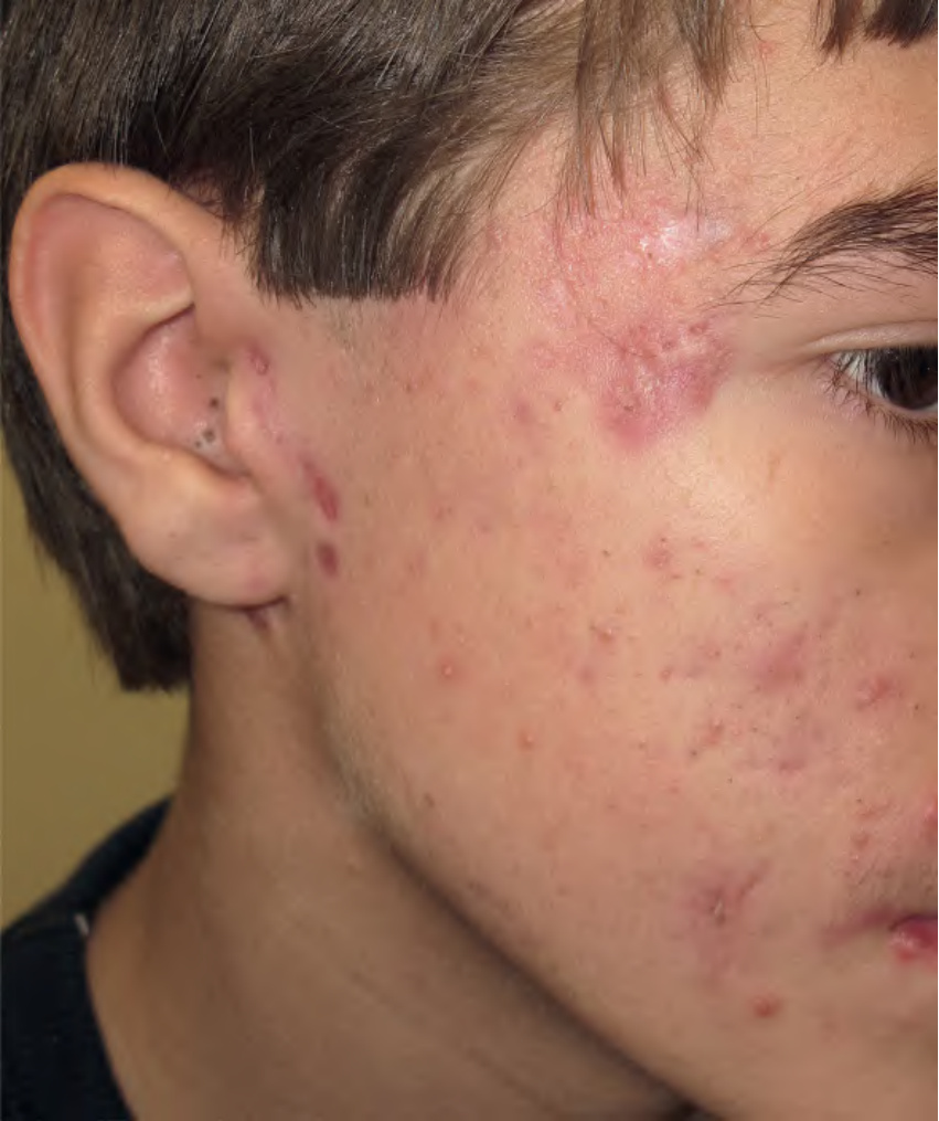
Fig. 36.8 Scarring secondary to acne.A Pitted scars on the cheeks. B, C Mixture of atrophic and hypertrophic scars on the back. B, Courtesy Julie V. Schaffer, MD.
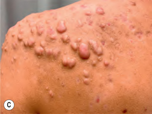
Fig. 36.8 Scarring secondary to acne.A Pitted scars on the cheeks. B, C Mixture of atrophic and hypertrophic scars on the back. B, Courtesy Julie V. Schaffer, MD.
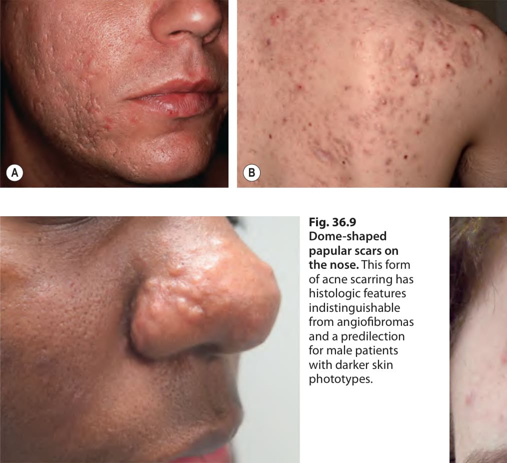
Fig. 36.9 Dome-shaped papular scars on the nose. This form of acne scarring has histologic features indistinguishable from angiofibromas and a predilection for male patients with darker skin phototypes.
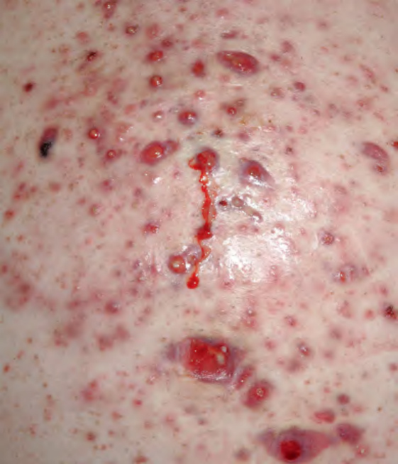
Fig. 36.10 Acne fulminans. Eruptive, friable papulopustules with erosions, oozing, and formation of granulation tissue.
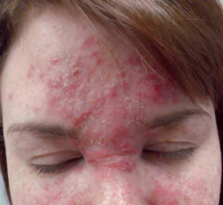
Fig. 36.11 Solid facial edema due to acne vulgaris. There is soft tissue swelling in the central portion of the face. Courtesy Boni Elewski, MD.
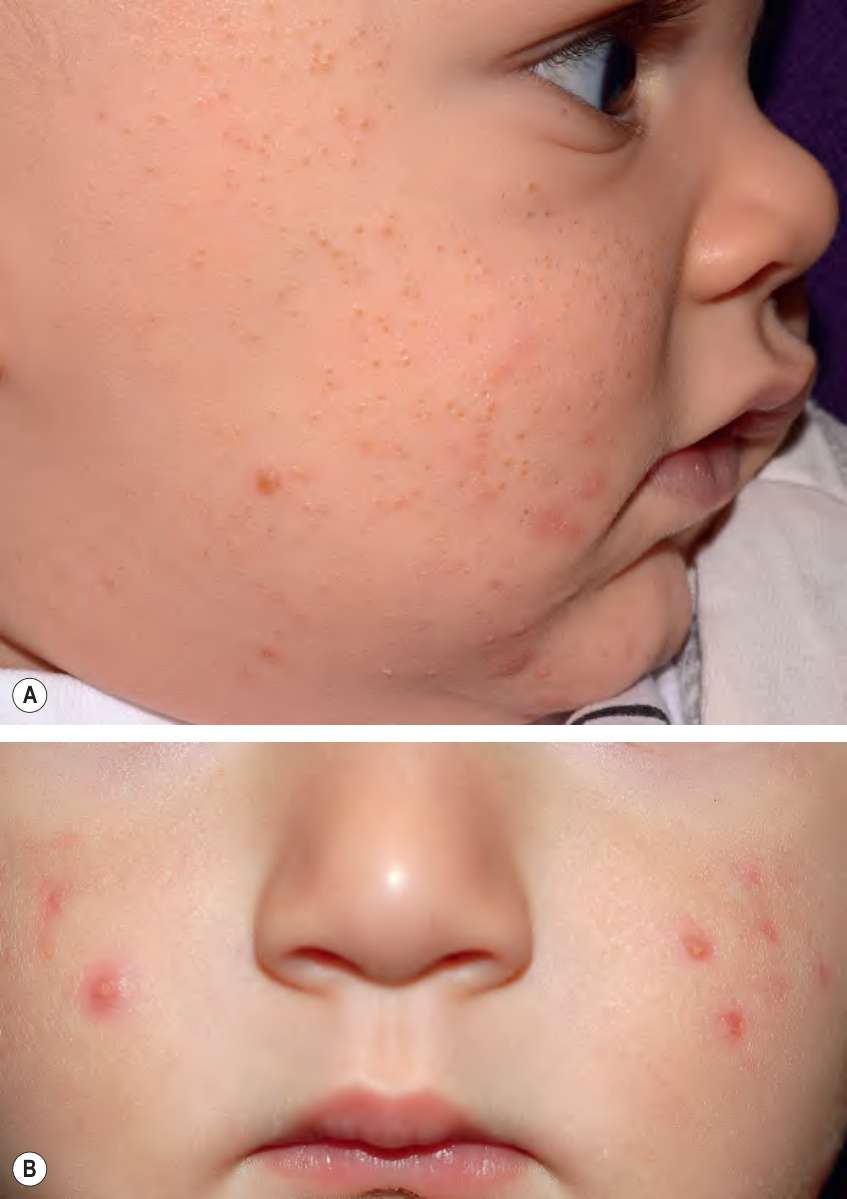
Fig. 36.12 Infantile acne. Presentations can range from numerous open comedones (A) to primarily papulopustules (B). A, Courtesy Julie V. Schaffer, MD; B, Courtesy Kalman Watsky, MD.
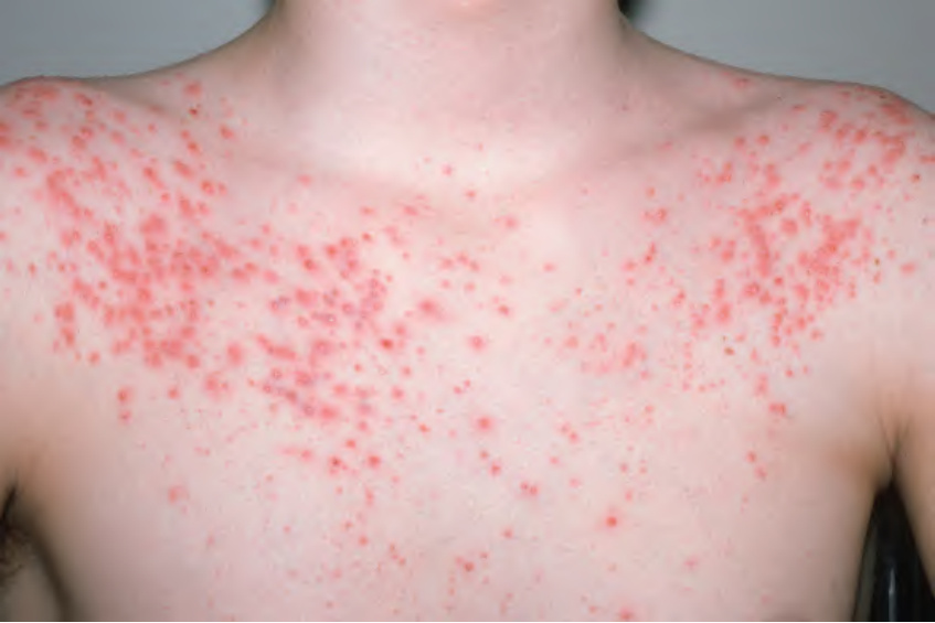
Fig. 36.13 Acneiform eruption secondary to high-dose dexamethasone.
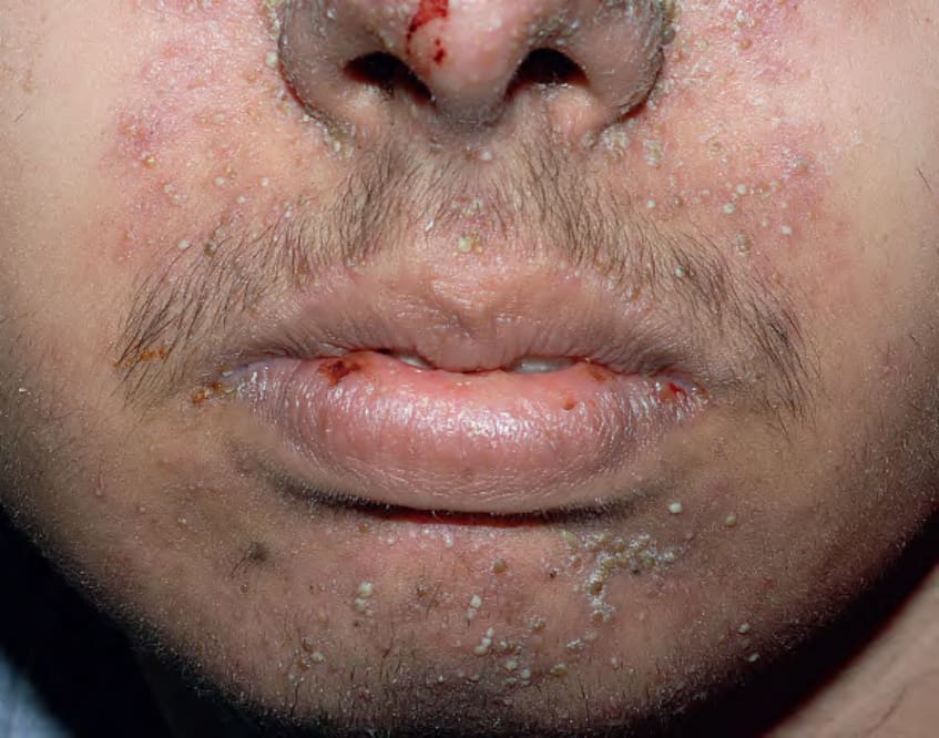
Fig. 36.14 Acneiform eruptions due to epidermal growth factor receptor inhibitors. Numerous monomorphous follicular pustules on the face of an adolescent boy treated with erlotinib.
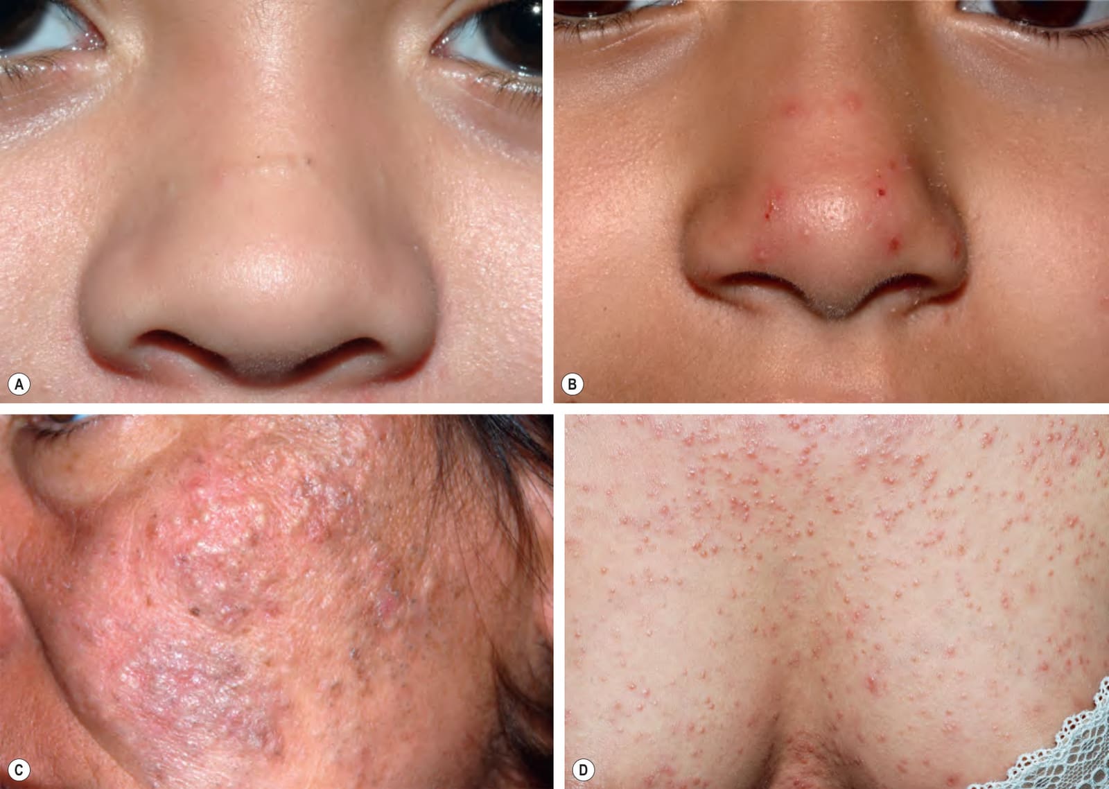
Fig. 36.15 Disorders in the differential diagnosis of acne vulgaris.A Pseudoacne of the transverse nasal crease in a young child. Note the milia and comedones located along this anatomical demarcation line. B Demodicosis presenting as pustules on the nose of a healthy child. An increased number of Demodex mites was observed in a scraping. C Acneiform follicular mucinosis on the cheek of a woman. D Follicular mycosis fungoides that presented as numerous lesions with a comedonal appearance on the chest, abdomen, and back. A, B, D, Courtesy Julie V. Schaffer, MD; C, Courtesy Lorenzo Cerroni, MD.
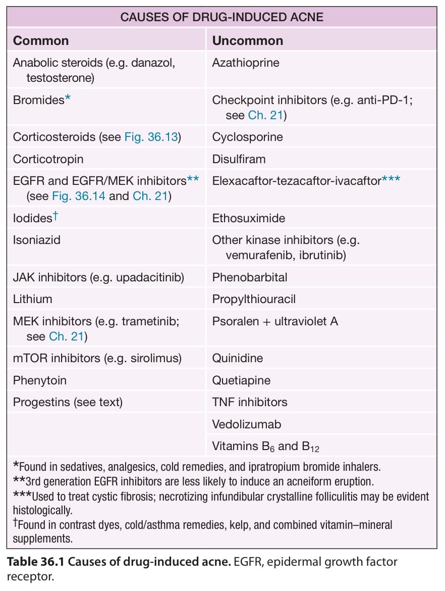
Table 36.1 Causes of drug-induced acne. EGFR, epidermal growth factor receptor.
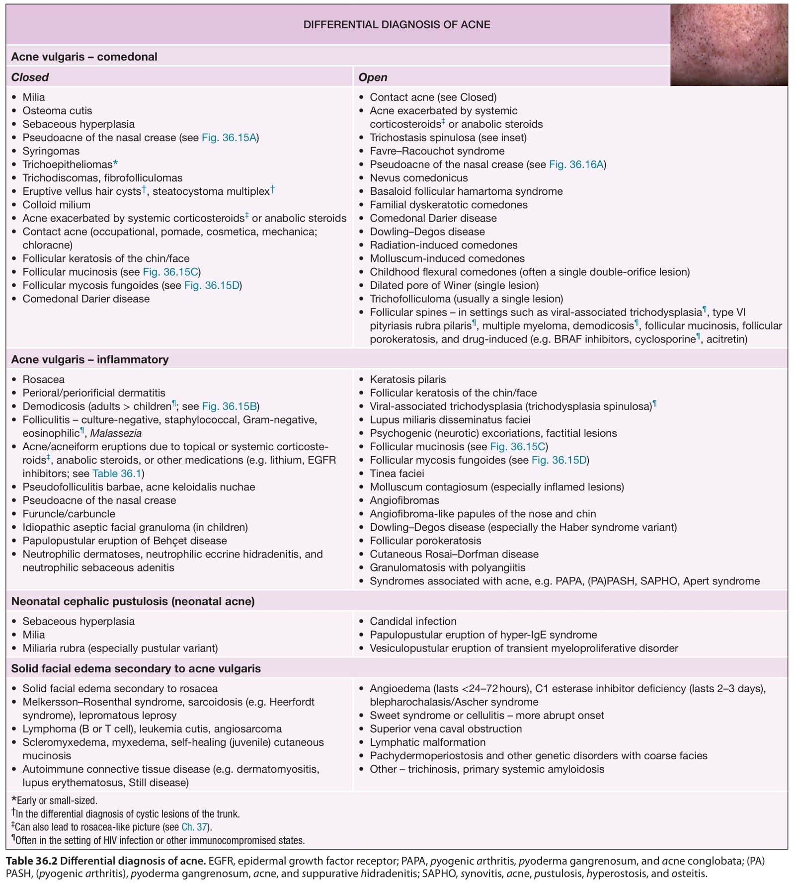
Table 36.2 Differential diagnosis of acne. EGFR, epidermal growth factor receptor; PAPA, pyogenic arthritis, pyoderma gangrenosum, and acne conglobata; (PA) PASH, (pyogenic arthritis), pyoderma gangrenosum, acne, and suppurative hidradenitis; SAPHO, synovitis, acne, pustulosis, hyperostosis, and osteitis.