PATHOLOGY
Histopathologic examination of acne lesions demonstrates the stages of acnegenesis that parallel the clinical findings (see Fig. 36.1). In early lesions, microcomedones are seen. A mildly distended follicle with a narrowed follicular opening is impacted by shed keratinocytes. The granular layer at this stage is prominent. In closed comedones, the degree of follicular distension is increased and a compact cystic structure forms. Within the cystic space, eosinophilic keratinaceous debris, hair, and numerous bacteria are present. Open comedones have broad, expanded follicular ostia and greater overall follicular distension. The sebaceous glands are typically atrophic or absent. A mild perivascular mononuclear cell infiltrate encircles the expanding follicle.
As the follicular epithelium distends, the cystic contents inevitably begin to rupture into the dermis (Fig. 36.16). The highly immunogenic cystic contents (keratin, hair, and bacteria) induce a marked inflammatory response. Neutrophils first appear, creating a pustule. As the lesion matures, foreign body granulomatous inflammation engulfs the follicle and end-stage scarring can result.
In acne fulminans, there is massive inflammation with varying degrees of overlying necrosis. Comedones are rarely observed. Severe scarring may be seen in resolving lesions.
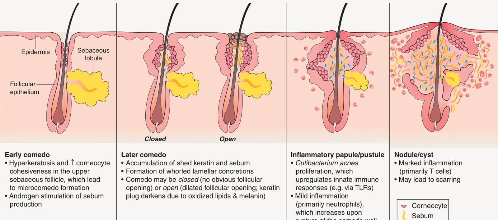
Fig. 36.1 Pathogenesis of acne.
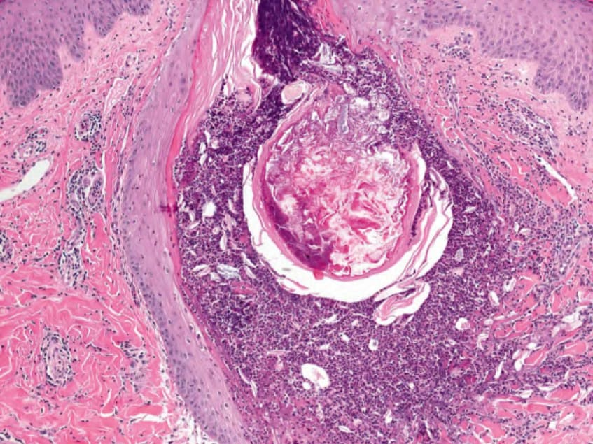
Fig. 36.16 Histology of an inflamed comedo. There is disruption of the pilosebaceous unit and secondary inflammation. Courtesy Lorenzo Cerroni, MD.