DIFFERENTIAL DIAGNOSIS
Although classic acne vulgaris is usually easily recognized clinically, the differential diagnosis of acneiform eruptions is broad and depends upon the age of onset, lesional morphology, and location (Table 36.2; see Fig. 36.15). During the neonatal period, acne must be differentiated from other common dermatoses. Sebaceous hyperplasia occurs in the majority of healthy neonates, presenting as transient yellowish papules on the cheeks, nose, and forehead. Miliaria rubra is also very common during the neonatal period, when overheating and bundling can cause temporary eccrine duct obstruction that leads to the formation of small inflammatory papulopustules. Small, white milia are often apparent on the cheeks and nose of neonates, but they generally resolve within a few months.
Predominantly comedonal acne vulgaris needs to be differentiated from comedonal eruptions caused by follicular occlusion or friction,
including acne mechanica, acne cosmetica, pomade acne, and occupational acne (see above); history as well as location can help to make the diagnosis of these forms of “contact acne”. Sebaceous hyperplasia, a very common finding in adults, is relatively uncommon in adolescents. These yellowish, lobulated papules arise primarily on the forehead and cheeks.
A solitary enlarged comedo is better classified as a dilated pore of Winer; such lesions rarely represent a large-pore basal cell carcinoma. Multiple open comedones are clustered in the lateral malar region in Favre–Racouchot syndrome (see Ch. 87) or appear in a linear array in nevus comedonicus (see Chs. 62 & 109). If multiple vellus hairs arise from a dilated follicular orifice in association with keratinous debris, trichostasis spinulosa is the likely diagnosis. The most common location is the nose.
Angiofibromas and appendageal tumors of follicular origin, e.g. trichoepitheliomas, trichodiscomas and fibrofolliculomas, often present as multiple facial papules (see Ch. 111). They are typically non-inflammatory, and trichoepitheliomas are concentrated in the nasolabial folds. Non-inflammatory, closed cystic papules and nodules on the central chest and back characterize steatocystoma multiplex (see Ch. 110). This autosomal dominant disorder must be differentiated from a related entity, eruptive vellus hair cysts. These smaller cysts may become inflamed and, as the name implies, contain multiple vellus hairs that can be easily visualized microscopically.
The follicle-based inflammatory papules and pustules of acne vulgaris must be distinguished from the many forms of folliculitis, including staphylococcal, Gram-negative, and eosinophilic variants (see Fig. 38.1). In folliculitis, the lesions are relatively monomorphous and comedones are not present. Gram-negative folliculitis can complicate acne vulgaris treated with oral antibiotics for a prolonged period; the inflammatory lesions typically appear on the central face, including the upper lip, and cheeks. In contrast, Pseudomonas (“hot tub”) folliculitis favors the lower trunk and other sites covered by a bathing suit. Malassezia (Pityrosporum) folliculitis, which may develop in acne patients receiving antibiotic therapy, presents with fine papules and pustules favoring the forehead and upper trunk. Eosinophilic folliculitis usually occurs in the setting of immunosuppression (e.g. HIV infection) and is markedly pruritic.
Pseudofolliculitis barbae and acne keloidalis nuchae most often affect men with Afro-textured hair (see Ch. 38). The papular component of rosacea favors the malar region, chin, and forehead; the presence of telangiectasias, an absence of comedones, and a history of easy flushing can aid in diagnosis (see Ch. 37). Rosacea typically occurs at a later age than acne, but both can develop in a single individual. Prolonged use of topical corticosteroids on the face may lead to rosacea-like lesions or periorificial dermatitis, and patients treated with oral corticosteroids can develop an eruption of monomorphous papulopustules that favors the trunk (“steroid folliculitis”; see Fig. 36.13). This can occur at any age and resolves upon discontinuation of the corticosteroid. Lastly, psychogenic (neurotic) excoriations and factitial dermatitis concentrated on the face, chest, and back can mimic acne, particularly acne excoriée. Linearity and the lack of clinically detectable primary lesions are clues.
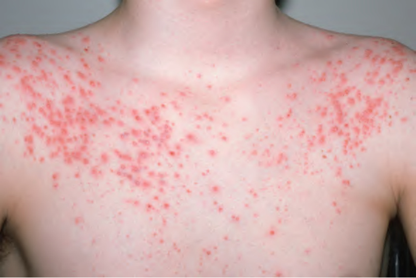
Fig. 36.13 Acneiform eruption secondary to high-dose dexamethasone.
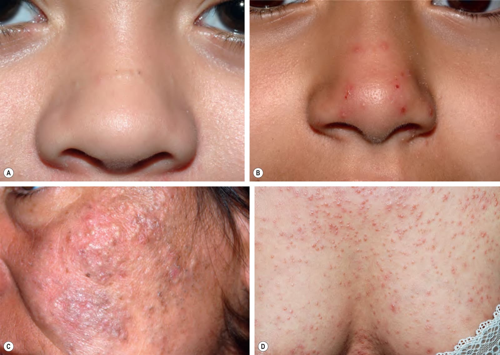
Fig. 36.15 Disorders in the differential diagnosis of acne vulgaris.A Pseudoacne of the transverse nasal crease in a young child. Note the milia and comedones located along this anatomical demarcation line. B Demodicosis presenting as pustules on the nose of a healthy child. An increased number of Demodex mites was observed in a scraping. C Acneiform follicular mucinosis on the cheek of a woman. D Follicular mycosis fungoides that presented as numerous lesions with a comedonal appearance on the chest, abdomen, and back. A, B, D, Courtesy Julie V. Schaffer, MD; C, Courtesy Lorenzo Cerroni, MD.
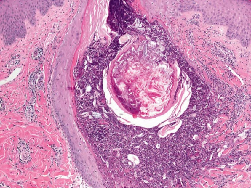
Fig. 36.16 Histology of an inflamed comedo. There is disruption of the pilosebaceous unit and secondary inflammation. Courtesy Lorenzo Cerroni, MD.
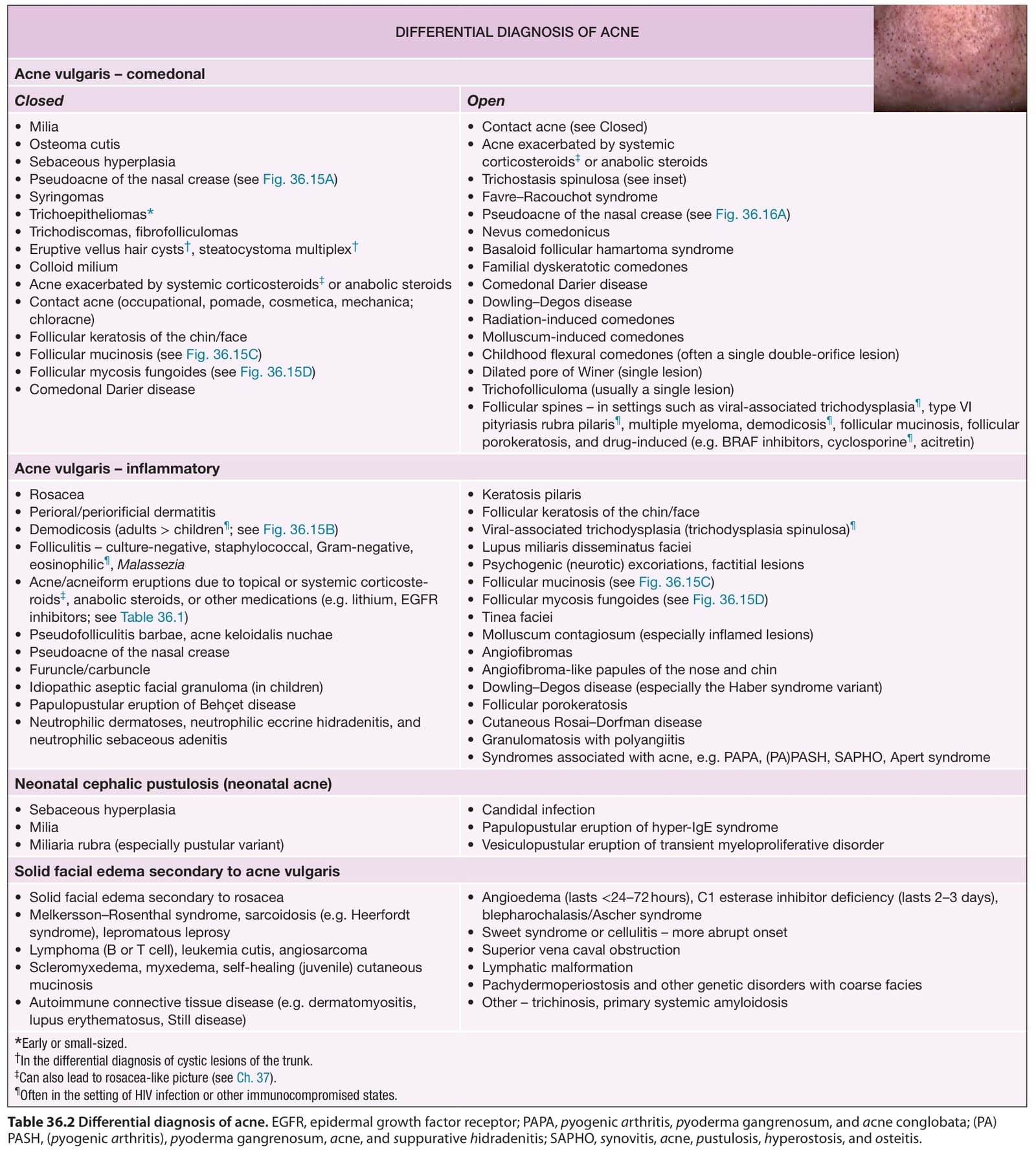
Table 36.2 Differential diagnosis of acne. EGFR, epidermal growth factor receptor; PAPA, pyogenic arthritis, pyoderma gangrenosum, and acne conglobata; (PA) PASH, (pyogenic arthritis), pyoderma gangrenosum, acne, and suppurative hidradenitis; SAPHO, synovitis, acne, pustulosis, hyperostosis, and osteitis.