PATHOGENESIS
Several different but interrelated pathomechanisms have been proposed for rosacea (Fig. 37.1), with predominant pathways reflecting clinical features.
Genetic Factors
Rosacea is more common in individuals of Northern European descent and concordance in monozygotic twins has been demonstrated. A genome-wide association study identified two single-nucleotide polymorphisms and three HLA alleles associated with rosacea.
Dysregulated Cutaneous Immunity
Dysregulation of the cutaneous innate immune system can lead to abnormal inflammatory responses to environmental stimuli and commensal microorganisms. Triggering this innate immune system leads to the release of antimicrobial peptides such as cathelicidin (LL-37). After LL-37 propeptide is secreted by keratinocytes and other epithelial cells, it is cleaved into its active form by proteases (e.g. kallikrein-5).
Active cathelicidin peptides then induce both proinflammatory and angiogenic activity. In the facial skin of individuals with rosacea, there are abnormally high levels of LL-37 (see Fig. 37.1). Also, LL-37 has been shown to contribute to inflammation via activation of the NLRP3 inflammasome (see Fig. 4.2).
Toll-like receptors (TLRs) are pathogen-associated molecular pattern sensors that are involved in cutaneous innate immunity (see Fig. 4.1). In rosacea, keratinocytes express elevated levels of TLR2, leading to increased expression and activity of kallikrein proteases and thus cathelicidins. In addition, matrix metalloproteinases, which activate kallikrein 5, are up-regulated in patients with rosacea. Mast cells, which also release cathelicidin, are increased in number within the dermis of rosacea patients. Lastly, in addition to dysregulated innate immunity, an abnormal adaptive immune response has been observed in rosacea, with Th1/Th17 polarized inflammation and macrophage infiltration.
Neurogenic Inflammation
Patients with rosacea often report sensations of stinging or burning of their skin, with affected individuals exhibiting lower heat pain thresholds when compared to controls. When cutaneous nerve endings expressing transient receptor potential vanilloid (TRPV) cation channels are stimulated by trigger factors (e.g. spicy food, heat, alcohol), this can lead to dysesthesia, flushing, and erythema. Heightened TRPV activity has been detected in the skin of rosacea patients and it leads to neurogenic inflammation, i.e. an inflammatory response induced by sensory nerves in which neuromediators are released at the site of inflammation. These neuromediators can result in vasodilation, extravasation of plasma proteins, and recruitment of inflammatory cells (see Fig. 37.1).
Vascular Alterations
Several clinical features of rosacea, including transient erythema, persistent centrofacial erythema, telangiectasias and flushing, point to the role the vascular system plays in its pathogenesis. An increase in blood flow within skin lesions of rosacea has been demonstrated, and patients with rosacea flush more readily in response to heat. Histopathologic studies of lesional skin found an elevated expression of vascular endothelial growth factor (VEGF), CD31, and the lymphatic endothelial marker podoplanin (D2-40), implying increased stimulation of vascular and lymphatic endothelial cells.
Ultraviolet Radiation (UVR)
Exposure to UVB can induce angiogenesis and increase secretion of angiogenic factors (e.g. VEGF) from keratinocytes. UVR also induces production of reactive oxygen species, which upregulate matrix metalloproteinases that lead to vascular and dermal matrix damage. Although clinically the erythema and telangiectasias of rosacea can resemble telangiectatic photoaging, a case–control observational study provided evidence that they are distinct entities, but as expected can overlap.
Epidermal Barrier Dysfunction
Several clinical features of rosacea imply skin barrier dysfunction. Rosacea patients often report facial dryness, and studies have confirmed a lowered threshold for skin irritancy. In rosacea skin, there is increased transepidermal water loss, a marker of epidermal barrier dysfunction. It has been suggested that disruption or abnormalities of the stratum corneum allow penetration of sensory irritants. In addition, patients with papulopustular rosacea have an abnormal skin surface fatty acid profile as well as reduced epidermal hydration levels. The latter have been shown to improve following treatment with minocycline and resolution of inflammatory lesions.
Microorganisms
Demodex mites (folliculorum and brevis) are normally present on the face as commensal microbes, but in rosacea, greater numbers of these mites are detected by skin surface biopsy techniques. In routine histologic sections, the mites often appear prominently within pilosebaceous follicles, and follicular infestation with multiple mites can be associated with an intense perifollicular infiltrate of predominantly CD4 T helper cells. In addition, antigenic proteins from a bacterium (Bacillus oleronius) isolated from Demodex mites can stimulate inflammation in patients with papulopustular rosacea. It has been suggested that Demodex mites and their associated bacteria upregulate local proteases, thereby potentiating dysregulation of the cutaneous innate immune response (see above).
Study results differ as to whether Helicobacter pylori infection of the upper gastrointestinal tract plays a role in the pathogenesis of rosacea. A meta-analysis of 14 observational studies did not find statistically significant associations between either H. pylori infection and rosacea or H. pylori eradication therapy and symptomatic improvement of rosacea.
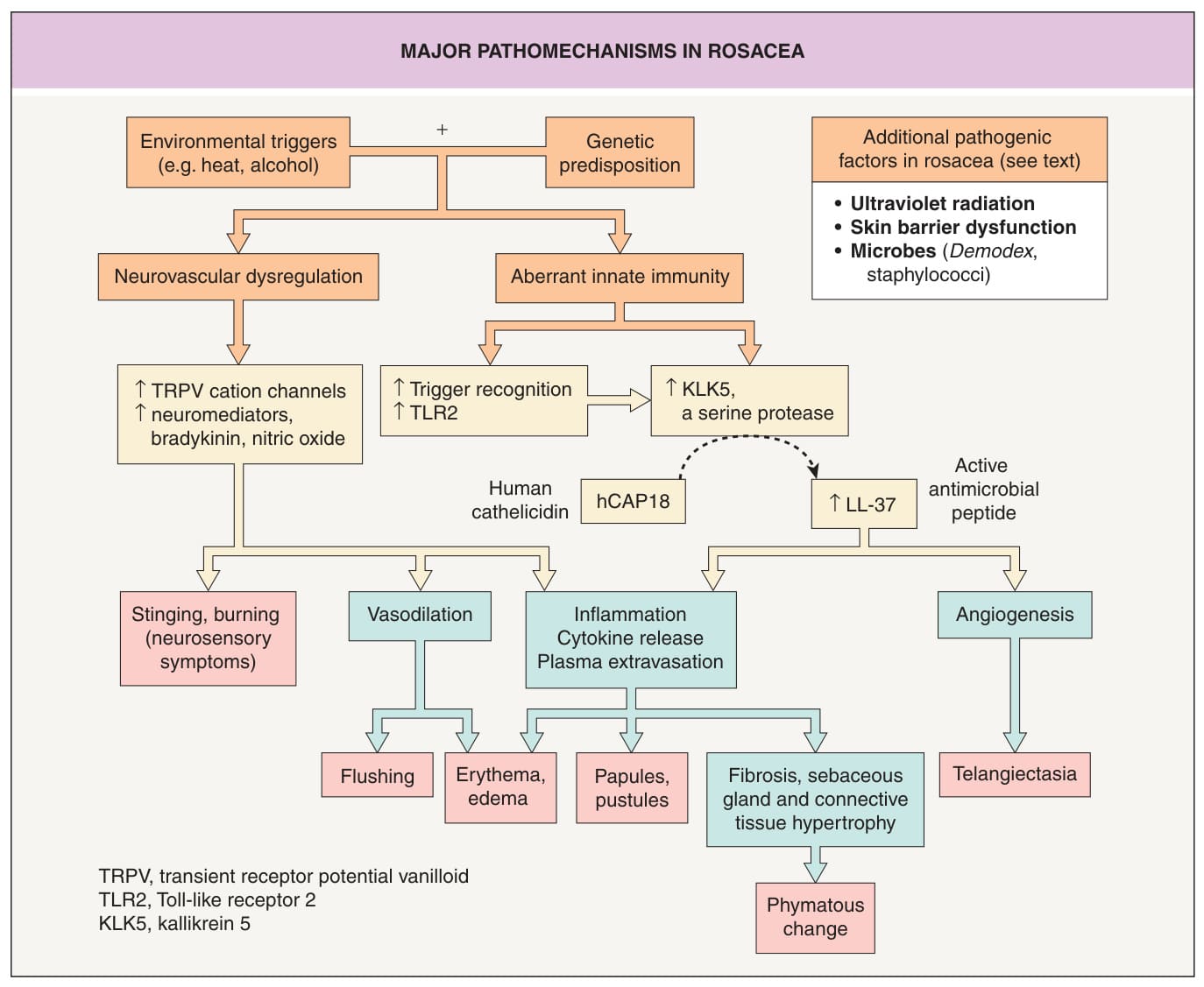
Fig. 37.1 Major pathomecha- nisms in rosacea. In genetically predisposed individuals (e.g. HLA-DRB103:01, HLA-DQA105:01, HLA-DQB1*02:01, SNP rs763035), environmental factors can trigger neurovascular dysregulation and an aberrant innate immune response, both of which can lead to cutaneous inflammation, including the clinical manifestations of rosacea. Adapted from Steinhoff M, Buddenkotte J, Aubert J, et al. Clinical, cellular, and molecular aspects in the pathophysiology of rosacea. J Invest Dermatol Symp Proc. 2011;15:2–11.