CLINICAL FEATURES
In 2002, a classification scheme divided rosacea into four main subtypes – erythematotelangiectatic, papulopustular, phymatous, and ocular – based upon the predominant lesion morphology. However, it did not account for the frequent co-occurrence of more than one subtype nor the potential progression of one subtype to another. Recognizing these potential shortcomings, a revised classification of rosacea was recommended by the global ROSacea COnsensus (ROSCO) panel in 2016 and by the National Rosacea Society Expert Committee in 2017 (Table 37.1).
Both groups agreed that either one of the following two phenotypes are diagnostic of rosacea: (1) fixed centrofacial erythema in a characteristic pattern that may periodically intensify; and (2) phymatous changes. They also concluded that in the absence of one of these two diagnostic phenotypes, the presence of two or more of the following four major phenotypes can be diagnostic: (1) papules and pustules; (2) flushing; (3) telangiectasia; and (4) ocular manifestations. Secondary signs and symptoms that may also occur in conjunction with one or more diagnostic or major phenotypes include burning and stinging, facial edema, dry appearance of the skin, and other ocular manifestations.
Diagnostic Phenotypes (Table 37.2)
Fixed centrofacial erythema in a characteristic pattern that
Persistent erythema of the central face is the most common presentation of rosacea in patients with skin phototypes I–III (Fig. 37.2). Of note, it may be more difficult to appreciate in darker skin phototypes. In addition, it can be challenging to differentiate from telangiectatic photoaging, although there are relative differences (see Table 37.4). In addition, other entities that can cause persistent facial erythema should be considered, e.g. lupus erythematosus, seborrheic dermatitis.
Phymatous changes
These changes include sebaceous gland hypertrophy, patulous follicles, a bulbous appearance on the distal portion of the nose, and eventual fibrosis. Rhinophyma is by far the most common clinical presentation, occurring primarily in men (Fig. 37.3). Involvement of other anatomic sites has been reported, but it is rare (Table 37.3). Patients with rhinophyma may have other features of rosacea, usually mild to moderately severe papules and pustules. However, phymatous changes may arise de novo without any preceding skin changes and therefore should not be viewed as “end-stage rosacea”.
The earliest clinical sign of rhinophyma is the appearance of patulous follicles on the distal portions of the nose. It has been suggested that telangiectatic vessels in this same location may predispose to hypertrophic changes (Fig. 37.4). In severe cases of rhinophyma, the tissue hypertrophy leads to nasal distortion, with soft fleshy nodular growths resulting in significant disfigurement. Although basal cell carcinomas have been reported to arise in skin affected by rhinophyma, there is insufficient evidence to suggest that this condition predisposes to malignant change.
Major Phenotypes
Papules and pustules
Patients typically have a centrofacial eruption of multiple small (<3 mm), dome-shaped, erythematous papules and/or pustules that
This hyperemia may predispose to the subsequent hypertrophic changes of rhinophyma. Note the early sign of patulous follicles (“dilated pores”). Courtesy Frank C. Powell, MD.
appear singly or in crops (Fig. 37.5 & 37.6). Individual lesions last about two weeks and are then replaced by blotchy postinflammatory erythema which gradually fades. Residual scarring is not a feature of papulopustular rosacea. A halo of erythema may surround larger inflammatory lesions and tiny telangiectatic vessels may be visible within this rim. Occasionally, when there is more severe disease, scaling or superficial crusting may be seen and this has been referred to as rosacea dermatitis (Fig. 37.7). Lastly, some patients will have some degree of persistent erythema of the cheeks that may represent a combination of postinflammatory erythema, telangiectasias, and vasodilation.
Flushing
Frequent flushing and blushing are common in patients with rosacea and occur in response to neurostimulation by various triggers (e.g. heat, alcohol consumption, certain social situations). Although quite noticeable in skin phototypes I–IV, flushing may be less visible in darker phototypes but rather experienced as a sensation of increasing warmth in the face. In addition, when patients complain of significant flushing, other causes of flushing should be considered as outlined in Tables 106.2 & 106.3.
Telangiectasia
Telangiectasias are common and involve primarily the central face, including the chin, in patients with lighter skin phototypes. They need to be distinguished from telangiectatic photoaging (see Table 37.4) and are less common as well as less perceptible in individuals with darker skin phototypes.
Ocular manifestations
Ocular manifestations of rosacea are discussed below.
Secondary Signs and Symptoms
Burning and stinging
Burning and stinging of the facial skin is a common complaint in patients with rosacea while pruritus is unusual. These sensations often occur within a background of erythema and sometimes scale. Patients also report significant sensitivity to and irritation from many products that are applied to the skin.
Facial edema
Facial edema may occur along with or result from prolonged erythema or flushing of the face and is thought to develop from post-capillary extravasation due to inflammation. The soft edematous changes that are sometimes seen in patients with severe inflammatory rosacea (Fig. 37.8) should not be confused with phymatous rosacea. Such changes, unlike those of phymatous rosacea, often improve following successful management of the inflammatory lesions. Solid facial edema is discussed below.
Dry appearance
The centrofacial skin may sometimes appear dry and scaly, almost resembling a type of eczematous dermatitis. Such a presentation is often accompanied by burning and stinging sensations as well as significant sensitivity to topical products. When present, consideration should be given to the concomitant diagnosis of seborrheic dermatitis, which often coexists (see Table 37.4).
Other ocular manifestations
Ocular manifestations of rosacea are discussed below.
Other Clinical Rosacea Presentations
Ocular rosacea
This entity may or may not be accompanied by cutaneous changes of rosacea. Without cutaneous manifestations, the diagnosis of ocular
rosacea can be difficult to establish with certainty (Fig. 37.9). Symptoms are often nonspecific and include dryness, a gritty sensation, an inability to wear contact lenses, tearing, and sometimes pruritus. Patients usually do not associate these ocular symptoms with their rosacea and may not volunteer such information unless specifically asked.
Signs that strongly suggest a diagnosis of ocular rosacea include eyelid margin telangiectasias, interpalpebral conjunctival injection, and on slit lamp examination, corneal spade-shaped infiltrates, scleritis, and sclerokeratitis (see Table 37.1). Other common, but less specific, signs include honey crusting and mild scaling of the eyelid margin, tiny concretions at the bases of the lashes (conical dandruff; see Fig. 37.9A), and irregularity of the eyelid margin. When the disease is more active, there is evidence of blepharitis, often with eyelid swelling and conjunctival injection (see Fig. 37.9C). Cysts arising from the meibomian glands (chalazia) present as firm non-tender swellings of the cutaneous tarsal surface, while hordeola (styes) are often painful. Severe ocular disease (e.g. keratitis, corneal neovascularization, uveitis, scleritis, iritis) is rarely seen in patients with rosacea.
Granulomatous rosacea
In granulomatous rosacea, there are monomorphic, persistent, skincolored to dull red–brown facial papules that are dome-shaped, favor the central face, and usually measure 1–3 mm in diameter (Fig. 37.10). This variant occurs in both children and adults and it may resolve spontaneously a few years after lesions first appear and not recur. Extrafacial lesions (e.g. on the scalp, neck, and upper trunk) are seen more often with the granulomatous variant. Histologically, non-caseating epithelioid granulomas are present within the dermis (see Pathology).
Some authors consider lupus miliaris disseminatus faciei (LMDF) to be a severe form of granulomatous rosacea, with a predilection for the periocular region (as well as the central face) and an “apple-jelly” appearance on diascopy. However, the dermal granulomas seen in biopsy specimens often have central caseation necrosis, thus explaining why this disorder was initially considered a tuberculid. More recently, facial idiopathic granulomas with regressive evolution (FIGURE) was proposed as an alternative term for LMDF. Unfortunately, significant facial scarring can be a permanent sequela. Occasionally, there is extrafacial involvement, which may be confused with other granulomatous
Monomorphous, discrete skin-colored to brown papules scattered on the face that are more persistent than the lesions of papulopustular rosacea. Histologically, granulomas with central caseation necrosis were observed, which led to the diagnosis of lupus miliaris disseminatus faciei.
diseases such as sarcoidosis, cutaneous tuberculosis, or the necrobiotic form of granuloma annulare.
Some authors consider both rosacea conglobata, which is characterized by an eruption of inflammatory cystic lesions that heal with scarring, and rosacea fulminans (pyoderma faciale) to be within the rosacea spectrum. In rosacea fulminans, an explosive onset of inflammatory papules and pustules is superimposed on a background of facial erythema, usually occurring in young women and sometimes during pregnancy.
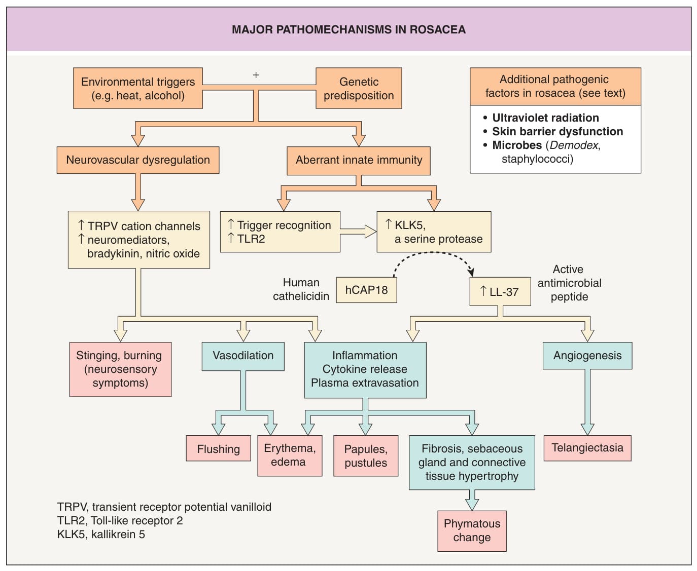
Fig. 37.1 Major pathomecha- nisms in rosacea. In genetically predisposed individuals (e.g. HLA-DRB103:01, HLA-DQA105:01, HLA-DQB1*02:01, SNP rs763035), environmental factors can trigger neurovascular dysregulation and an aberrant innate immune response, both of which can lead to cutaneous inflammation, including the clinical manifestations of rosacea. Adapted from Steinhoff M, Buddenkotte J, Aubert J, et al. Clinical, cellular, and molecular aspects in the pathophysiology of rosacea. J Invest Dermatol Symp Proc. 2011;15:2–11.
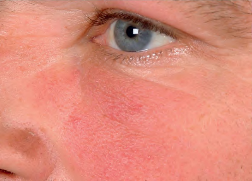
Fig. 37.2 Fixed facial erythema due to rosacea. There was greater involvement of the medial cheek. Courtesy Frank C. Powell, MD.
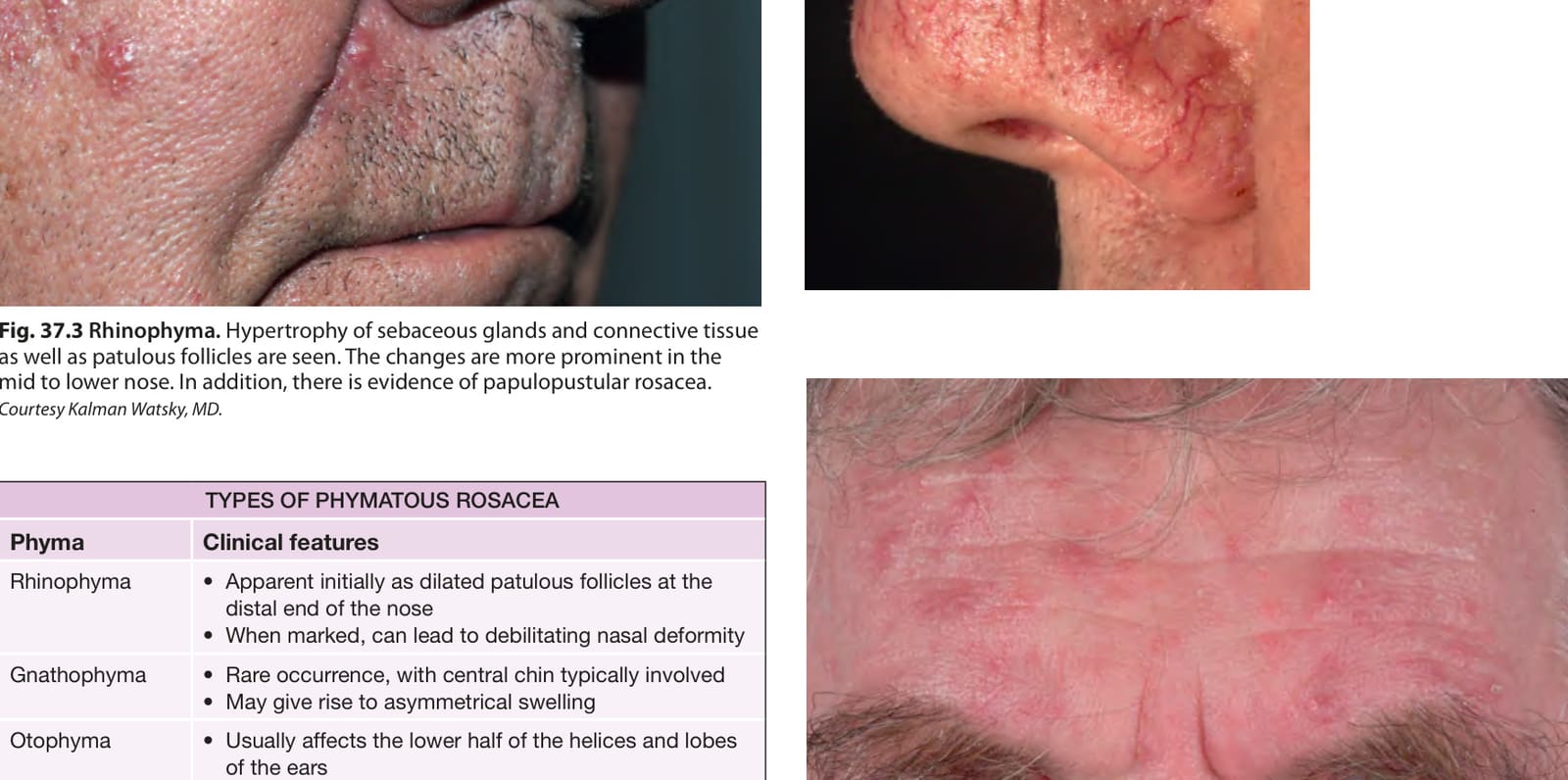
Fig. 37.3 Rhinophyma. Hypertrophy of sebaceous glands and connective tissue as well as patulous follicles are seen. The changes are more prominent in the mid to lower nose. In addition, there is evidence of papulopustular rosacea. Courtesy Kalman Watsky, MD.
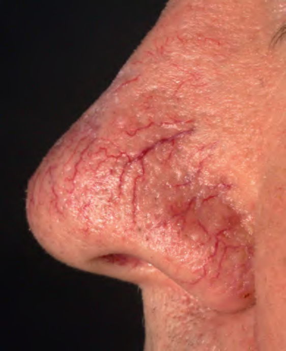
Fig. 37.4 Tortuous, telangiectatic vessels on the distal aspect of the nose contribute to its hyperemic appearance.
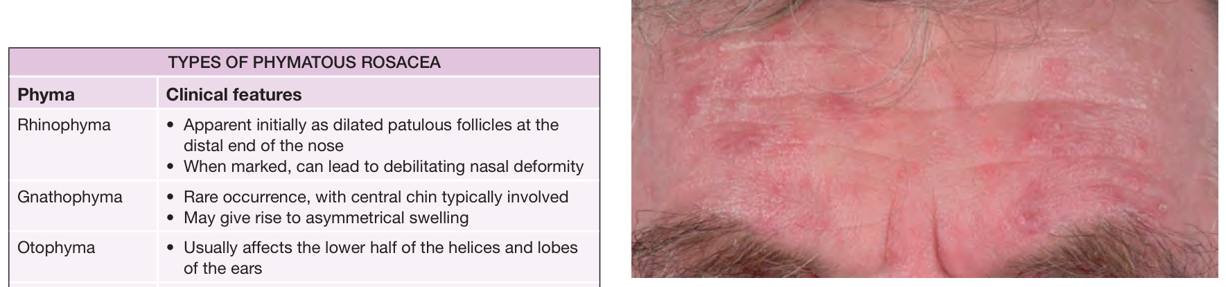
Fig. 37.5 Moderate papulopustular rosacea of the forehead. Note the super-ficial nature of the inflammatory lesions. Courtesy Frank C. Powell, MD.
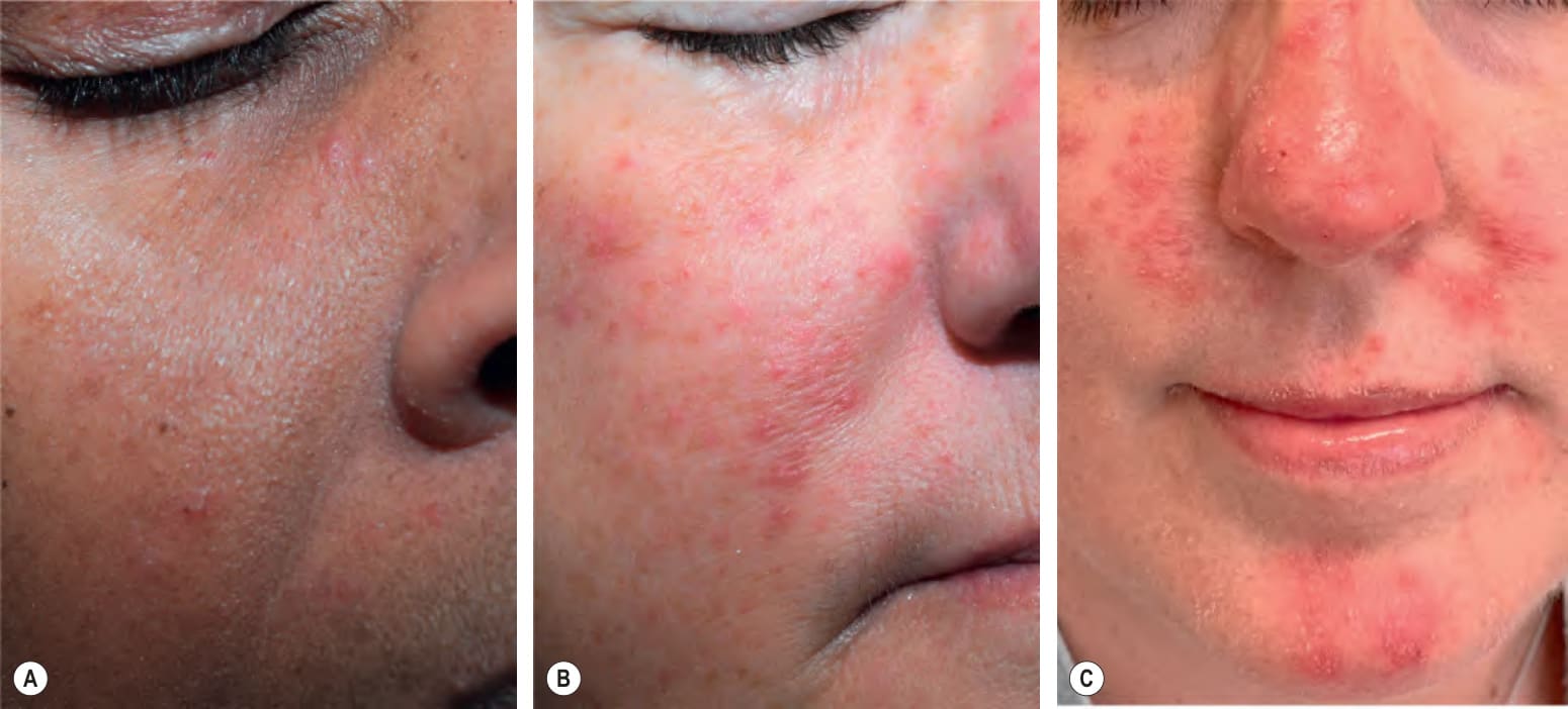
Fig. 37.6 Papulopustular rosacea – spectrum of severity in different skin phototypes.A Mild disease with scattered lesions on the cheek. B Moderate disease with increased number of lesions and areas of background erythema. C Severe disease with characteristic centrofacial distribution. Note the associated xerotic scale. A, B, Courtesy Kalman Watsky, MD.
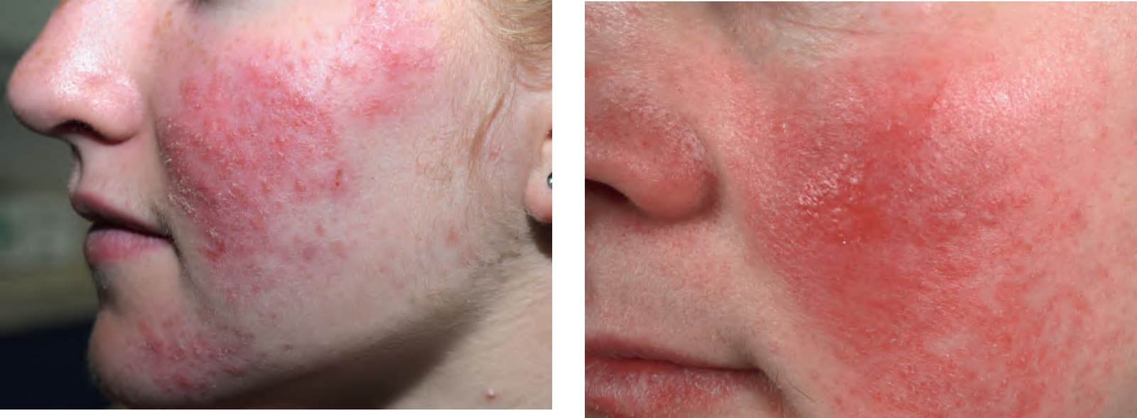
Fig. 37.7 Rosacea dermatitis. When there is more severe disease, scaling and superficial crusting may be seen as on the cheek of this woman. Courtesy Kalman Watsky, MD.
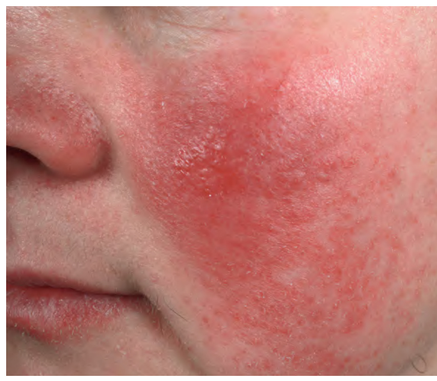
Fig. 37.8 Inflammatory rosacea with edematous changes. An intensely erythematous plaque is present on the medial aspect of the cheek. This may improve once the underlying inflammation is treated appropriately. Courtesy Frank C. Powell, MD.
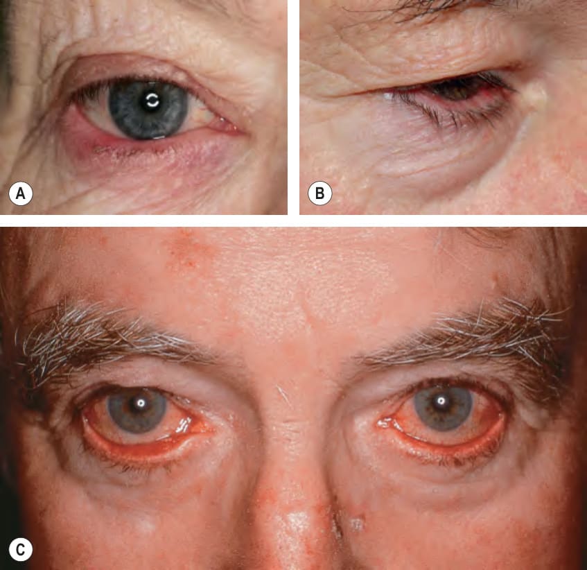
Fig. 37.9 Ocular rosacea.A Tiny concretions of keratin (conical dandruff) are visible at the bases of some of the eyelashes of the lower eyelid. There is also evidence of blepharitis of the lower eyelid and conjunctival injection. B Erythema of the mucosal portion of the lower eyelid and ectropion. C Marked injection of the conjunctivae, leading to the appearance of red eyes. Ectropion is also present. A, Courtesy Frank C. Powell, MD.
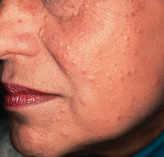
Fig. 37.10 Granulomatous rosacea, lupus miliaris disseminatus faciei variant.
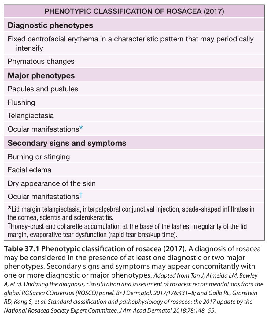
Table 37.1 Phenotypic classification of rosacea (2017). A diagnosis of rosacea may be considered in the presence of at least one diagnostic or two major phenotypes. Secondary signs and symptoms may appear concomitantly with one or more diagnostic or major phenotypes. Adapted from Tan J, Almeida LM, Bewley A, et al. Updating the diagnosis, classification and assessment of rosacea: recommendations from the global ROSacea COnsensus (ROSCO) panel. Br J Dermatol. 2017;176:431–8; and Gallo RL, Granstein RD, Kang S, et al. Standard classification and pathophysiology of rosacea: the 2017 update by the National Rosacea Society Expert Committee. J Am Acad Dermatol 2018;78:148–55.
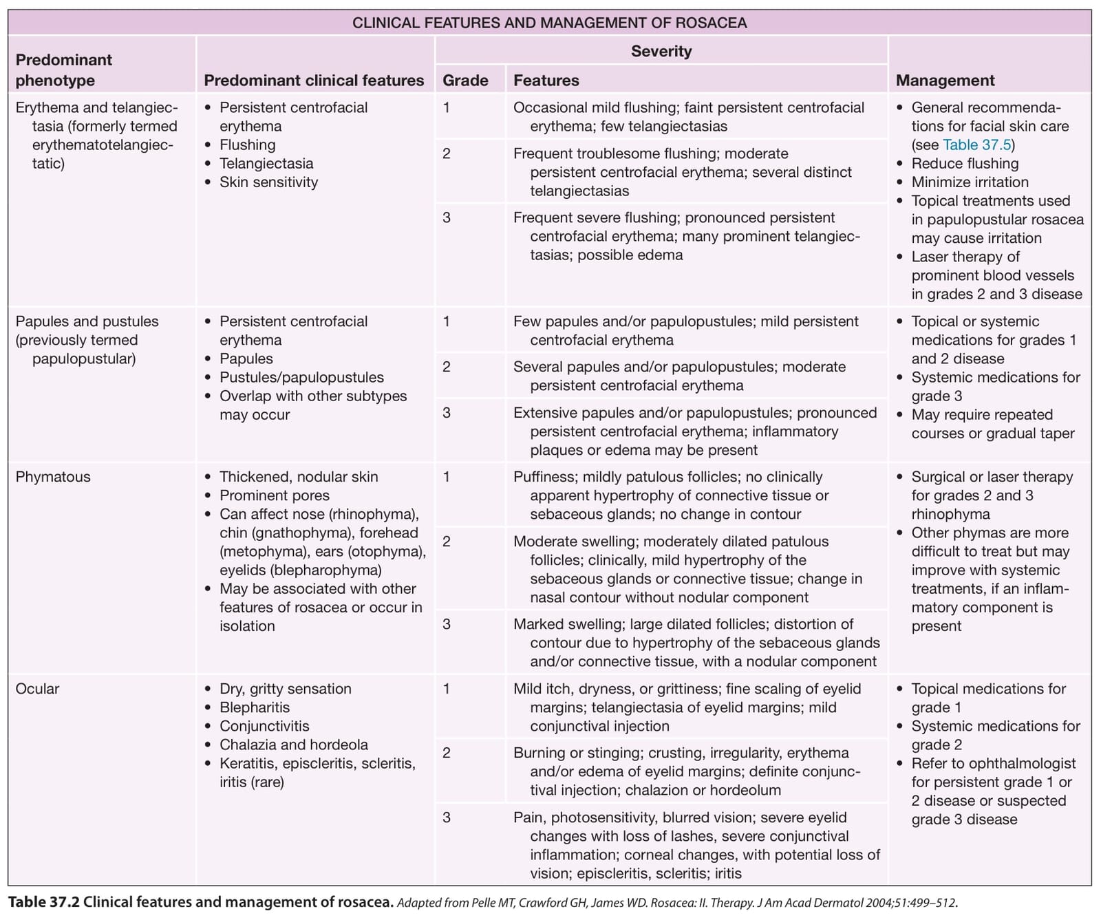
Table 37.2 Clinical features and management of rosacea.Adapted from Pelle MT, Crawford GH, James WD. Rosacea: II. Therapy. J Am Acad Dermatol 2004;51:499–512.
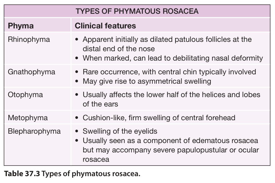
Table 37.3 Types of phymatous rosacea.
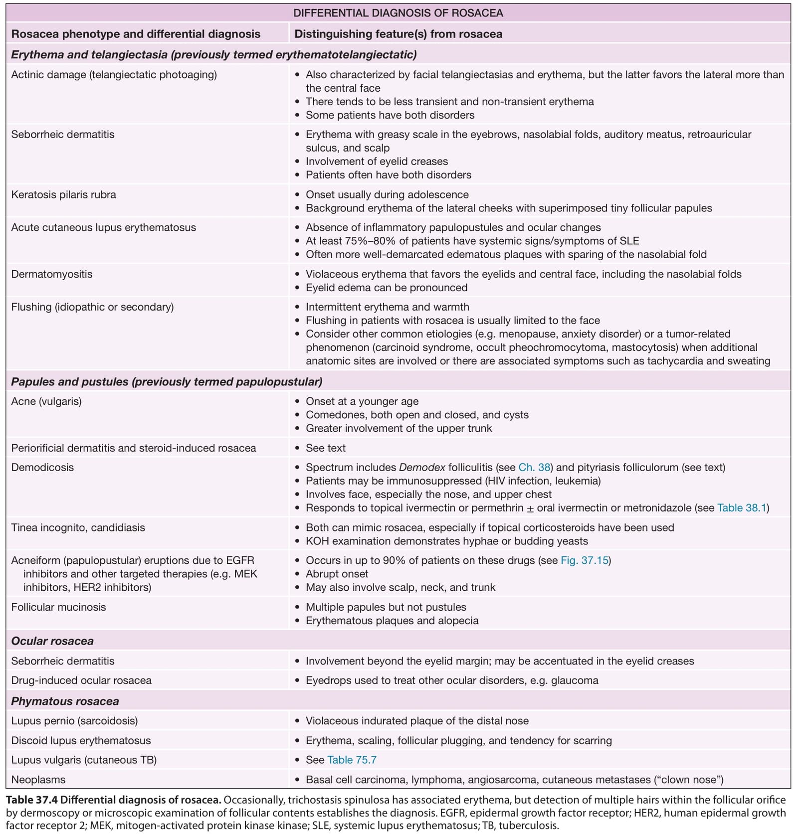
Table 37.4 Differential diagnosis of rosacea. Occasionally, trichostasis spinulosa has associated erythema, but detection of multiple hairs within the follicular orifice by dermoscopy or microscopic examination of follicular contents establishes the diagnosis. EGFR, epidermal growth factor receptor; HER2, human epidermal growth factor receptor 2; MEK, mitogen-activated protein kinase kinase; SLE, systemic lupus erythematosus; TB, tuberculosis.