PATHOGENESIS
The pathogenesis of cutaneous LE is complex, and it involves an interaction between genetic and environmental factors. The latter include ultraviolet radiation (UVR), medications, cigarette smoking, and possibly viruses. This interplay triggers a complex inflammatory cascade of cytokine, chemokine, and inflammatory cell responses that include cells residing within as well as recruited to the skin. Overall, the lichenoid tissue reaction, defined as epidermal basal cell damage and a band-like lymphocytic infiltrate in the upper dermis, characterizes most subsets of cutaneous LE. It involves activation of keratinocytes, endothelial cells, and skin dendritic cells plus the production of type I interferons (IFNs), followed by the recruitment and activation of CD4+ and CD8+ cytotoxic T cells. The end result is cytotoxic keratinocyte damage.
Genes that can affect overall immunoreactivity include those whose protein products are involved in B and T cell function, innate immunity, immune complex clearance, apoptosis, ubiquitination, DNA methylation, or cellular adhesion. Examples from all these gene classes, in addition to genes with unknown function, have been implicated in some way in human or animal models of SLE (Table 41.1; see Ch. 4).
Both genetic background based upon ancestry and mutations in specific genes contribute to the clinical heterogeneity in cutaneous LE. For example, the incidences of photosensitivity and discoid LE (DLE) differ in those of northern versus southern European ancestry, and SCLE is associated with the HLA-B8-DR3 extended haplotype (including TNF2), as well as C2 and C4 deficiencies (see Table 60.9). Genes previously associated with SLE, e.g. TYK2, IRF5, CTLA4, also confer an increased risk for developing DLE and SCLE, while mutations in TREX1, which encodes a DNA exonuclease, are associated with familial chilblain lupus. In the latter patients, dysfunction of the exonuclease leads to an accumulation of IFN-stimulatory nucleic acids.
Autoantibodies clearly play a role in SCLE and neonatal lupus, where anti-SSA/Ro (more specifically, anti-SSA/Ro60 and anti-SSA/Ro52) and anti-La autoantibodies are frequently observed (see Ch. 40); in
the case of neonatal lupus, these antibodies are transmitted transplacentally. Blocking the function of either SSA/Ro60 or SSA/Ro52 presumably predisposes one to these diseases. Of note, SSA/Ro60 plays an important role in cell survival following UVR, possibly via binding to misfolded non-coding RNAs and targeting them for degradation, and mice which lack SSA/Ro60 develop a lupus-like syndrome. SSA/ Ro52 has a known regulatory role in inflammation, targeting both interferon regulatory factor 3 (IRF3) and IRF8 for degradation; thus, antibodies to SSA/Ro52 may be proinflammatory. Alternatively, or in addition, these autoantibodies could lead to disease via activation of proteins and cells of the immune system, following immune complex formation.
Both ultraviolet B (UVB) and ultraviolet A radiation have been implicated in exacerbation of cutaneous LE, although UVB is a more efficient cause of photo-induced changes in the skin. UVR induces apoptosis, which leads to translocation of cellular and nuclear antigens, and there may also be a reduction in the clearance of apoptotic cells. In addition, UVR increases keratinocyte production of SSA/Ro52 and type I IFNs.
A number of proinflammatory cytokines, including tumor necrosis factor (TNF), interleukin (IL)-1, IFN-γ, HMGB1 (high-mobility group box 1), and IL-18 are induced by UVR. There is also upregulation of antimicrobial peptides within the skin as well as a release of extracellular traps (ETs) by dying neutrophils (NETosis); these traps are composed of a lattice of DNA, histones, and cytoplasmic proteins (Fig. 41.1). Following UV irradiation of keratinocytes, a complex array of chemokines, including chemokine (C-C motif) ligand 5 (CCL5), CCL20, CCL22, and chemokine (C-X-C motif) ligand 8 (CXCL8), is produced (see Table 11.3). Of note, there is an increase in these chemokines within lesions of cutaneous LE, likely leading to leukocyte recruitment to the skin. These effects require adhesion molecules, with activation of endothelial cells (increased expression of ICAM-1, VCAM-1, and E-selectin) and induction of ICAM-1 on basal keratinocytes.
IFN is another cytokine that plays a role in the pathogenesis of cutaneous LE. Increased secretion of IFN can result from: binding of apoptotic cells to Fcγ receptors on macrophages; binding of viral pathogens, DNA, RNA, or immune complexes to Toll-like receptors on plasmacytoid dendritic cells; and single nucleotide polymorphisms (SNPs) in genes that encode proteins in the IFN pathway. Notably, an increase in proteins upregulated by IFN, including the chemokines CXCL9 and CXCL10, has been observed in the skin of patients with
cutaneous LE (see Fig. 41.1). IFN-α further drives the differentiation of monocytes to plasmacytoid dendritic cells, which are potent producers of IFN-α/β, thus forming an amplification loop. Increased numbers of plasmacytoid (CD123+) and myeloid dendritic cells have been found within cutaneous LE lesions. Epidermal and dermal dendritic cells can acquire keratinocyte-derived antigens and prime CD8+ T cells to these antigens within the skin-draining lymph nodes. Corroborating evidence for the role of IFN-α is provided by the induction of cutaneous LE in patients receiving IFN-α for other medical conditions. The increased IFN signature seen in SLE has also been detected in peripheral blood mononuclear cells from patients with DLE and SCLE, but not those with LE tumidus.
The IFN-upregulated chemokines can recruit CXCR3-positive CD4+ and CD8+ T cells to the skin, as well as immature plasmacytoid dendritic cells, contributing to the characteristic interface infiltrate of cutaneous LE. In addition, via the production of IFN-α, plasmacytoid dendritic cells drive the activation and expansion of T cells. There is also evidence for the presence of granzyme B and TIA1 (poly(A)-binding protein), two cytotoxic granule-associated proteins involved in apoptosis, in the skin of all subsets of cutaneous LE, although somewhat less so in SCLE, suggesting that there are fewer CD8+ T cells in SCLE. In patients with disseminated, scarring DLE, high numbers of circulating CCR4+ cytotoxic T cells were detected. One study found decreased numbers of Foxp3+CD4+CD25+ T regulatory cells in the skin, but not the blood, of cutaneous LE patients, and Treg cells are known to downregulate the immune response.
A scenario that integrates current theories regarding the pathogenesis of several subsets of cutaneous LE is shown in Fig. 41.1. In the proposed model, a response to UVR triggers cytokine, chemokine, and antimicrobial peptide production by keratinocytes as well as endothelial cell activation, thereby initiating the immune response. In the context of genetic and environmental risk factors, a complex cascade ensues that includes activation of dendritic cells, release of IFN, activation of T cells, and production of chemokines; a positive feedback loop ultimately results in a lichenoid tissue reaction. In some patients with hydroxychloroquine-refractory disease, myeloid dendritic cells play a role and are associated with a TNF signature.
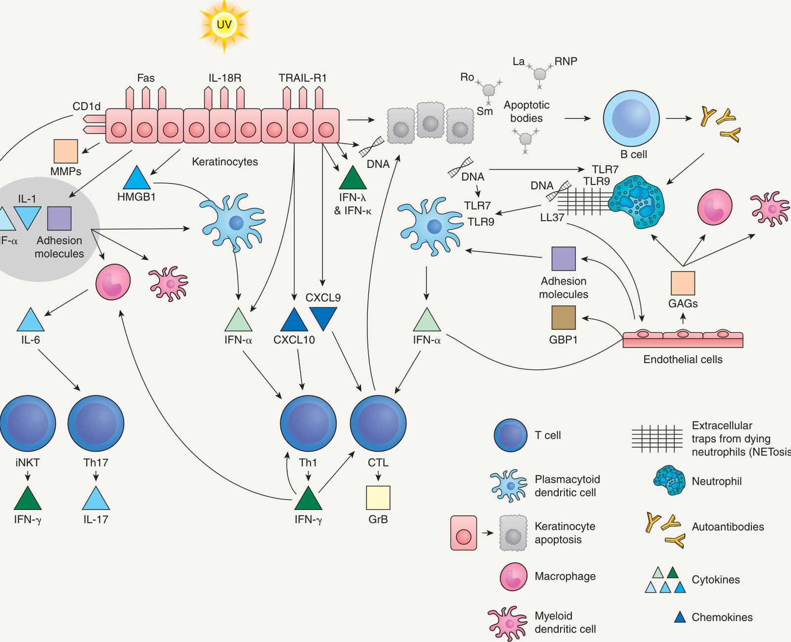
Fig. 41.1 Pathogenesis of lupus erythematosus. In photosensitive cutaneous LE, ultraviolet radiation triggers cytokine and chemokine production, innate immune proteins, NETosis of neutrophils, and apoptosis of cells leading to release of DNA. A lichenoid tissue reaction is the endpoint of a complex cascade that includes activation of dendritic cells, in part due to activation of the TLR7/MyD88 pathway, release of interferon (IFN), production of chemokines, and activation of T cells. CTL, cytotoxic T lymphocyte; CXCL, chemokine (C-X-C motif) ligand; GAGs, glycosaminoglycans; GBP1, guanylate binding protein-1; GrB, granzyme B; HMGB1, high-mobility group box 1; IL, interleukin; iNKT cells, invariant natural killer T cells; LL-37, cathelicidin; MMPs, matrix metalloproteinases; RNP, ribonucleoprotein; Th, T helper cell; TLR, Toll-like receptor; TNF, tumor necrosis factor; TRAIL-R1, tumor necrosis factor-related apoptosis-inducing ligand-receptor 1. Adapted from Achtman JC, Werth VP. Pathophysiology of cutaneous lupus erythematosus. Arthritis Res Ther 2015;17:182.

Table 41.1 Genes associated with systemic lupus erythematosus. For detailed information on the function of these protein products, see www. genecards.org. The most strongly associated genes are in bold. Adapted from Moser KL, Kelly JA, Lessard CJ, et al. Recent insights into the genetic basis of systemic lupus erythematosus. Genes Immun 2009;10:373–9.