CLINICAL FEATURES
Cutaneous Lupus – Three Major Forms
Classification
The most commonly used classification of cutaneous lesions in LE is that of Dr. James Gilliam. He segregated skin lesions into those that are specific for cutaneous LE and those that are not specific based upon whether the histopathology demonstrated an interface dermatitis or not, respectively. Within the category of specific cutaneous lesions, he subdivided these into acute cutaneous LE, subacute cutaneous LE, and chronic cutaneous LE (Fig. 41.2). This choice of terms was based upon the observation that three distinct types of cutaneous LE are commonly seen: the often transient lesions of acute cutaneous LE (ACLE) typified by malar erythema; the frequently long-lived, intensely inflammatory discoid LE (DLE) lesions which can lead to permanent disfiguring scars; and a photosensitive eruption characteristically more long-lasting than ACLE but without the potential for atrophy or scarring, for which the term subacute cutaneous LE (SCLE) was coined. Typically grouped within the chronic cutaneous LE category are the less common disorders LE tumidus, lupus panniculitis, and chilblain lupus (see Fig. 41.2), despite the observation that characteristic lesions frequently lack an interface dermatitis, especially LE tumidus.
Formal studies have not been done comparing duration of disease activity for the various types of cutaneous LE, and there is a considerable range in the duration of activity within each subtype. Patients with SCLE lesions may have a chronic, relapsing course, while DLE or LE tumidus lesions may have a relatively short duration due to resolution or effective disease control. In fact, some authorities have proposed that LE tumidus should be removed from the chronic category and be given its own category, intermittent cutaneous LE. These controversies aside, Gilliam’s classification schema has proven utility in the organization of the various types of cutaneous LE and it is the basis for the classification herein, accompanied by a focus on the morphology and histopathology of lesions (Fig. 41.3). The three major forms of cutaneous LE will be discussed first, followed by the remaining entities outlined in Fig. 41.2.
Discoid lupus erythematosus (DLE)
Discoid lesions represent one of the most common skin manifestations of lupus and they are most frequently found on the face, scalp, and ears (Fig. 41.4), but may be present in a more widespread distribution (Fig. 41.5). It is unusual for discoid lesions to be present below the neck without lesions also being present above the neck41a. Occasionally, discoid lesions develop on mucosal surfaces, including the lips, nasal mucosa, conjunctivae, and genital mucosa.
Some patients with discoid lesions exhibit a photodistribution, and sun exposure appears to have a role in lesional development. However, many patients have discoid lesions on sun-protected skin, and there is no clear association between sun exposure and their development.
(LE). Based upon the classification system originally proposed by Gilliam and Sontheimer.
Discoid lesions have the potential for scarring, and, over time, a substantial proportion of patients develop disfiguring scarring. Active lesions are intensely inflammatory, with a pronounced superficial and deep dermal inflammatory infiltrate. As a result, on palpation, active lesions typically feel thicker and firmer than surrounding uninvolved skin. The adnexa are prominently involved, with follicular plugging and scarring alopecia commonly observed. Dyspigmentation is a common sequela noted in longstanding lesions, typically with hypopigmentation in the central area and hyperpigmentation at the periphery (Fig. 41.6), but sometimes with vitiligo-like depigmentation. Rarely, squamous cell carcinoma develops in a longstanding discoid lesion.
Patients who present with discoid lesions may have associated arthralgias, but, over time, only ~10%–20% of these patients eventually meet the classification criteria for SLE (see below). Many of these patients meet criteria based primarily upon mucocutaneous disease and/or mild hematologic abnormalities (e.g. leukopenia) rather than serious internal disease, and the majority of those who progress do so within five years. The risk may be higher in patients with widespread
(disseminated) discoid lesions, defined as having lesions below the neck, and in children.
An unusual variant of DLE is hypertrophic (verrucous) DLE, characterized by thick scaling overlying the discoid lesion or occurring at the periphery of the discoid lesion. The intensely hyperkeratotic lesions are often prominent on the extensor surface of the arms (see Fig. 41.5J), but the face and upper trunk may also be involved. Frequently, there are characteristic lesions of discoid LE present in other locations.
Subacute cutaneous lupus erythematosus (SCLE)
Patients with SCLE typically report photosensitivity and lesions of SCLE most frequently occur on sun-exposed skin. It is notable that the midface is usually spared, while the sides of the face, upper trunk, and extensor aspects of the upper extremities are commonly involved (Fig. 41.7; see Fig. 41.4). In some patients, the disease may be mild, with only a few small scaly patches appearing after sun exposure.
Lesions of SCLE may have an annular configuration, with raised pink– red borders and central clearing (Fig. 41.8), or a papulosquamous presentation with a chronic psoriasiform or eczematous appearance. SCLE
lesions characteristically have a relatively sparse, superficial inflammatory infiltrate, and, consequently, they are minimally palpable. Lesions often result in dyspigmentation, particularly hypopigmentation or even depigmentation, but rarely develop scarring or dermal atrophy as sequelae.
In about a third of patients with SCLE, the disease is linked to medications, either at its onset or as a drug-induced exacerbation of previously well-controlled disease (Table 41.2). The first drug associated with the development of SCLE was hydrochlorothiazide, but at least
100 agents have been reported subsequently to induce or exacerbate SCLE. A population-based study from Sweden identified (in decreasing order) terbinafine, TNF inhibitors, anti-epileptics, and proton pump inhibitors as the most frequently associated medications. More recently, immune checkpoint inhibitors, in particular pembrolizumab and nivolumab, have been linked to the development or exacerbation of SCLE. The cutaneous lesions may or may not clear once the medication is discontinued.
Approximately half of patients presenting with SCLE fulfill classification criteria for SLE. However, most do so on the basis of laboratory and mucocutaneous findings. In a study of 85 SCLE patients, only six had renal and three had neurologic disease. Since anti-SSA/Ro autoantibodies are associated with Sjögren syndrome as well as SCLE, it is not surprising that some patients have features of both conditions, and some may have serious internal manifestations of Sjögren syndrome such as pulmonary or neurologic disease.
An important feature of SCLE, from the standpoint of understanding the pathogenesis of lupus, is its regular association with anti-SSA/Ro autoantibodies (see Ch. 40). Although investigators differ in their opinions as to the prevalence of anti-SSA/Ro autoantibodies in SCLE, it is likely that a substantial majority of patients with this condition (~70% in a large series, reported range of 60%–100%) have anti-SSA/Ro antibodies.
Acute cutaneous lupus erythematosus (ACLE)
The lesions of ACLE are exemplified by the development of bilateral malar erythema (“butterfly rash”; Fig. 41.9). These lesions tend to be transient, follow sun exposure, and resolve without scarring (but sometimes with dyspigmentation). An association with anti-dsDNA antibodies and lupus nephritis has been proposed and is plausible, although some patients with a malar rash have neither anti-dsDNA antibodies nor lupus nephritis. Patients presenting with this type of eruption must be evaluated carefully for evidence of internal disease.
This patient had ACLE lesions on the arms as well as the face.
The morphology of the lesions ranges from mild erythema to intense edema. The presence of telangiectasias, erosions, dyspigmentation and epidermal atrophy (i.e. poikiloderma) may help to distinguish the malar erythema of ACLE from that of common facial eruptions such as seborrheic dermatitis and the erythematotelangiectatic type of rosacea. Occasionally, there is a papular component, and occasionally lesions develop scaling (see Fig. 41.9C). The duration may range from a few hours to several weeks. The face, particularly the malar area, is most commonly affected, and there is often sparing of the nasolabial fold; sometimes lesions may be more widespread in distribution (Fig. 41.10; see Fig. 41.4). When lesions occur on the hands, the knuckles are typically spared. It is not unusual for patients with ACLE to also have oral ulcerations.
The presence of erythema multiforme-like lesions in lupus patients has been termed Rowell syndrome. Rarely, patients develop an acute eruption clinically similar to toxic epidermal necrolysis or erythema multiforme major (see below). These lesions may represent a severe variant of ACLE or, in some cases, SCLE.
The three major types of cutaneous LE are not mutually exclusive. In a given patient, more than one type of cutaneous lesion may occur.
Cutaneous Lupus – Additional Variants (See Fig. 41.2)
Lupus erythematosus tumidus
Lesions are typically firm erythematous plaques that lack scale or follicular plugging. Although the epidermis appears to be uninvolved in the disease process, there is an intense perivascular and periadnexal inflammatory infiltrate within the dermis, as well as mucin deposition. LE tumidus lesions may be the same as the “urticarial plaques” described in lupus patients. However, these fixed plaques should not be confused with urticarial vasculitis (see Ch. 24). Some authors state that the lesions most commonly occur on the face, but they are often
observed on the trunk as well (Fig. 41.11). Morphologically, the lesions are similar to those of lymphocytic infiltrate of Jessner and may have central clearing (see Ch. 121); some clinicians believe that lymphocytic infiltrate of Jessner and LE tumidus are either very closely related or exist along the same disease spectrum.
In patients reported to have LE tumidus, the very low prevalence of SLE, the relative lack of serologic abnormalities, and the very low prevalence of immunoglobulin deposition within the cutaneous lesions have made it difficult to determine whether LE tumidus is actually a variant of LE or an independent entity. However, the presence of LE tumidus lesions in patients with other specific types of cutaneous LE is evidence in favor of its being classified as a form of cutaneous LE. In the majority of patients, LE tumidus has been reported to be reproducible by phototesting. The lesions tend to resolve without scarring, atrophy, or dyspigmentation.
Lupus panniculitis
Intense inflammation in the fat leads to indurated plaques that can evolve into disfiguring, depressed areas. Lesions of lupus panniculitis have a distinctive distribution, occurring predominantly on the upper arms (Fig. 41.12), face, scalp, upper trunk, thighs, buttocks, and breasts (see Fig. 41.4). Some patients may have discoid lesions overlying the panniculitis, and, in those cases, the condition is sometimes referred to as lupus profundus. For further discussion of lupus panniculitis, see Chapter 100.
Chilblain lupus
Chilblain lupus (SLE pernio; Fig. 41.13) consists of red or dusky purple papules and plaques on the toes, fingers, and sometimes the nose,
elbows, knees, and lower legs. The lesions are brought on or exacerbated by cold, particularly moist cold climates. These lesions may represent the concurrence of ordinary chilblains in a patient with LE (see Ch. 88), although, with time, the lesions may develop a gross and microscopic appearance consistent with that of a discoid lesion.
Heterozygous mutations in TREX1 (see above) or SAMHD1 can lead to a familial form of chilblain lupus with an onset during childhood. Affected individuals may have arthralgias and a positive ANA, but otherwise do not develop internal disease. Mutations in these same genes, as well as ADAR1, IFIH1, and RNASEH2A/B/C, can result in the Aicardi–Goutières syndrome, a primarily autosomal recessive autoinflammatory disorder characterized by recurrent sterile fevers, progressive developmental delay, and chilblains (see Table 45.7, Type I interferonopathies). Mutations in these seven genes lead to an accumulation of nucleic acids, e.g. dsDNA, RNA:DNA duplexes.
Discoid lupus erythematosus/lichen planus overlap
A discoid lupus erythematosus/lichen planus overlap syndrome has been described, in which lesions with features of both conditions are present (see Ch. 11).
Neonatal lupus erythematosus (NLE)
A neonatal form of SCLE may occur in infants whose mothers have anti-SSA/Ro autoantibodies. In babies who have neonatal lupus erythematosus (NLE), the SCLE-like lesions are histologically identical to
those of SCLE in adults, and there is a strong association with anti-SSA/ Ro antibodies. Indeed, almost 100% of babies with NLE have anti-SSA/ Ro antibodies, and less frequently anti-URNP autoantibodies are present. Unlike SCLE in adults, lesions have a predilection for the face, especially the periorbital region and scalp (Fig. 41.14).
Photosensitivity is very common in NLE, but sun exposure is not required for lesions to form, as it is possible for lesions to be present at birth. Neonatal lupus skin lesions typically resolve completely, although perhaps one-fourth to one-third of children have cutaneous residua of telangiectasias, dyspigmentation, and/or atrophic scarring. Atrophic scarring may be more likely in infants whose lesions began in utero.
Infants who have the cutaneous lesions of NLE may also exhibit internal manifestations. The major extracutaneous findings are congenital heart block (with or without cardiomyopathy), hepatobiliary disease, and cytopenias, in particular thrombocytopenia. The heart block is almost always present by birth, but on rare occasions has developed after birth. Clinically significant cardiomyopathy occurs concurrently in a small percentage of babies who have heart block. Usually, the cardiomyopathy is apparent during the neonatal period, but it is possible for it to become apparent only after several months have elapsed. Cardiac NLE has a mortality rate of approximately 20%, and approximately two-thirds of children require pacemakers.
Hepatobiliary disease and cytopenias, especially thrombocytopenia, may be present at birth, or they may develop within the first few months of life. Hepatobiliary disease can vary in severity and may present as liver failure during gestation or in the neonatal period, conjugated hyperbilirubinemia during the first few weeks of life, or mild elevations of aminotransferases at 2–3 months of life. There are also reports of hydrocephalus, microangiopathic hemolysis, and disseminated intra-vascular coagulation.
Although most children with cutaneous NLE do not have significant internal involvement, a systemic evaluation and counseling is recommended (Table 41.3). Mothers who have had a baby with cutaneous NLE have an increased likelihood of having a subsequent pregnancy complicated by NLE, including cardiac involvement. When pregnant, these women should be cared for by a perinatologist who has specialized training in high-risk prenatal care.
Bullous lesions
In the clinical setting of lupus, bullous lesions may appear for several reasons. On occasion, bullous or crusted lesions occur simply as a result of the intensity of the basal cell damage in lesions of ACLE or SCLE (see Fig. 41.8A) or, possibly, DLE. Rarely, a dramatic, acute eruption
similar to erythema multiforme major or toxic epidermal necrolysis (TEN) may occur in patients with preexisting ACLE or SCLE, or it may appear de novo (Fig. 41.15). Blisters occurring within ACLE and SCLE lesions and erythema multiforme-like and TEN-like cutaneous lupus fit within the category of lupus-specific skin lesions.
The term bullous eruption of SLE, or bullous SLE, has been used to describe an acquired blistering eruption in patients who fulfill the criteria for SLE. It consists of vesicles and bullae whose histopathology often resembles dermatitis herpetiformis, with a primarily neutrophilic infiltrate and microabscesses within the dermal papillae (Fig. 41.16). In some patients, the clinical and histopathologic features may resemble neutrophil-rich bullous pemphigoid or epidermolysis bullosa acquisita. Immunoreactants are often found at the basement membrane zone and antibodies to type VII collagen have been detected in several patients. This eruption may represent the concurrence of lupus with an autoimmune blistering disease due to autoantibodies to a component of the basement membrane zone. There are also a number of case reports of other autoimmune bullous diseases appearing in patients with lupus (reviewed in reference 65).
Systemic Lupus Erythematosus
Lupus erythematosus is potentially a multi-organ disease, although in individual patients often only one or a few organs are significantly involved. Organ systems most commonly affected are joints, skin, hematologic, renal, and CNS as well as pleural and pericardial serosal surfaces. Nonspecific signs and symptoms such as fever, weight loss, fatigue, myalgias, and lymphadenopathy are also common in SLE. Additional less common manifestations are quite varied and include sensorineural hearing loss and vision loss. Although ACLE is the cutaneous phenotype with the strongest association with systemic disease, patients with any type of cutaneous LE may develop internal involvement.
Since SLE is complex and variable, classification criteria have been developed to ensure that patients in clinical studies have SLE rather than other conditions that may mimic it. Historically, the American College of Rheumatology (ACR) criteria, developed in 1982 and modified in 1997, have been most commonly used. In 2012, a new set of criteria from the Systemic Lupus International Collaborating Clinics (SLICC) aimed to improve the sensitivity of the ACR criteria (Table 41.4). In 2019, the ACR, in concert with the European League Against Rheumatism (EULAR), released a new classification system (Table 41.5). Some of its notable features are as follows: 1.Presence of ANA (titer ≥1 : 80) is an entry criterion. Patients with persistently negative ANA tests are excluded. 2.Individual criteria have different weights and need not occur simultaneously. 3.Within a particular domain (e.g. renal), only the highest weighted criterion counts. 4.A criterion is counted only if SLE is the most likely explanation for the finding.
Nonspecific Cutaneous Lesions
Vascular lesions are common in patients with LE, particularly in those who have systemic disease (Table 41.6). These lesions include Raynaud phenomenon (see Ch. 43), livedo reticularis, palmar erythema, and nail-fold telangiectasia. Purpura, urticarial papules, or ulcerations due to vasculitis may occur, as well as cutaneous infarctions resembling Degos disease or atrophie blanche. Patients with cutaneous LE who have any of these findings should be evaluated for systemic disease. Livedo reticularis, thromboses, ulcerations, and lesions resembling Degos disease have each been associated with antiphospholipid antibodies (see Ch. 23). The association of livedo reticularis with ischemic CNS disease has been called Sneddon syndrome (see Ch. 23), and the phenotype of the antiphospholipid syndrome has been called Hughes syndrome.
Alopecia often occurs as a result of scarring discoid lesions. However, non-scarring diffuse alopecia can appear in patients with systemic disease. It has been reported that patients with lupus have an increased likelihood of alopecia areata compared with the general population.
Sclerodactyly, calcinosis, and rheumatoid nodules, findings more consistent with systemic sclerosis or rheumatoid arthritis, have been observed in some patients with lupus, although many of the patients with these findings may have overlap syndromes rather than classic lupus. Other skin findings reported to occur in patients who have lupus include erythromelalgia, papulonodular mucinosis (of Gold; see Ch. 46), and anetoderma (see Ch. 99). Of note, ~75% of the patients with papulonodular mucinosis have SLE. Rarely, significant periorbital edema can develop secondary to dermal mucin. A Sweet syndrome-like presentation can also be seen in patients with SLE and this has been given several names including neutrophilic dermatosis in conjunction with LE and non-bullous neutrophilic LE as well as simply Sweet syndrome in association with SLE.
Patients with SLE have more subtle nail-fold capillary abnormalities than do patients with systemic sclerosis or dermatomyositis. Prominent large, tortuous capillaries and areas of marked avascularity are not characteristic of SLE.
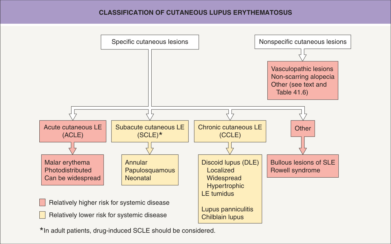
Fig. 41.2 Classification of cutaneous lupus erythematosus
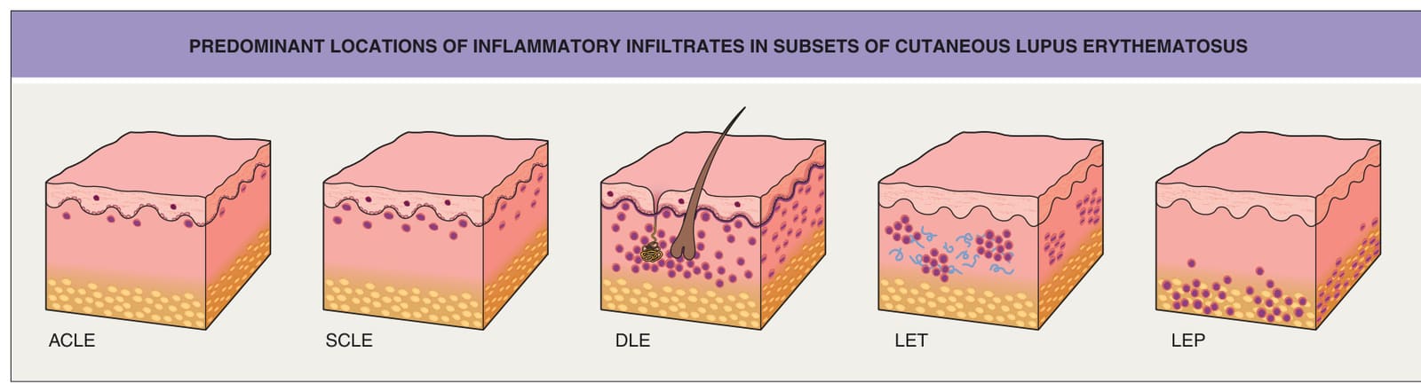
Fig. 41.3 Predominant locations of inflammatory infiltrates in subsets of cutaneous lupus erythematosus. The types of cutaneous lupus erythematosus are: acute cutaneous lupus erythematosus (ACLE), subacute cutaneous lupus erythematosus (SCLE), discoid lupus erythematosus (DLE), lupus erythematosus tumidus (LET), and lupus panniculitis (LEP); the latter three are forms of chronic cutaneous lupus erythematosus (see Fig. 41.2). The primary locations of the infiltrates are as follows: superficial dermis, ACLE and SCLE; superficial and deep dermis perivascular and periadnexal, DLE; superficial and deep dermis perivascular and periadnexal, LET; and subcutaneous fat, LEP. The final diagnosis requires clinicopathologic correlation.
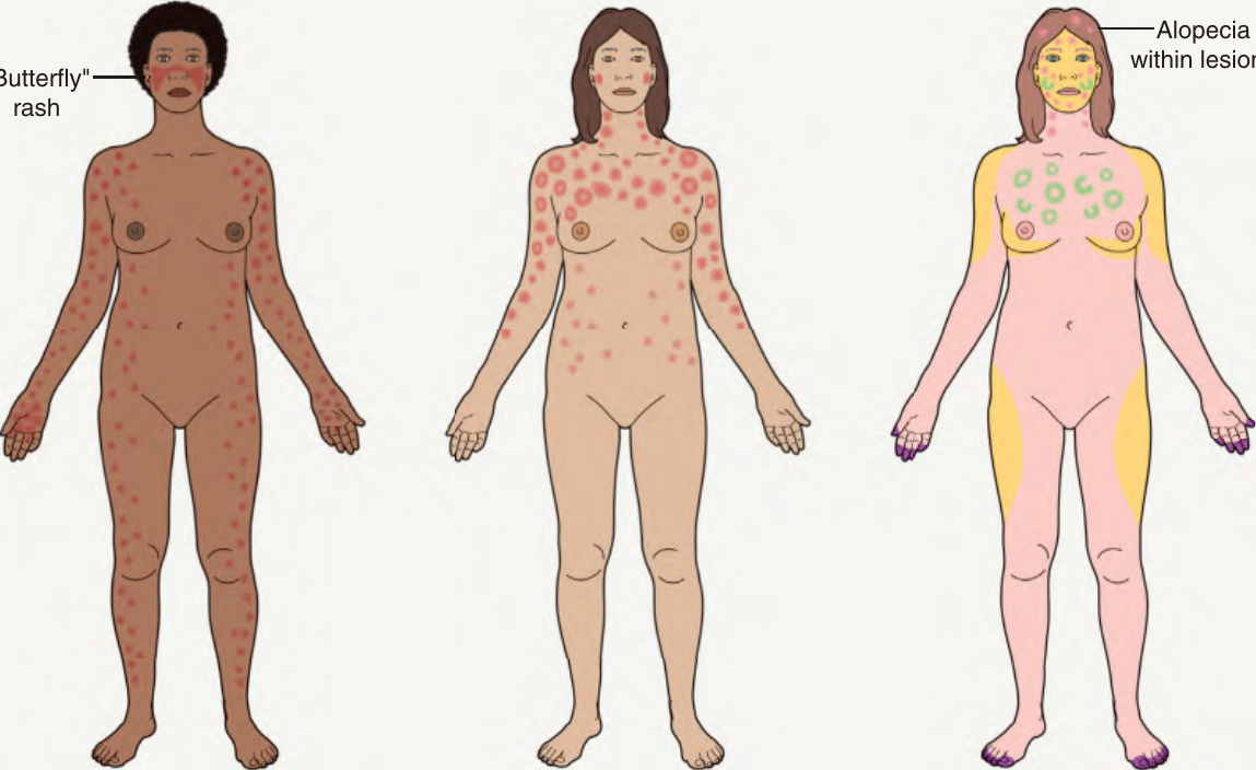
Fig. 41.4 Characteristic sites of involvement for the three major forms of cutaneous lupus erythematosus (LE).
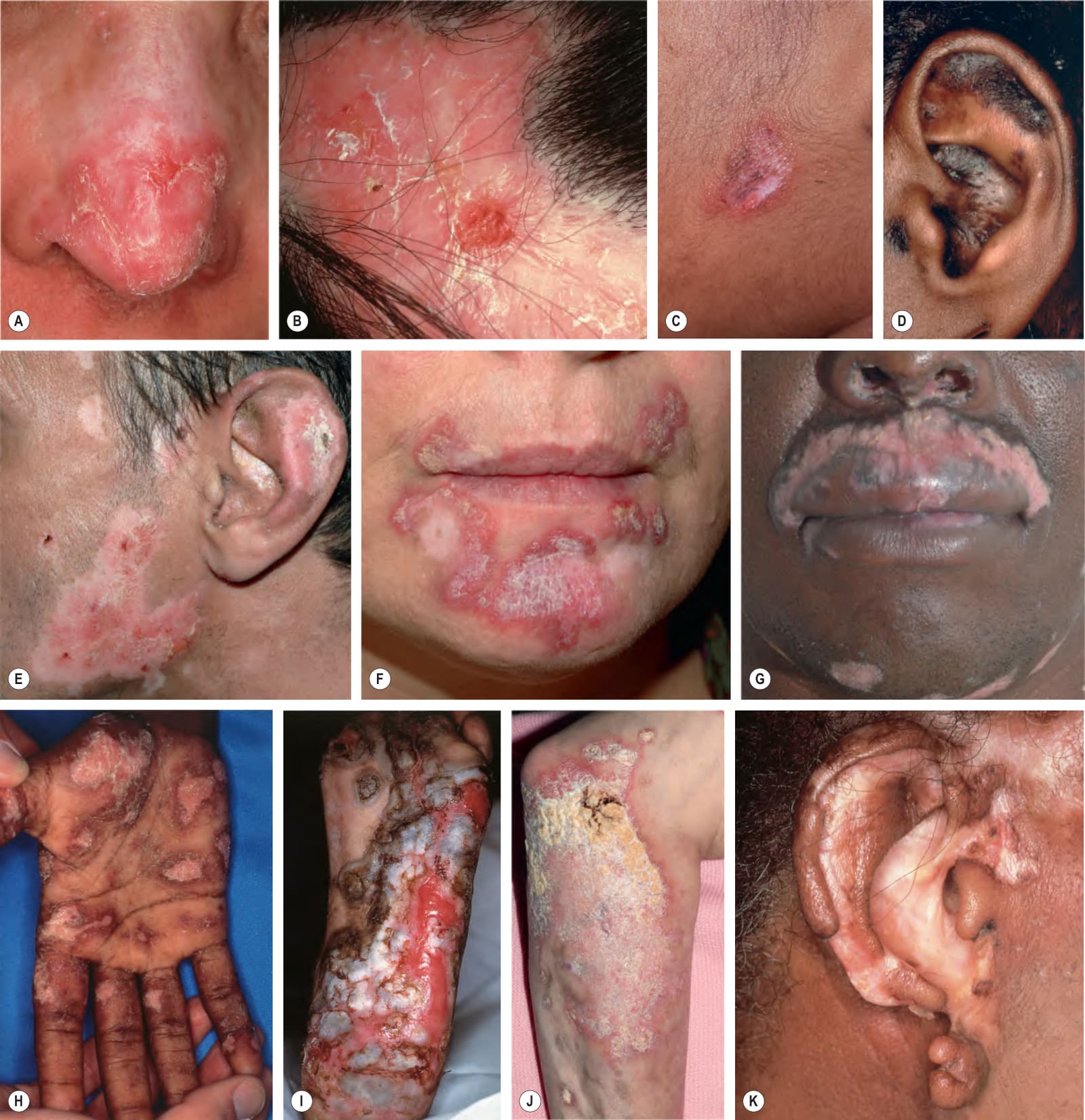
Fig. 41.5 Various presentations of discoid lesions of lupus erythematosus.A–C Lesions, which favor the head and neck region, may show erythema, scaling, atrophy, and dyspigmentation in addition to scarring (and alopecia). D Note the patulous follicular openings in addition to hyperpigmentation and scale. E–G The plaques are well-demarcated and both hypopigmentation and scarring can be seen in a range of skin phototypes. H, I Less common sites include the palms and soles, where lesions can be keratotic or ulcerative as in lichen planus. The patient with plantar involvement had systemic lupus erythematosus and responded well to isotretinoin. J Occasionally, hypertrophic lesions develop with significant hyperkeratosis. K The scarring process may be destructive. C, Courtesy Kalman Watsky, MD; E–G, Courtesy Luis Requena, MD; J, Courtesy Julie V. Schaffer, MD.
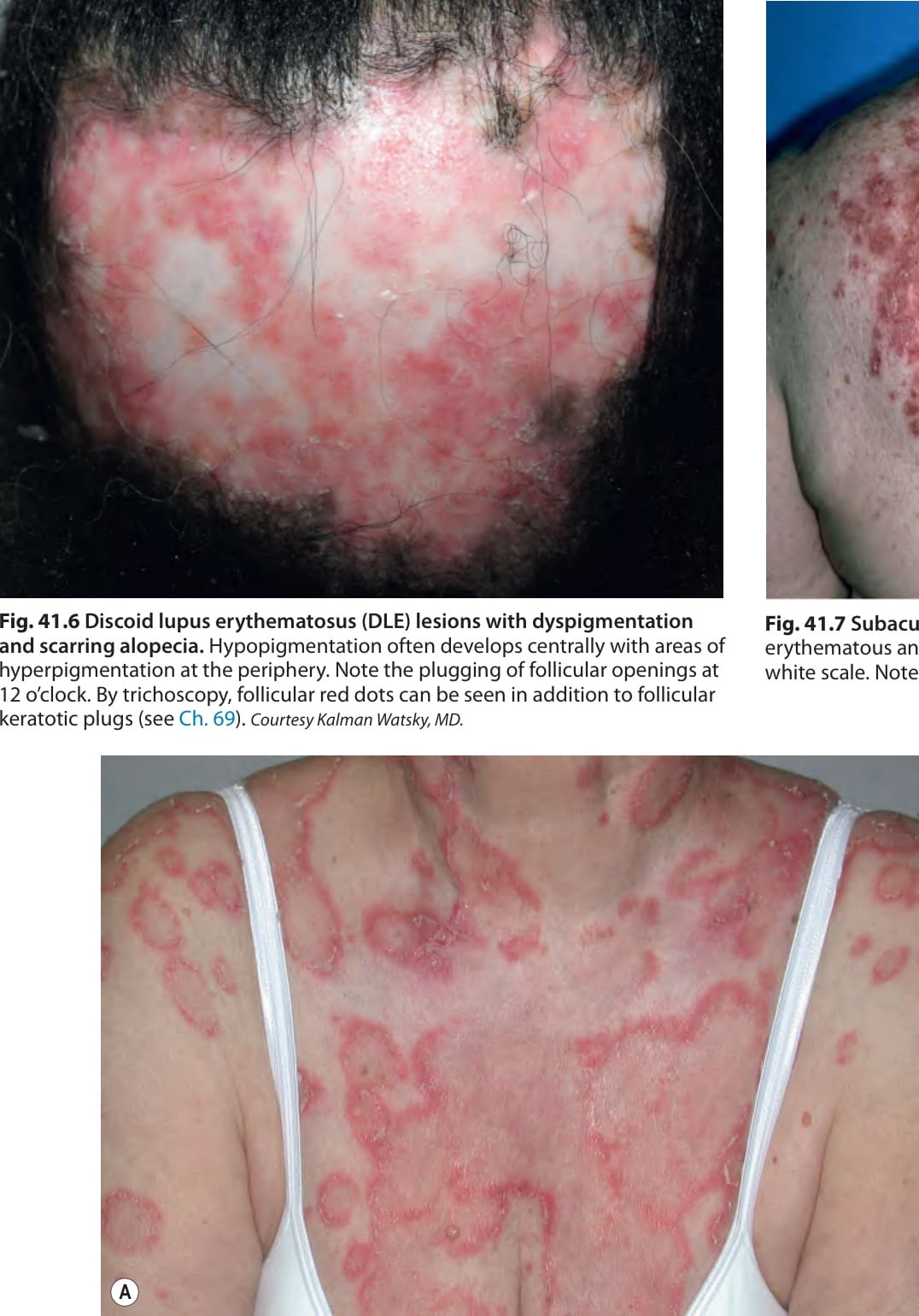
Fig. 41.6 Discoid lupus erythematosus (DLE) lesions with dyspigmentation and scarring alopecia. Hypopigmentation often develops centrally with areas of hyperpigmentation at the periphery. Note the plugging of follicular openings at 12 o’clock. By trichoscopy, follicular red dots can be seen in addition to follicular keratotic plugs (see Ch. 69). Courtesy Kalman Watsky, MD.
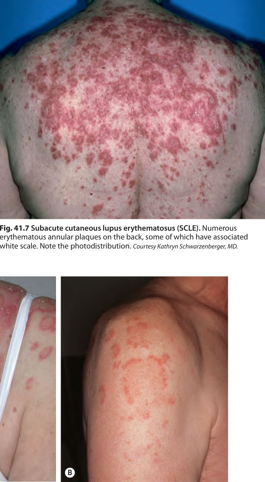
Fig. 41.7 Subacute cutaneous lupus erythematosus (SCLE). Numerous erythematous annular plaques on the back, some of which have associated white scale. Note the photodistribution. Courtesy Kathryn Schwarzenberger, MD.
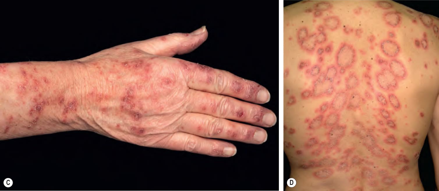
Fig. 41.8 Subacute cutaneous lupus erythematosus (SCLE). Lesions are most commonly seen on the upper trunk and sun-exposed aspects of the upper extremities. The margins of the annular lesions may have scale-crust (A) or be composed of multiple papules (B). C Note the peripheral scale and relative sparing of the proximal interphalangeal joints. D Numerous annular lesions with scale-crust in the inner borders in a patient with terbinafine-induced SCLE. C, Courtesy Lorenzo Cerroni, MD; D, Courtesy Luis Requena, MD.

Fig. 41.9 Acute cutaneous lupus erythematosus (ACLE). The facial erythema, often referred to as a “butterfly rash” may be variable (A), edematous (B), or have associated scale (C). The presence of small erosions can aid in the clinical differential diagnosis. A, Courtesy Kalman Watsky, MD.
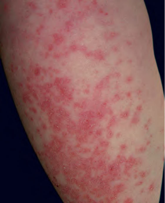
Fig. 41.10 Acute cutaneous lupus erythematosus (ACLE).
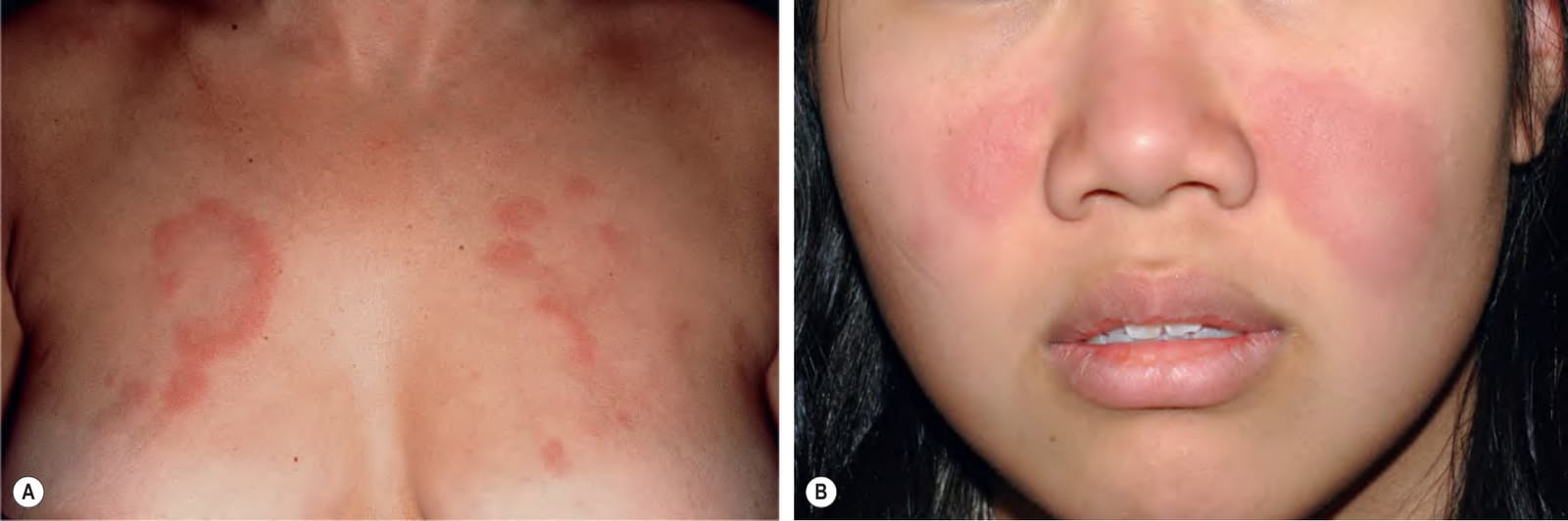
Fig. 41.11 Lupus erythematosus tumidus. Annular pink plaques on the chest (A) and pink–violet plaques on the face (B). None of the lesions have epidermal change. B, Courtesy Julie V. Schaffer, MD.
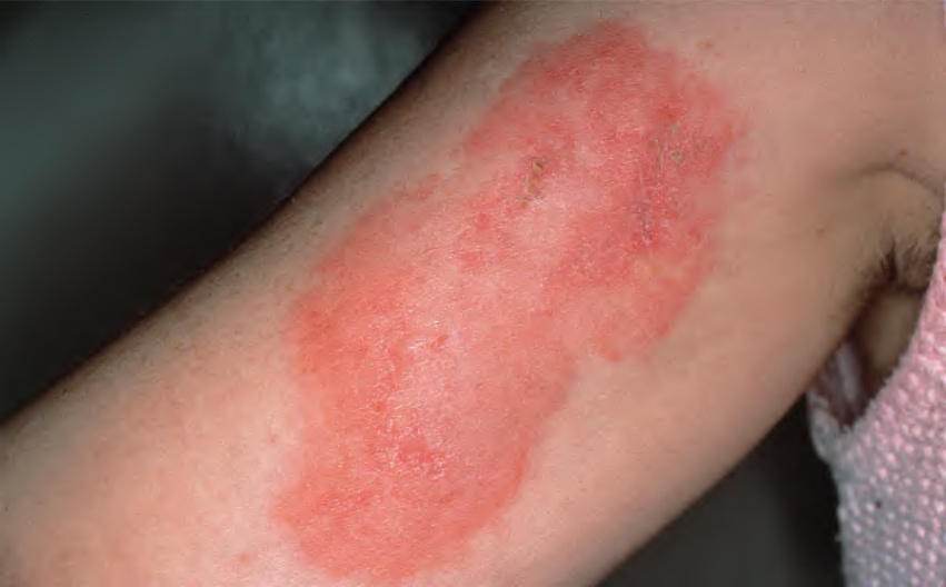
Fig. 41.12 Lupus panniculitis. Erythematous plaque on the upper arm. The lesions may resolve with lipoatrophy.
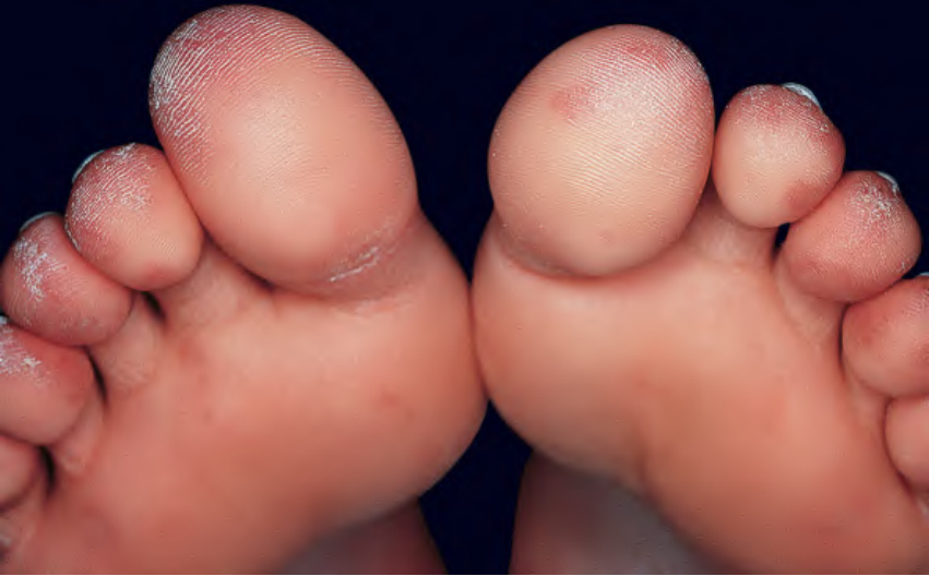
Fig. 41.13 Chilblain lupus. Violaceous plaques, some with scale, on toes. If there is a family history of this disorder, the possibility of mutations in TREX1, which encodes a DNA exonuclease, or SAMHD1, which encodes a host restriction nuclease that plays a role in the innate immune response, can be considered.
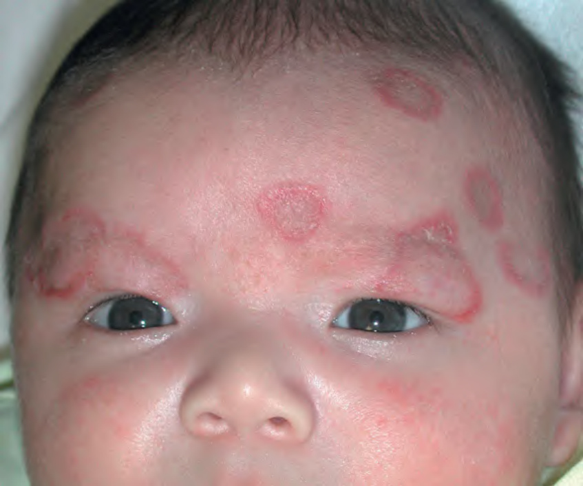
Fig. 41.14 Neonatal lupus erythematosus. Annular erythematous plaques on the forehead and scalp. Note the resemblance to the annular form of subacute cutaneous lupus erythematosus. Courtesy Julie V. Schaffer, MD.
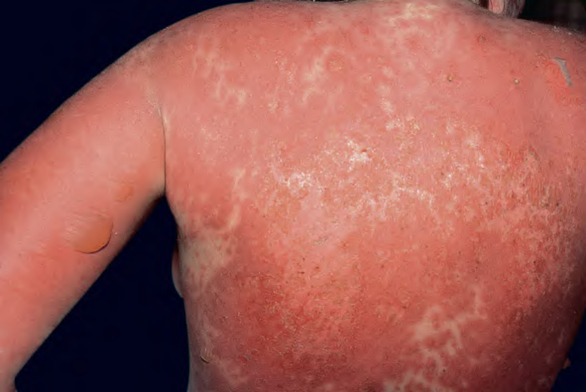
Fig. 41.15 Toxic epidermal necrolysis-like eruption of acute lupus erythema- tosus. This presentation has also been referred to as a form of acute syndrome of apoptotic pan-epidermolysis (ASAP).
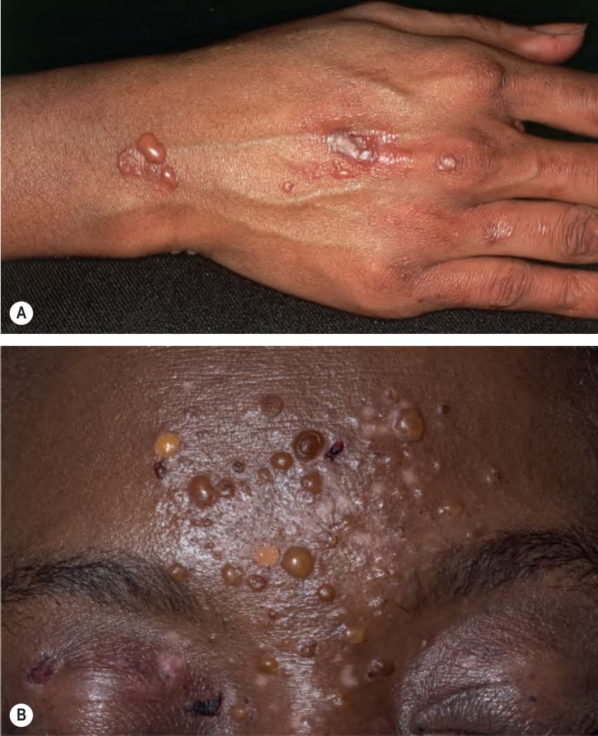
Fig. 41.16 Bullous eruption of systemic lupus erythematosus.A Vesicles and bullae due to autoantibodies against type VII collagen can develop in patients with systemic disease. B Multiple vesicles and bullae as well as erosions and hypopigmentation at sites of previous lesions in a woman with SLE. B, Courtesy Edward Cowen, MD.
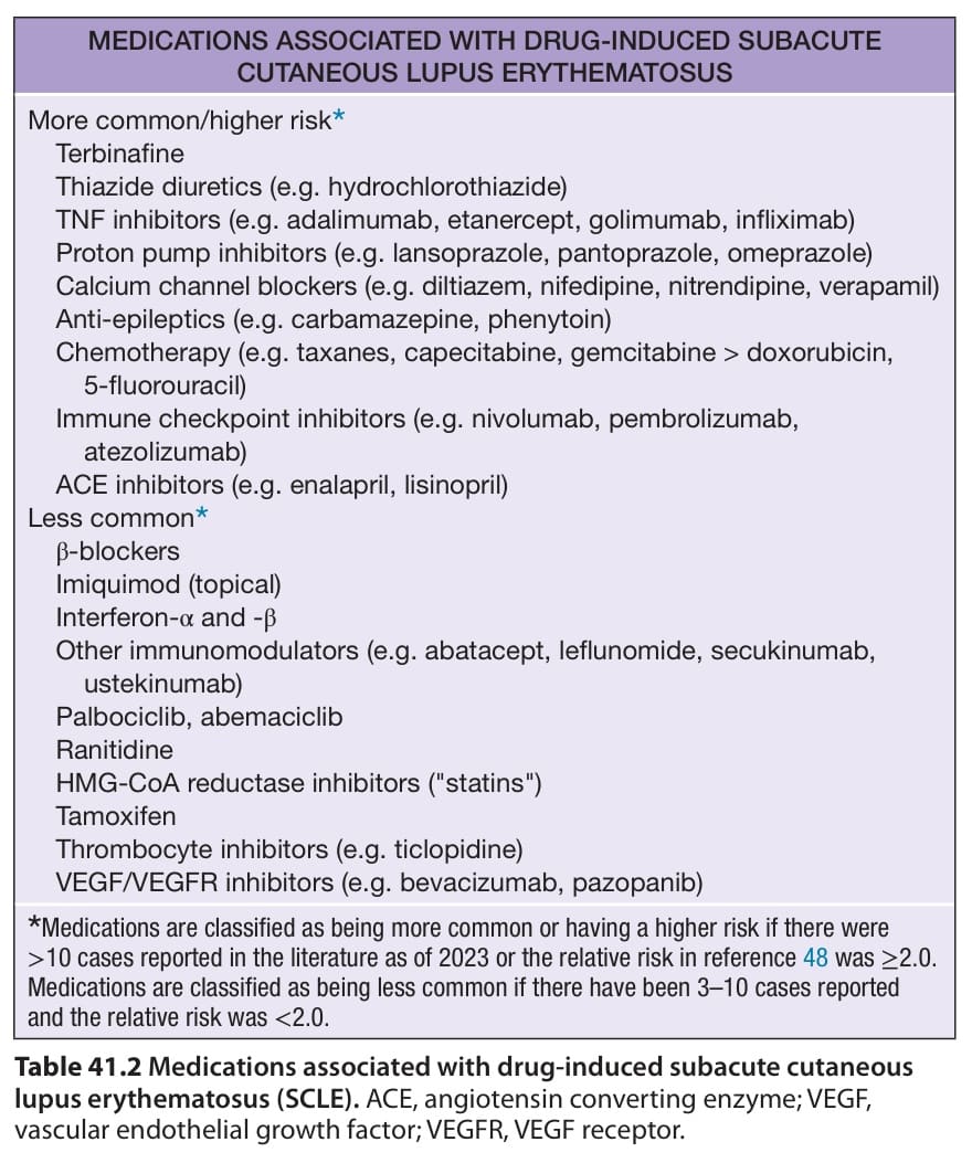
Table 41.2 Medications associated with drug-induced subacute cutaneous lupus erythematosus (SCLE). ACE, angiotensin converting enzyme; VEGF, vascular endothelial growth factor; VEGFR, VEGF receptor.
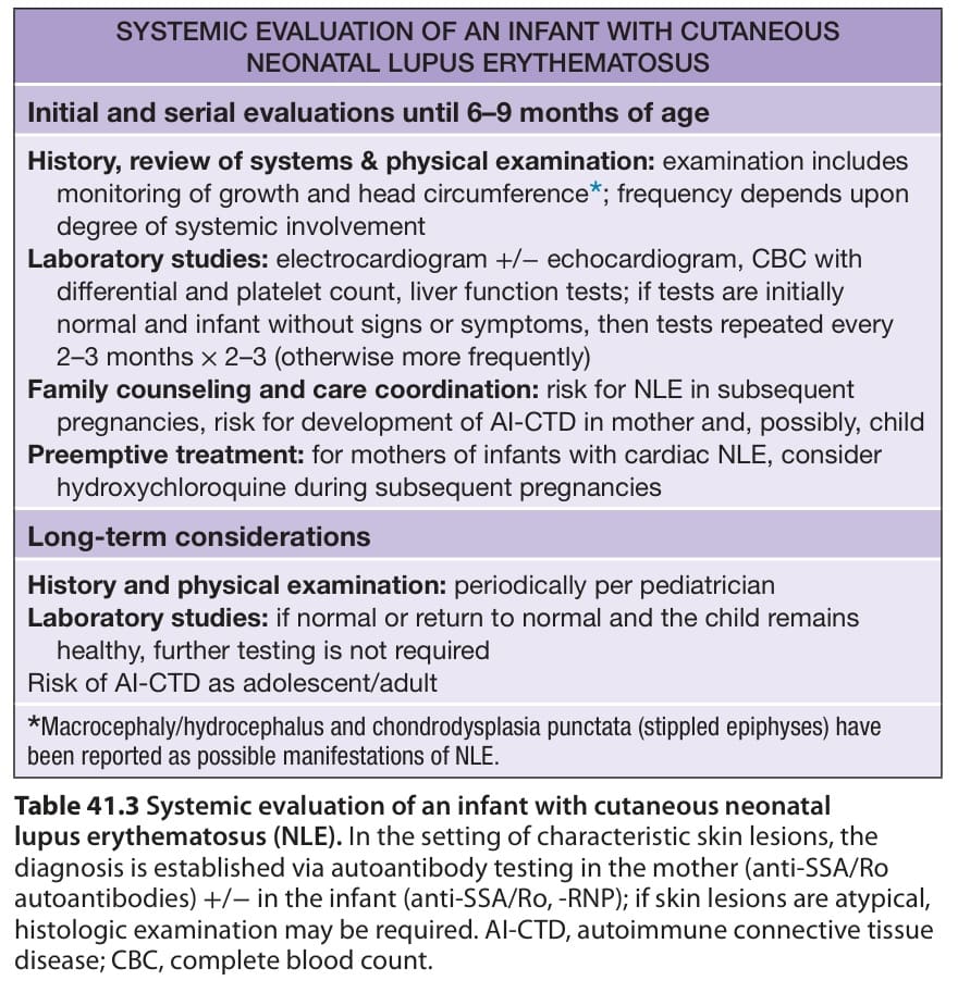
Table 41.3 Systemic evaluation of an infant with cutaneous neonatal lupus erythematosus (NLE). In the setting of characteristic skin lesions, the diagnosis is established via autoantibody testing in the mother (anti-SSA/Ro autoantibodies) +/− in the infant (anti-SSA/Ro, -RNP); if skin lesions are atypical, histologic examination may be required. AI-CTD, autoimmune connective tissue disease; CBC, complete blood count.
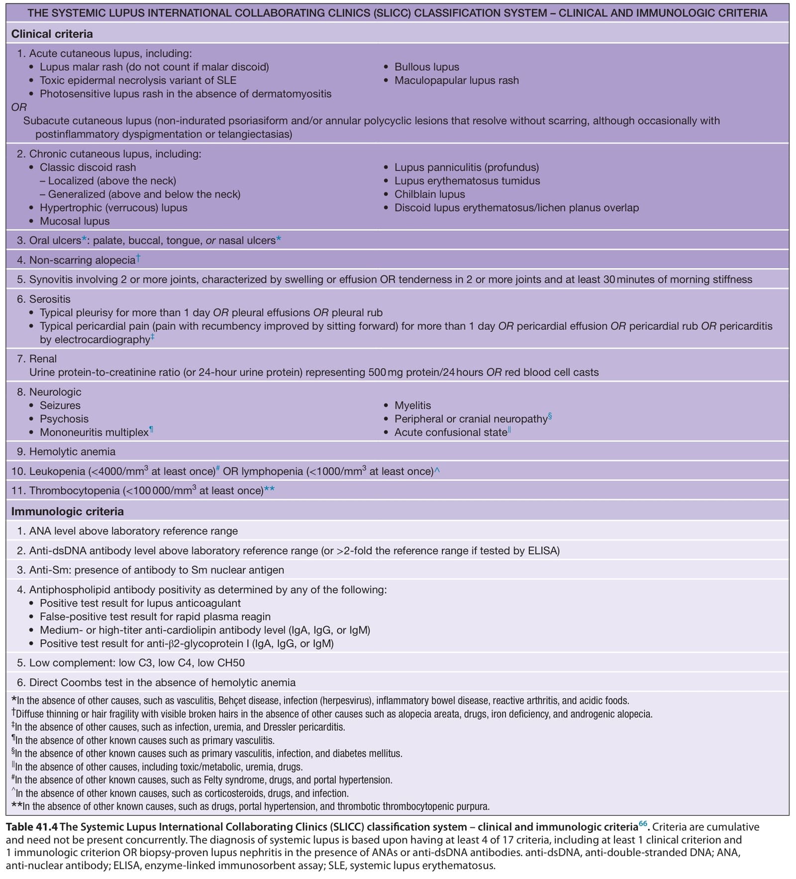
Table 41.4 The Systemic Lupus International Collaborating Clinics (SLICC) classification system – clinical and immunologic criteria. Criteria are cumulative and need not be present concurrently. The diagnosis of systemic lupus is based upon having at least 4 of 17 criteria, including at least 1 clinical criterion and 1 immunologic criterion OR biopsy-proven lupus nephritis in the presence of ANAs or anti-dsDNA antibodies. anti-dsDNA, anti-double-stranded DNA; ANA, anti-nuclear antibody; ELISA, enzyme-linked immunosorbent assay; SLE, systemic lupus erythematosus.
Table 41.5 The European League Against Rheumatism/ American College of Rheumatology (EULAR/ACR) classification criteria for systemic lupus erythematosus
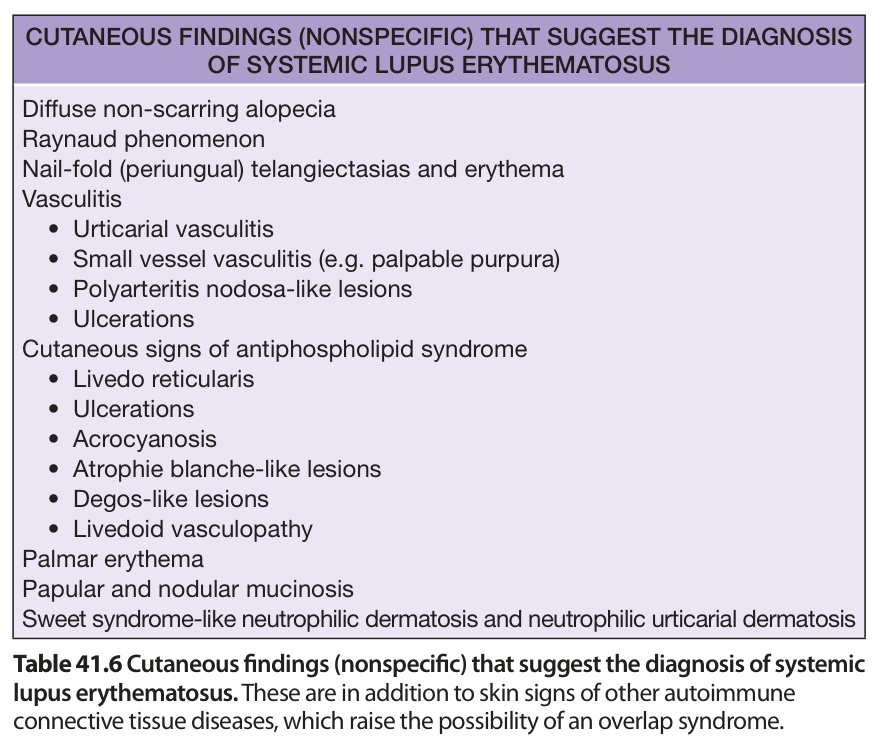
Table 41.6 Cutaneous findings (nonspecific) that suggest the diagnosis of systemic lupus erythematosus. These are in addition to skin signs of other autoimmune connective tissue diseases, which raise the possibility of an overlap syndrome.