PATHOLOGY
Histopathology
Histologic findings in cutaneous LE depend in large part on the subtype (Fig. 41.17; see Fig. 41.3). Characteristic findings in ACLE, SCLE, discoid lesions, LE tumidus, and lupus panniculitis are outlined in Table 41.7. However, in practice, an overlap in histologic findings occurs among the various clinical phenotypes, particularly ACLE, SCLE, and discoid lesions. Some of the more distinctive histologic features of cutaneous LE are basal cell damage (also referred to as vacuolar degeneration, hydropic change, or interface dermatitis), lymphohistiocytic inflammatory infiltrates admixed with CD123+ plasmacytoid dendritic cells, and, primarily in discoid lesions, periadnexal inflammation, follicular plugging, and scarring. In lesions of ACLE, dermal changes can be relatively subtle although basal cell damage may be pronounced. In SCLE, epidermal changes and a superficial lymphocytic infiltrate are common. In contrast to discoid lesions, SCLE lesions tend to have little or no hyperkeratosis, basement membrane thickening, periadnexal infiltrate, follicular plugging, deep dermal infiltrate, or scarring. LE tumidus has prominent dermal mucin deposition and dermal perivascular and periadnexal lymphocytic infiltrates with a lack of epidermal change. While changes of lupus panniculitis are most prominent in the subcutis, there may be overlying changes of DLE. Occasionally, cutaneous LE presents with a neutrophil-rich infiltrate mimicking that of a neutrophilic dermatosis.
The inflammatory infiltrates of cutaneous lupus typically contain plasmacytoid dendritic cells that produce interferons and may play a role in the induction of lesions. Immunohistochemical staining for CD123, a marker for plasmacytoid dendritic cells, has been explored as a means to distinguish cutaneous lupus from other inflammatory diseases (e.g. lichen planopilaris, polymorphous light eruption) and from cutaneous lymphoma. Although positive CD123-staining is not specific for lupus and its sensitivity and specificity remain to be definitively established, the pattern and intensity of staining may help to distinguish lupus from other conditions.
Antibody Deposits in Lesional Skin
Examination of the skin for deposits of immunoreactants is called direct immunofluorescence (DIF). DIF of lesional skin does not replace routine histologic staining as the method of choice for establishing a diagnosis of cutaneous LE. However, in those cases where the routine histopathology is equivocal, DIF can be a valuable asset in establishing a diagnosis.
The most characteristic DIF finding in cutaneous LE is antibody deposition at the dermal–epidermal junction and around hair follicles.
These deposits are typically granular, and they are composed primarily of IgG and/or IgM (Fig. 41.18), although IgA can also occasionally be seen. In addition, deposits of complement proteins are to be expected. Some investigators have reported that in SCLE, granular deposits of IgG and IgM are observed primarily within the epidermis rather than at the dermal–epidermal junction. There is evidence that the epidermal deposits are due to anti-SSA/Ro autoantibodies depositing directly within the skin.
In active lesions of ACLE, SCLE, and DLE, DIF of lesional skin is positive in the majority of cases. In general, a positive DIF supports
(SLE). plts, platelets. Modified from Aringer M, Costenbader K, Daikh D, et al. 2019 European League Against Rheumatism/American College of Rheumatology Classification Criteria for Systemic Lupus Erythematosus. Arthritis Rheumatol 2019;71:1400–12.
Anti-nuclear antibodies (ANA) at a titer of ≥1:80 on HEp-2 cells or an equivalent positive test (ever)
• SLE classification requires at least one clinical criterion and ≥10 points
the diagnosis of cutaneous LE, but a negative DIF does not exclude the diagnosis. It has been noted that DIF is most likely to be positive in well-established, active lesions. DIF is often negative or nonspecific in LE tumidus. In lupus panniculitis, DIF may show immunoreactants around dermal vessels, but granular deposits at the dermal–epidermal junction are not uniformly present.
Antibody Deposits Within Normal-Appearing Skin
In normal-appearing skin, the presence of antibody deposits at the dermal–epidermal junction correlates reasonably well with systemic disease. The antibody deposits are typically granular and are sometimes referred to as a “lupus band”, with examination for the deposits referred to as the “lupus band test”. The terminology is confusing, because some authors use the term “lupus band” to apply to the antibody deposits at the dermal–epidermal junction, whether the skin tested is normal-appearing or lesional, while other authors reserve the term “lupus band” to describe antibody deposits in normal-appearing skin. It has been proposed that, if this terminology is used, the investigator should modify the term “lupus band” by a preceding adjective of “lesional” or “non-lesional”, so that the subject of discussion is clearly identified.
Weak, discontinuous deposits may be seen in persons who do not have LE, including healthy adults, particularly when chronically sun-exposed skin is examined. For this reason, many investigators do not consider a non-lesional lupus band test to be positive unless the deposition of immunoreactants is strong and continuous. A true positive non-lesional lupus band test occurs in three-quarters or more
of patients with SLE if sun-exposed skin is examined, and in about one-half of patients with SLE if sun-protected skin is examined. A positive non-lesional lupus band test is unlikely to occur in patients who do not have SLE, but there are instances where the non-lesional lupus band test has been positive in patients with other autoimmune diseases.
In most cases, clinical evaluation and serologic testing for specific autoantibodies provide the required information, and the non-lesional lupus band test is superfluous. In cases where the clinical presentation or laboratory findings are atypical, the non-lesional lupus band test may have diagnostic value.
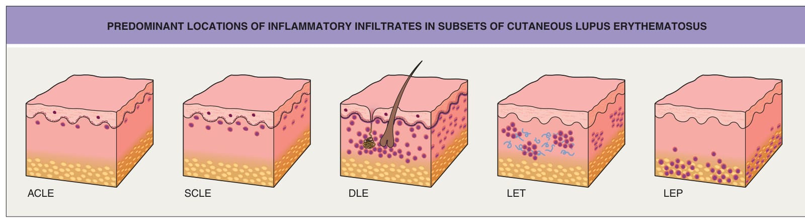
Fig. 41.3 Predominant locations of inflammatory infiltrates in subsets of cutaneous lupus erythematosus. The types of cutaneous lupus erythematosus are: acute cutaneous lupus erythematosus (ACLE), subacute cutaneous lupus erythematosus (SCLE), discoid lupus erythematosus (DLE), lupus erythematosus tumidus (LET), and lupus panniculitis (LEP); the latter three are forms of chronic cutaneous lupus erythematosus (see Fig. 41.2). The primary locations of the infiltrates are as follows: superficial dermis, ACLE and SCLE; superficial and deep dermis perivascular and periadnexal, DLE; superficial and deep dermis perivascular and periadnexal, LET; and subcutaneous fat, LEP. The final diagnosis requires clinicopathologic correlation.
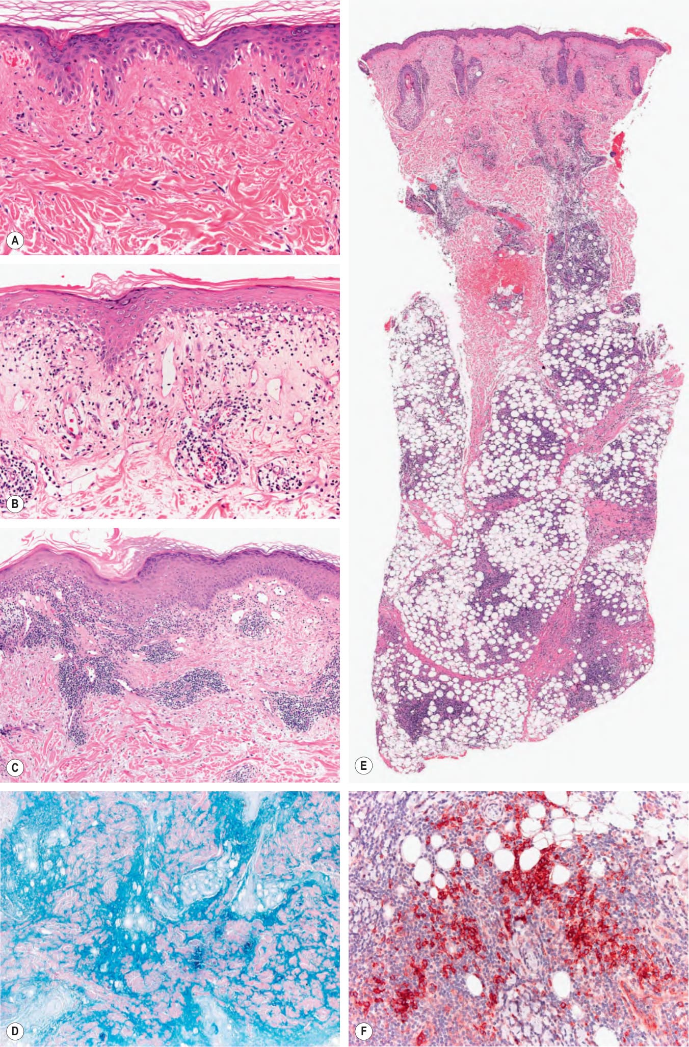
Fig. 41.17 Histopathologic features of cutaneous lupus erythematosus (LE).A Acute cutaneous LE showing mild interface dermatitis with vacuolization of basal keratinocytes and sparse superficial lymphoid infiltrates. B Subacute cutaneous LE with more obvious interface dermatitis and perivascular inflammation. C Discoid LE showing focal interface dermatitis and dense perivascular lymphoid infiltrates throughout the dermis. D Mucin deposition within the dermis highlighted by a colloidal iron stain. E Lupus panniculitis with prominent inflammatory infiltrates within the lobules of the subcutaneous fat. Perivascular and periadnexal lymphocytic infiltrates are also present in the dermis. F The pattern and intensity of immunohistochemical staining for CD123 (plasmacytoid dendritic cells) may help to distinguish cutaneous lupus, including lupus panniculitis, from other disorders. Courtesy Lorenzo Cerroni, MD.
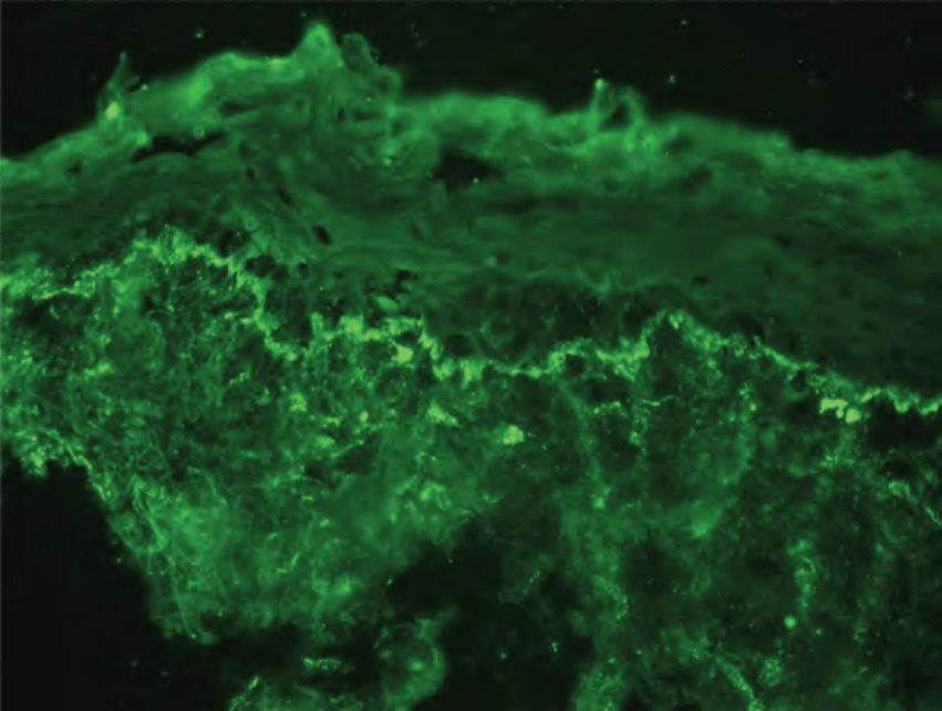
Fig. 41.18 Direct immunofluorescence of cutaneous lupus. Granular deposits of IgM are present at the dermal–epidermal junction within lesional skin. Antibody deposits at the dermal–epidermal junction are the most characteristic immunohistologic finding in lesions of cutaneous lupus and normal skin of patients with systemic lupus erythematosus. Courtesy Janet Fairley, MD.
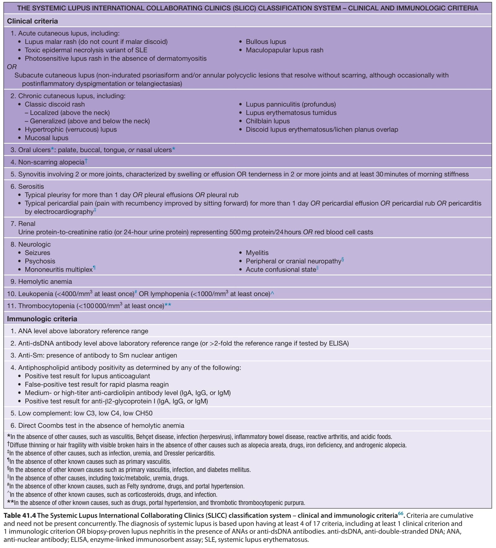
Table 41.4 The Systemic Lupus International Collaborating Clinics (SLICC) classification system – clinical and immunologic criteria. Criteria are cumulative and need not be present concurrently. The diagnosis of systemic lupus is based upon having at least 4 of 17 criteria, including at least 1 clinical criterion and 1 immunologic criterion OR biopsy-proven lupus nephritis in the presence of ANAs or anti-dsDNA antibodies. anti-dsDNA, anti-double-stranded DNA; ANA, anti-nuclear antibody; ELISA, enzyme-linked immunosorbent assay; SLE, systemic lupus erythematosus.

Table 41.5 The European League Against Rheumatism/ American College of Rheumatology (EULAR/ACR) classification criteria for systemic lupus erythematosus

Table 41.6 Cutaneous findings (nonspecific) that suggest the diagnosis of systemic lupus erythematosus. These are in addition to skin signs of other autoimmune connective tissue diseases, which raise the possibility of an overlap syndrome.
Table 41.7 Characteristic histologic findings in cutaneous lupus erythematosus (LE). The table is intended as a broad overview only. Not every feature will be present in every lesion, some features may be present that are not indicated as characteristic, and overlap exists amongst subtypes. [−], not a defining feature; [+/−], a feature that may be present in some lesions; [+], a feature that is typically present; [++], a defining feature that may be prominent.