TREATMENT
Topical Therapy
Topical or intralesional corticosteroids are a mainstay of therapy (Table 41.11). They offer a high degree of safety as well as the potential for a relatively rapid response. The systemic side effects of
(DI-SLE). CNS, central nervous system.
corticosteroids are largely avoided, although the cutaneous side effects are not. It is frequently the case that topical corticosteroids need to be of high potency in order for a response to occur, and discoid lesions are one of the few instances where it may be appropriate to use high-potency corticosteroids on the face. Patients should be instructed about the risks and benefits of therapy, the need to limit the application to affected areas, and the need for monitoring for cutaneous side effects. Particularly in active discoid lesions and LE tumidus lesions, intra lesional triamcinolone, often given in a concentration of 2.5–5 mg/ml, depending upon anatomic location, can be very effective. The injections may be repeated monthly while the lesions are active. There are also reports of the successful use of topical immunomodulators (e.g. tacrolimus, pimecrolimus) for cutaneous lesions.
Systemic Therapy
Antimalarial therapy has been used for more than a half-century for cutaneous LE, and it remains the gold standard for systemic therapy. Hydroxychloroquine sulfate is the most commonly chosen antimalarial, as it is usually well tolerated. Chloroquine and quinacrine (mepacrine) are alternatives, but quinacrine is currently not readily available in many parts of the world. In patients who are not responsive to hydroxychloroquine sulfate, quinacrine may be added to the regimen. Quinacrine can turn the skin yellow, although it does not invariably do so. The dose of hydroxychloroquine sulfate chosen is usually 200 mg once or twice per day. It has been reported that if the dose does not exceed 5 mg/kg actual body weight/day, eye toxicity is quite unlikely. For chloroquine, the usual dose is 125–250 mg/day, with the eye toxicity-minimizing dose being no more than 2.3 mg/kg actual body weight/day. Quinacrine is thought by most not to cause eye toxicity. For patients on hydroxychloroquine sulfate or chloroquine, periodic eye examinations should be done by a physician knowledgeable in antimalarial eye toxicity.
The response to antimalarials is relatively slow. It may take 2 or 3 months for efficacy to be appreciated, and sometimes several more months to achieve maximal efficacy. Consequently, for patients who are beginning therapy for cutaneous LE, topical or intralesional therapy should usually be given along with antimalarial therapy. However, some patients do not respond either to single antimalarial therapy or to the combination of quinacrine with either hydroxychloroquine sulfate or chloroquine78a.
Disease that is refractory to antimalarials is often refractory to other therapies as well. Nonetheless, it is reasonable to seek a therapy that will work well, if the risks of therapy are deemed to be worth the potential benefits. For antimalarial-resistant patients, therapeutic options include oral retinoids, dapsone, immunosuppressive agents such as mycophenolate mofetil, azathioprine or methotrexate, sulfasalazine, thalidomide or lenalidomide, and systemic corticosteroids. Dapsone is used occasionally for cutaneous LE prior to advancing to immunosuppressive therapy, but it is most often employed for the rare bullous eruption of SLE. Both retinoids and thalidomide are potent teratogens, making them difficult choices in women of childbearing potential. Thalidomide also frequently causes a peripheral neuropathy. For that reason, some clinicians have advocated giving thalidomide in low or intermittent dosing in an attempt to minimize toxicity. Lenalidomide, a secondgeneration thalidomide derivative, has led to improvement of refractory cutaneous LE and there may be a lower risk of developing peripheral neuropathy.
Responses to both belimumab and rituximab have been disappointing for patients with DLE and mixed for patients with SCLE, suggesting that the underlying pathogenesis, at least for DLE, is not dependent on B cells or that depletion of regulatory B cells by rituximab might be deleterious. Of note, rituximab may be helpful for refractory bullous LE. In the future, other immune response modifiers such as anifrolumab (anti-IFN-α receptor antibody), drugs that inactivate or deplete plasmacytoid dendritic cells (e.g. litifilimab, daxdilimab), iberdomide (promotes degradation of transcription factors Ikaros and Aiolos which regulate lymphocyte differentiation), anti-IL-33 antibody, or Janus kinase (JAK) inhibitors may play a role in the treatment of cutaneous LE.
In patients with systemic LE but no major organ involvement (i.e. potentially life-threatening involvement of visceral organs), treatment for mild disease includes NSAIDs, while corticosteroids and immunosuppressives (e.g. mycophenolate mofetil, azathioprine, methotrexate, leflunomide) are usually prescribed for moderate to severe disease. When there is a mild degree of major organ involvement, corticosteroids are the primary treatment, but for moderate to severe involvement, pulse cyclophosphamide or mycophenolate mofetil +/− pulse corticosteroids are often recommended. For a comprehensive review of future biologic therapies for refractory disease, see reference 79.
Adjunctive Therapy
Sun protection is a vital part of therapy for many patients because the sun exacerbates or initiates their skin lesions. For others, sun protection is important for cancer prevention, particularly in hypopigmented skin or in chronic discoid lesions, where the risk of skin cancer development may be higher. Cancer prevention is also essential for patients who are on immunosuppressive therapy. Lastly, it has been reported that sun exposure can exacerbate systemic disease in patients who have SLE. Therefore, there are a variety of reasons why sun protection should be emphasized, even in persons whose skin lesions are not induced or exacerbated by sun exposure.
Sunscreens should be applied to exposed skin daily, and more often if sun sensitivity is great or sun exposure is intense or prolonged. Broad-spectrum, high-SPF sunscreens are preferred (see Ch. 132). In some cases, the addition of a physical sunblock such as titanium dioxide or zinc oxide is useful. Protective clothing is important to emphasize, as the proper protective clothing is often significantly more effective than sunscreens. Sun avoidance is even more effective than protective chemicals and clothing. Midday sun is particularly high in UVB, a spectrum of UV radiation to which many patients are susceptible. It is more difficult to minimize UVA exposure, as UVA is present in substantial quantities throughout the day and can penetrate some types of window glass. Education on the optimal use of sunscreens and protective clothing and effective approaches to sun avoidance is important for most patients with lupus. Careful attention should be given to vitamin D and calcium intake.
For some patients, cosmetic cover-up is the most helpful therapeutic intervention (see Ch. 153). Particularly in situations where the disease activity has subsided but dyspigmentation remains,
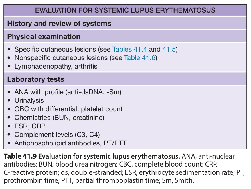
Table 41.9 Evaluation for systemic lupus erythematosus. ANA, anti-nuclear antibodies; BUN, blood urea nitrogen; CBC, complete blood count; CRP, C-reactive protein; ds, double-stranded; ESR, erythrocyte sedimentation rate; PT, prothrombin time; PTT, partial thromboplastin time; Sm, Smith.
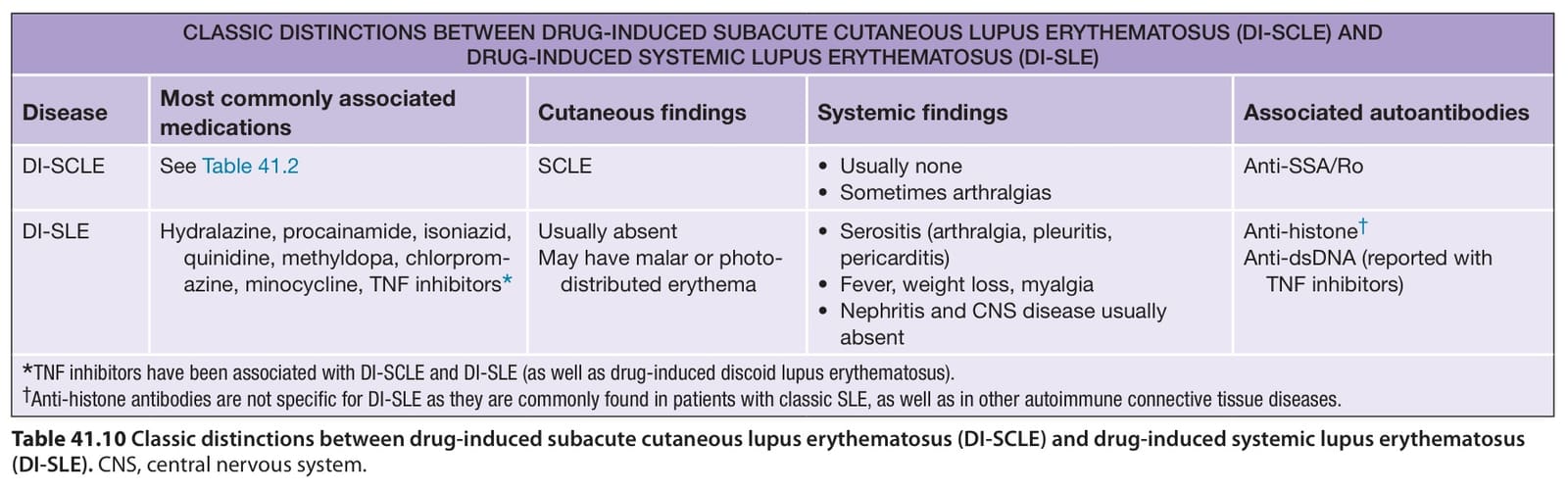
Table 41.10 Classic distinctions between drug-induced subacute cutaneous lupus erythematosus (DI-SCLE) and drug-induced systemic lupus erythematosus
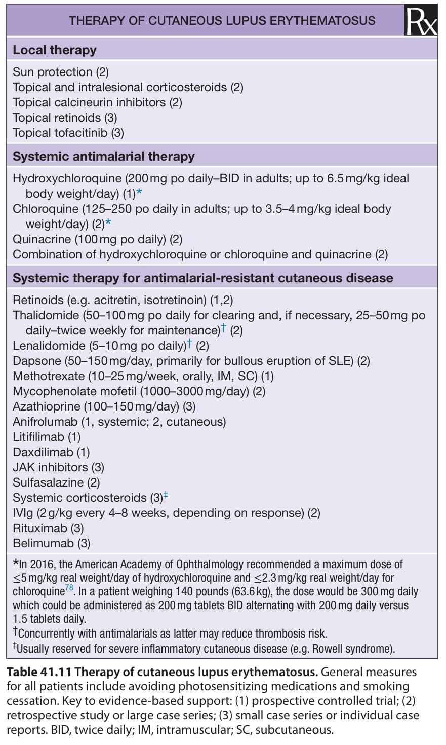
Table 41.11 Therapy of cutaneous lupus erythematosus. General measures for all patients include avoiding photosensitizing medications and smoking cessation. Key to evidence-based support: (1) prospective controlled trial; (2) retrospective study or large case series; (3) small case series or individual case reports. BID, twice daily; IM, intramuscular; SC, subcutaneous.
cosmetic camouflage of the hyper- or hypopigmentation may be the best approach.
It has been observed that a larger-than-expected percentage of cigarette smokers are represented amongst patients who present with cutaneous lupus and that smokers may have more extensive cutaneous disease and may be more refractory to therapy. Thus, smoking cessation represents a useful adjunctive therapy in some individuals.
Additional figures and table, 1997 Update of the 1982 American College of Rheumatology revised criteria for classification of systemic lupus erythematosus, available in our eBook (see inside front cover for access code).