CLINICAL FEATURES
Cutaneous Disease
The features that are classically considered pathognomonic for dermatomyositis include the heliotrope sign and Gottron papules (Fig. 42.1). The former is characterized by a pink–violet color, primarily of the eyelids and periorbital skin, which may have associated edema (Fig. 42.2). The heliotrope sign can be quite subtle, with only mild erythema of the eyelid margins, and it may wax and wane in intensity. Some patients will have more widespread facial erythema (see Fig. 42.2B,C) or midfacial erythema that tends to involve the nasolabial folds (Fig. 42.3).
The cutaneous lesions of dermatomyositis are accentuated on extensor surfaces, including the elbows, knees, metacarpophalangeal joints, and both the proximal and distal interphalangeal joints (knuckles; Fig. 42.4A). When papules on the knuckles develop a secondary lichenoid quality, they are termed Gottron papules (Fig. 42.4B,C); involvement of the elbows and/or knees is known as Gottron sign (Fig. 42.5).
A critical diagnostic feature of the cutaneous eruption of dermatomyositis is poikiloderma. This feature may occur both in patients with dermatomyositis, where it is frequently characterized by a pink–violet color, and in patients with lupus erythematosus, where the poikiloderma is typically more red in color. However, in patients with skin phototypes I and II, the hue of the poikiloderma of dermatomyositis may be pink– red rather than truly violet (Fig. 42.6A). Photodistributed poikiloderma is very characteristic of dermatomyositis, often involving the upper chest (V-neck sign) and the upper back (shawl sign). Poikiloderma can also affect photoprotected areas, such as the lateral thigh, referred to as the holster sign (Fig. 42.6B,C). If the clinician misses the poikiloderma, the eruption of dermatomyositis may occasionally be misdiagnosed as psoriasis, especially when there are well-defined plaques on the elbows and knees with fine silvery scale.
The cutaneous lesions of dermatomyositis are often intensely pruritic (Fig. 42.7), and this can significantly affect patients’ quality of life. IL-31 may play a role as it does in other pruritic disorders. Of note, pruritus is a feature that can occasionally help to distinguish dermatomyositis from LE.
An additional clinical clue to the diagnosis of dermatomyositis is nail-fold changes. Cuticular dystrophy (i.e. “ragged” cuticles and cuticular hypertrophy) is quite characteristic, as are nail-fold telangiectasias in which dilated capillary loops alternate with capillary dropout (Fig. 42.8). If the photodistribution and nail-fold changes are missed, the eruption may be misdiagnosed as other conditions characterized by poikiloderma, such as cutaneous T cell lymphoma. Often, the dermatologist correctly notices the photodistribution but considers the diagnosis of photodrug eruption or lupus erythematosus rather than dermatomyositis.
Calcinosis cutis is more prevalent in juvenile dermatomyositis, affecting 25%–70% of pediatric patients, and it is usually associated
A Inflammation of the upper eyelid can be more subtle in darkly pigmented skin; note involvement of the lateral nasal root and the cheeks. The eyebrow alopecia is due to chemotherapy. B The characteristic pink–violet color is seen with involvement of the hairline, lower forehead, upper eyelids, and cheeks; the edema is striking, especially of the upper eyelids and nasal root. C Heliotrope with marked lower eyelid edema. B, Courtesy Jean L. Bolognia, MD; C, Courtesy Kalman Watsky, MD.
with a delay in diagnosis, a delay in the institution of systemic corticosteroids, and/or advanced, therapy-resistant disease. In addition to the skin, calcium deposits may develop within the deep fascia and intra-muscular connective tissue. Calcinosis cutis presents as hard, irregular
papules, plaques, or nodules that occasionally drain a chalky material (Fig. 42.9A). Lesions favor sites of trauma, such as the elbows and knees (Fig. 42.9B), but can occur anywhere and may be painful and interfere with function.
Additional cutaneous manifestations range from poikiloderma and scaling of the scalp (often accompanied by non-scarring alopecia) to centripetal flagellate erythema (Fig. 42.10A) to erosions and ulcerations (Fig. 42.10B; Table 42.5). “Mechanic’s hands” can also be seen, more commonly in patients with anti-synthetase syndrome (Fig. 42.10C). Occasionally, especially in the setting of more severe disease, there can be edema of the extremities or even anasarca. Wong-type dermatomyositis is a variant that occurs more commonly in Asians in which the cutaneous findings mimic pityriasis rubra pilaris. Lastly, because of the frequency of overlap syndromes, it is important to look for the skin signs of other autoimmune connective tissue diseases in patients with dermatomyositis.
Patients may present with the classic cutaneous manifestations of dermatomyositis but no muscle disease. Upon careful patient evaluation and longitudinal assessment, some of these patients will be found to have skin disease alone (amyopathic dermatomyositis) while others eventually evolve into classic dermatomyositis (with muscle disease) or develop hypomyopathic dermatomyositis, i.e. no muscle weakness clinically but characteristic laboratory or radiographic abnormalities. The clinical characteristics of patients with classic and amyopathic dermatomyositis have been compared, including an examination of differences in malignancy risk. Although it was generally thought that the malignancy risk was greater in the setting of classic dermatomyositis, there have been multiple studies that have observed associated malignancies in patients with amyopathic dermatomyositis. Fulminant lung disease has also been reported in patients with amyopathic dermatomyositis. Therefore, both malignancy screening and evaluation for pulmonary involvement are recommended, irrespective of muscle involvement.
Systemic Disease
In dermatomyositis, the dermatologic manifestations frequently precede the onset of objective muscle disease; however, when present, the muscle disease is clinically indistinguishable from that seen in polymyositis. The myopathy affects proximal muscle groups, especially the extensor groups (triceps and quadriceps), in a symmetric fashion. In advanced disease, all muscle groups can be affected. Patients may be unable to complete simple tasks such as combing their hair or rising to their feet from a sitting position. Muscle involvement is often accompanied by malaise and fatigue.
Muscle strength should be graded on sequential visits by muscle groups (e.g. right and left triceps 3/5, right and left quadriceps 4/5,
etc.), although other more formal methods of quantification have been described. Assessment of neck flexor weakness can also be used to gauge muscle involvement. Symptoms of dysphagia should prompt investigation for overlap with systemic sclerosis, although patients with advanced muscle disease can have cricopharyngeal muscle dysfunction and consequently difficulty initiating swallowing. Distal esophageal involvement can present as gastroesophageal reflux disease.
Pulmonary disease occurs in ~15%–30% of patients with dermatomyositis, and generally presents as a diffuse interstitial fibrosis that is similar to that seen in patients with rheumatoid arthritis or systemic sclerosis. Symptoms include a dry cough and progressive breathlessness. In one US-based study, there was no significant difference in the prevalence of interstitial lung disease when amyopathic dermatomyositis was compared to the classic form. All adult patients should have screening pulmonary function tests (PFTs) that include diffusion capacity for carbon monoxide (DLCO). In those who have reduced DLCO, a high-resolution chest CT should then be performed. Patients with a high-risk phenotype for pulmonary involvement (e.g. anti-MDA5 dermatomyositis) often have a high-resolution chest CT performed at baseline as well as PFTs with DLCO.
In Eastern Asian populations, patients with anti-MDA5 (CADM) antibodies have a high risk of rapidly progressive lung disease and a poor prognosis despite a lack of muscle disease (see Table 42.4). Furthermore, a novel MDA5 antibody-associated dermato-pulmonary syndrome has been reported in non-Asian populations in which patients develop tender palmar papules and cutaneous ulcerations, primarily on the elbows, dorsal hands, and periungual region (Fig. 42.11). Additional characteristic features of anti-MDA5 dermatomyositis include arthritis, oral ulcerations, and severe non-scarring alopecia. Recognition of this constellation of cutaneous findings alerts the clinician to the increased risk of associated and potentially fatal pulmonary disease.
Pulmonary involvement is also a notable feature of the anti-synthetase syndrome. For example, if anti-Jo-1 antibodies are present, there is a 70% likelihood that the patient will develop pulmonary disease. Because pulmonary disease associated with dermatomyositis is resistant to corticosteroid therapy, it follows a course independent from that of the muscle disease and can adversely affect prognosis.
Cardiac disease is usually not symptomatic. When it is, it commonly presents as arrhythmias or as conduction defects. Patients with
dermatomyositis are often not thoroughly evaluated for possible cardiac disease, and prompt treatment of the muscle disease may mask the true frequency of cardiac involvement. In one retrospective review, cardiac involvement was shown to be the major prognostic factor for death in patients with dermatomyositis, in the absence of an under-lying malignancy. Additional reported indicators of poor prognosis (other than malignancy) include older age, progressive disease, initiation of therapy after 24 months of muscle weakness, longer duration of symptoms before diagnosis, pulmonary disease, dysphagia, and extensive cutaneous involvement of the trunk.
For the patient with dermatomyositis, a thorough review of systems should include a discussion of arthritis, gastrointestinal disease,
Photodistributed poikiloderma is very characteristic of dermatomyositis and often involves the upper chest (V-neck sign). In this patient with skin phototype I, the color is pink– red. B Broken-up pink–violet plaque on the upper lateral thigh (holster sign). C Holster sign with a pink–brown color in a patient with more darkly pigmented skin. A, Courtesy Edward Cowen, MD; B, Courtesy Jonathan Leventhal, MD; C, Courtesy Sara Perkins, MD.
The cuticles are “ragged” and within the proximal nail fold, dilated capillary loops alternate with vessel dropout (inset). Atrophy, telangiectasias, and hypopigmentation are present on the fingers. Courtesy Julie V. Schaffer, MD.
pulmonary disease, cardiac disease, and other manifestations such as Raynaud phenomenon. The presence of additional clinical findings and/ or systemic symptoms may signify an overlap with other connective tissue diseases – in particular, systemic LE, rheumatoid arthritis, and systemic sclerosis. One dermatology-based case series observed a 19% overlap rate in adult patients with dermatomyositis.
Malignancy
The reported frequency of an internal malignancy in adults with dermatomyositis varies from <10% to >50%; however, the best data point to a probable risk of 15%–25%. The malignancy association occurs with adult dermatomyositis (both classic and amyopathic), but not with the juvenile form or with polymyositis. Genitourinary
malignancies, especially ovarian cancer, and colon cancer may be overrepresented; in some Southeast-Asian populations, nasopharyngeal carcinoma is overrepresented. Other commonly detected malignancies include breast, lung, gastric, pancreatic, and lymphomas (including non-Hodgkin). The risk of malignancy may return to a relative baseline after 3 years.
Callen has suggested that the best approach to patient management in this setting is ongoing vigilance with frequent and thorough medical histories, repeated review of systems, complete physical examinations, and screening laboratory tests. He has also advocated for additional cancer-screening investigations, including CT of the chest, abdomen, and pelvis as well as transvaginal pelvic ultrasound in women (Table 42.6). Patients should be up-to-date with regard to recommended screenings such as colonoscopy and mammography.
Several myositis-associated autoantibodies have been described (see Table 42.4), and presence of the anti-TIF1-γ (p155) autoantibody in particular has been associated with an increased risk of malignancy. Indeed, in one study, at least 80% of patients with cancer-associated dermatomyositis had antibodies against either TIF1-γ (transcription intermediary factor 1-γ) or nuclear matrix protein NXP-2. In a recent systematic review and meta-analysis, older age, male sex, dysphagia,
cutaneous ulceration, anti-TIF1-γ antibodies, and classic dermatomyositis (i.e. without anti-synthetase syndrome or clinically amyopathic dermatomyositis) were associated with a significantly higher risk of an underlying malignancy. This study also concluded that CT
of the chest, abdomen, and pelvis appeared to be effective in identifying otherwise asymptomatic malignancies, further supporting this screening strategy.
With regard to long-term survival, as expected, patients with cancerassociated dermatomyositis have an inferior survival curve when compared to patients with juvenile or non-cancer-associated adult myopathies.
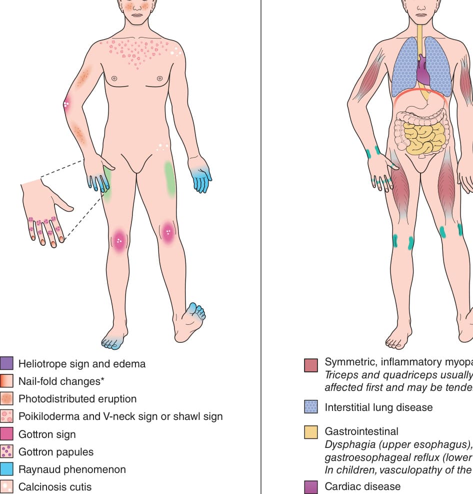
Fig. 42.1 Cutaneous and systemic findings in dermatomyositis.A Cutaneous. B Systemic.

Fig. 42.2 Dermatomyositis – eyelid edema and heliotrope sign.
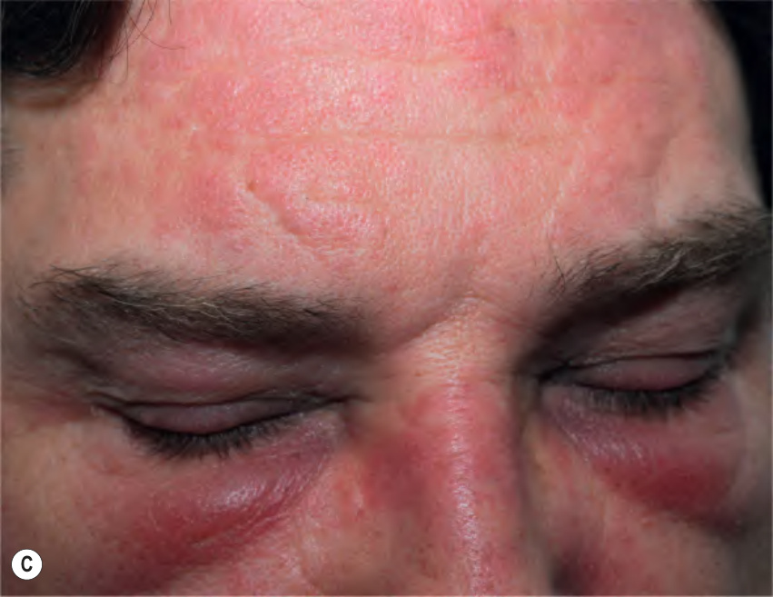
Fig. 42.2 Dermatomyositis – eyelid edema and heliotrope sign.
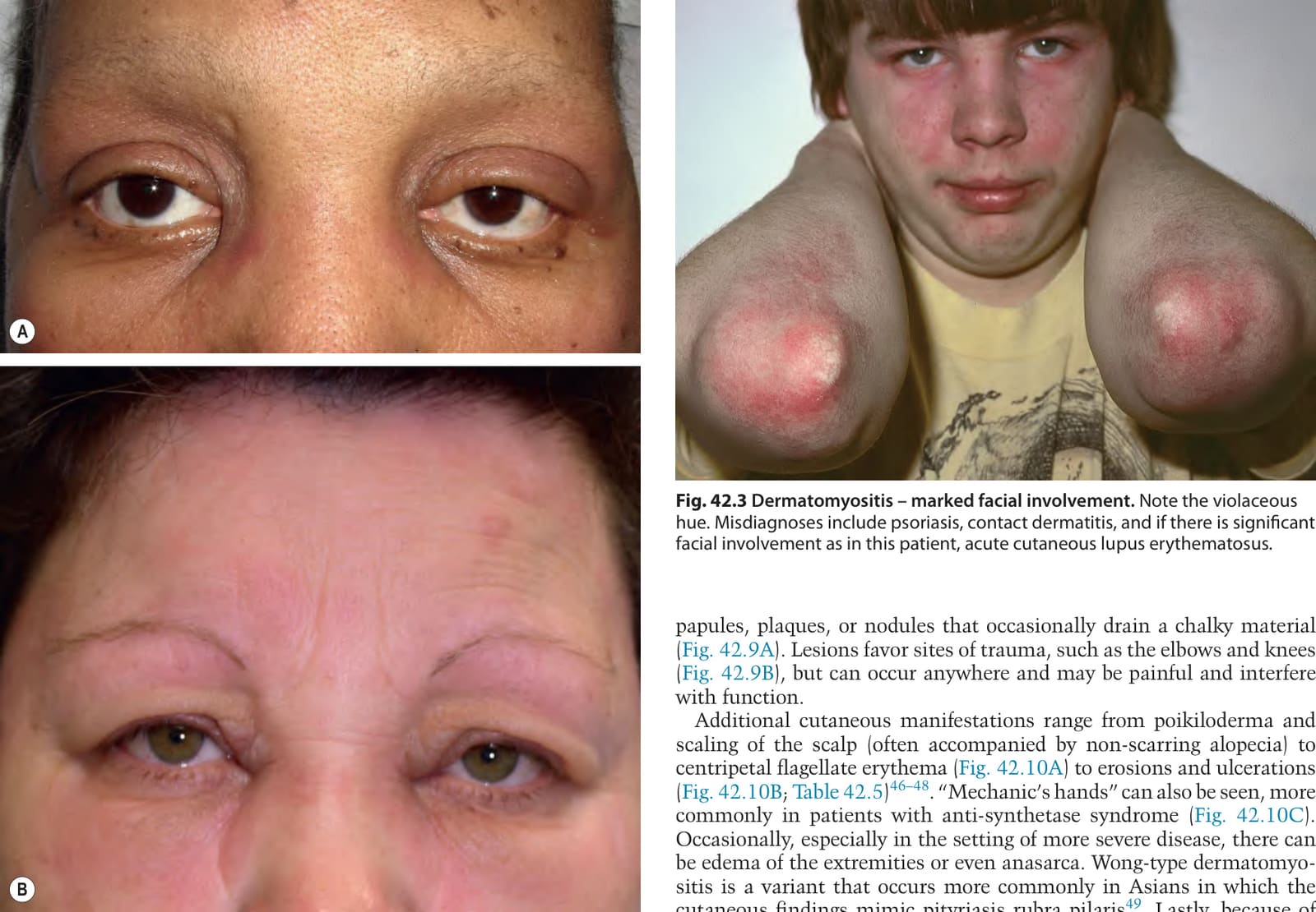
Fig. 42.3 Dermatomyositis – marked facial involvement. Note the violaceous hue. Misdiagnoses include psoriasis, contact dermatitis, and if there is significant facial involvement as in this patient, acute cutaneous lupus erythematosus.
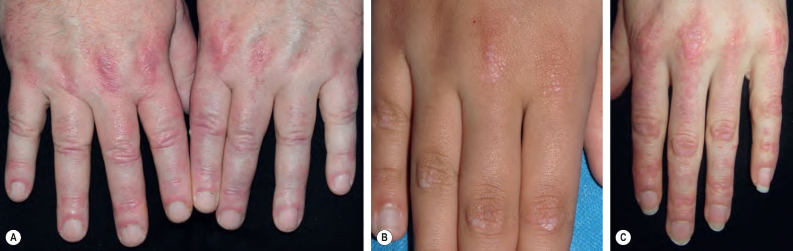
Fig. 42.4 Dermatomyositis – Gottron papules.A Only a few Gottron papules are present, but there is definite accentuation of the pink–violet inflammation over the metacarpophalangeal (MCP) joints and proximal and distal interphalangeal (PIP, DIP) joints as well as the proximal nail folds. Note the ragged cuticles. B The flat-topped (lichenoid) papules overlying the DIP, PIP, and MCP joints (knuckles) are subtle and were misdiagnosed as verrucae vulgares in this child. C More obvious disease in which the multiple pink–violet lichenoid papules are coalescing and there is some involvement of the interphalangeal skin. A, Courtesy Kalman Watsky, MD; B, Courtesy Julie V. Schaffer, MD.
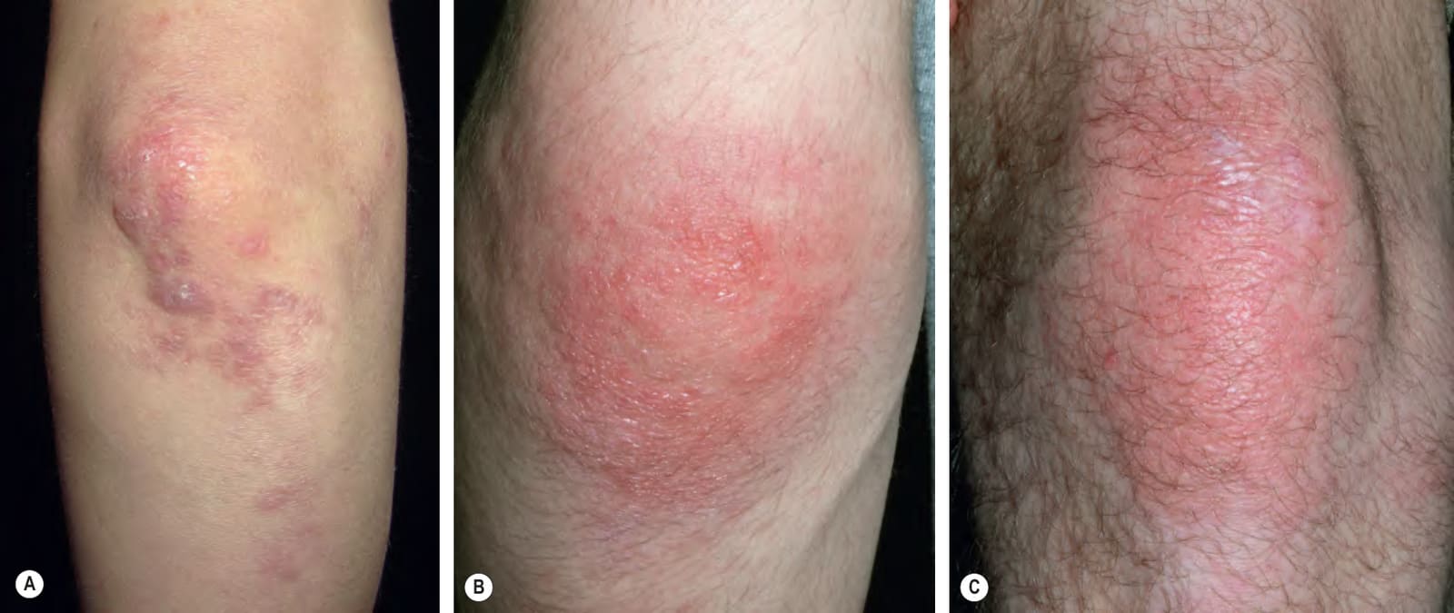
Fig. 42.5 Dermatomyositis – Gottron sign. Pink–violet papules (A) and thin pink plaques (B) of the elbows and knee (C). Some of the papules on the elbow are flattopped (lichenoid) and others have white scale. The presence of plaques on the elbows and knees can lead to the misdiagnosis of psoriasis. B,C, Courtesy Julie V. Schaffer, MD.
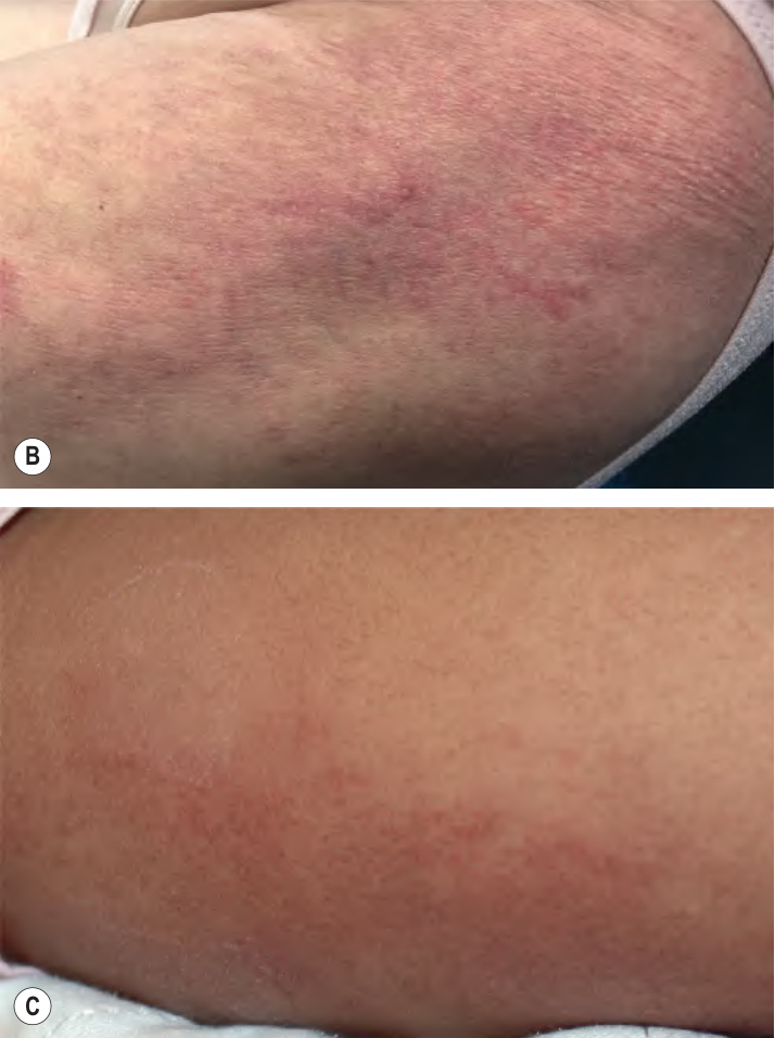
Fig. 42.6 Dermatomyositis – poikiloderma and holster sign.A
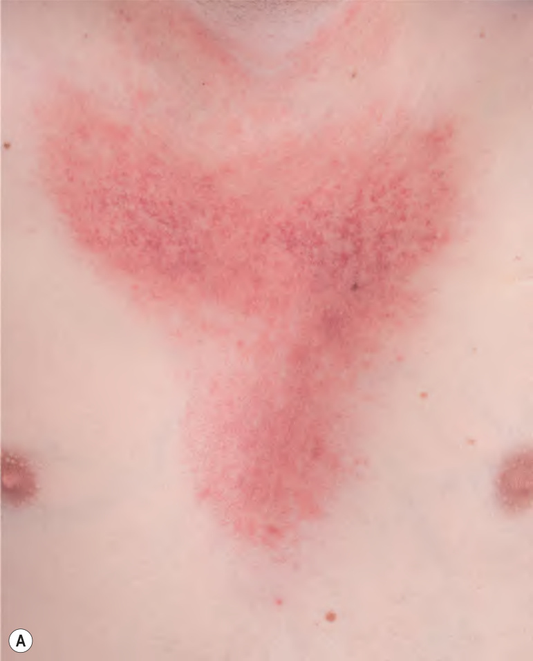
Fig. 42.7 Dermatomyositis – involvement of the upper back. The pink–violet plaques, some with associated scale, were very pruritic, as evidenced by multiple excoriations. Linear streaks of erythema are also seen. Courtesy Jean L. Bolognia, MD.
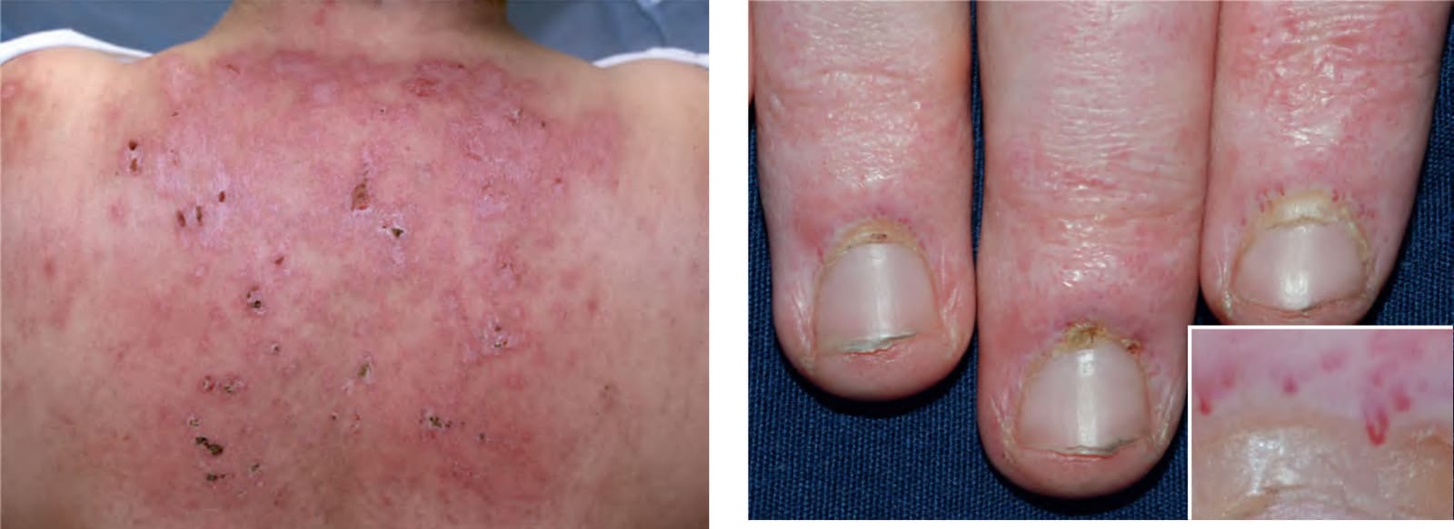
Fig. 42.8 Dermatomyositis – cuticular dystrophy and nail-fold telangiectasias.
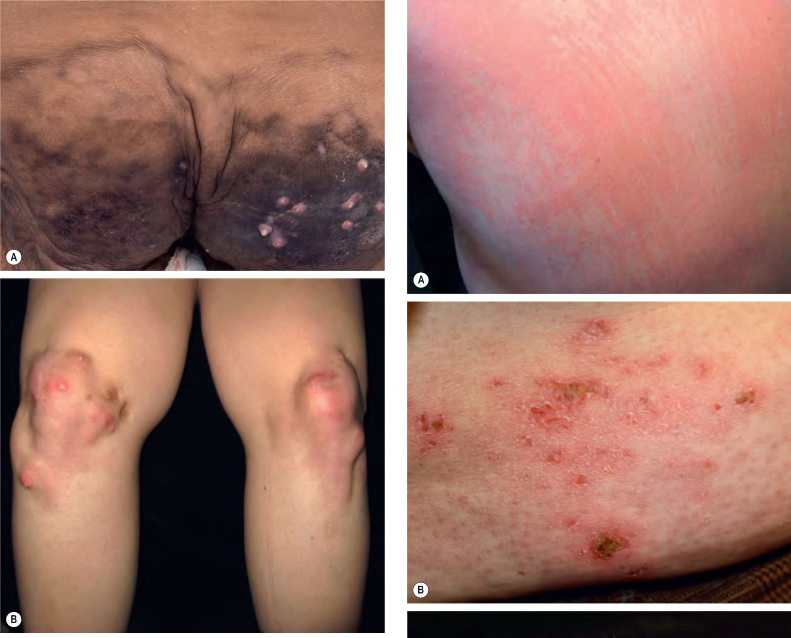
Fig. 42.9 Calcinosis cutis.A Rock-hard hyperpigmented plaques on the buttocks with multiple sites of drainage in an adult with longstanding dermatomyositis. B In this patient with juvenile dermatomyositis, the nodules favored the extensor surface of the knees and resulted in a decrease in joint mobility. A, Courtesy Edward Cowen, MD.
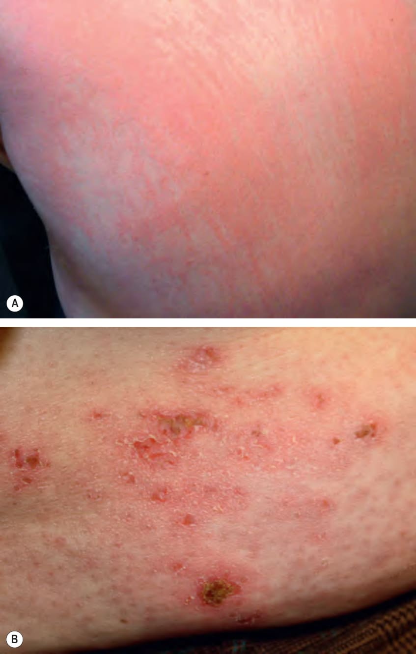
Fig. 42.10 Dermatomyositis – less common presentations.A Flagellate erythema of the posterior trunk. B Cutaneous erosions and ulcerations can also occur. C Hyperkeratosis and mild erythema of the lateral and palmar surfaces of the fingers in a patient with “mechanic’s hands”.
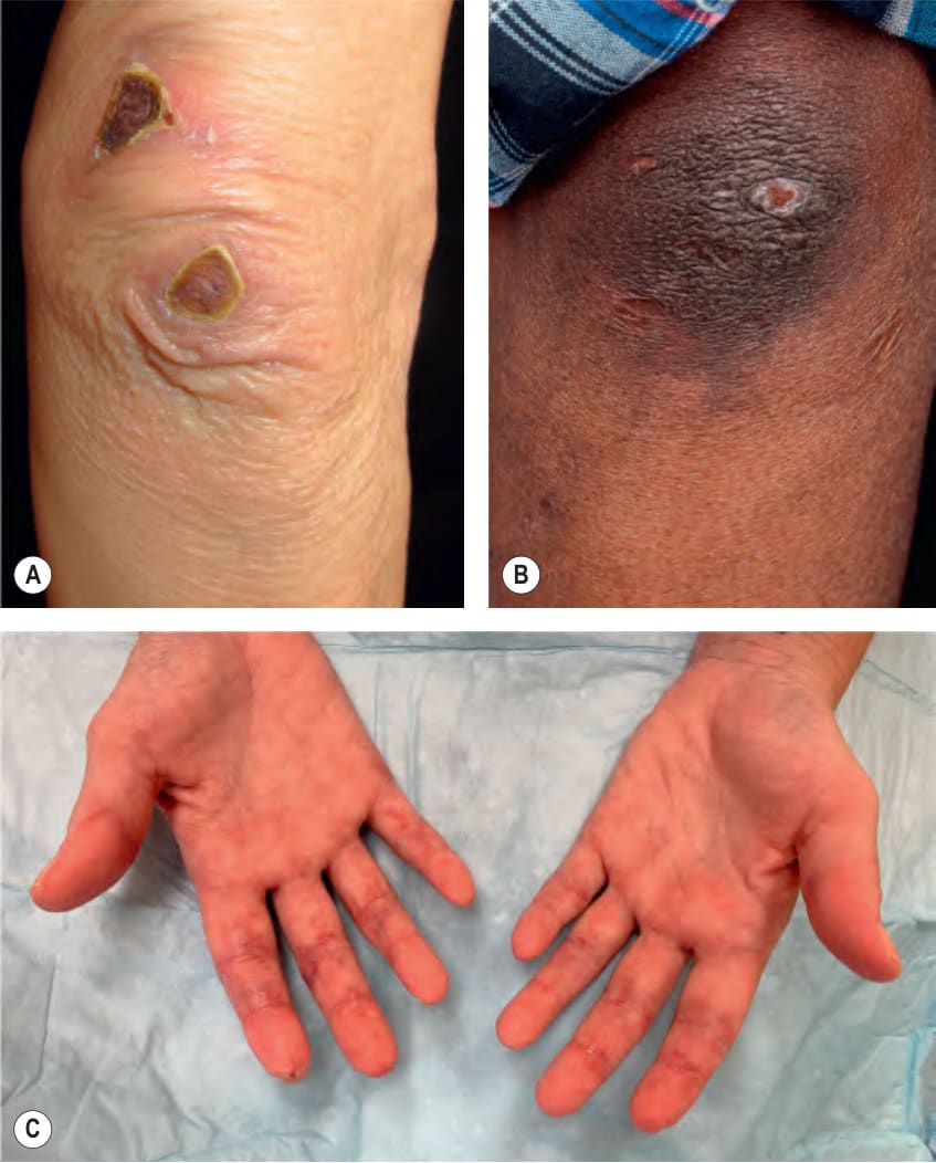
Fig. 42.11 Three patients with anti-MDA5 dermatomyositis.A, B Vasculopathic ulcerations favor the elbows, dorsal hands, and periungual region; they can be associated with Gottron sign and Gottron papules. C Painful violaceous macules and papules with an irregular outline that favor the digital creases.
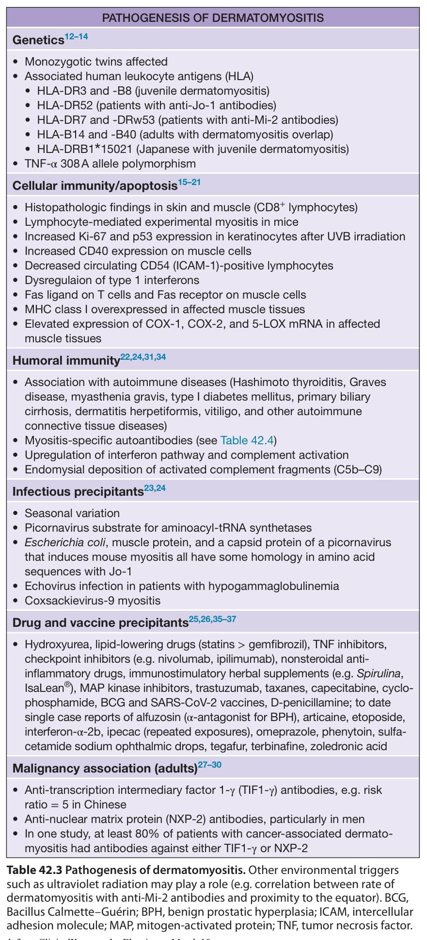
Table 42.3 Pathogenesis of dermatomyositis. Other environmental triggers such as ultraviolet radiation may play a role (e.g. correlation between rate of dermatomyositis with anti-Mi-2 antibodies and proximity to the equator). BCG, Bacillus Calmette–Guérin; BPH, benign prostatic hyperplasia; ICAM, intercellular adhesion molecule; MAP, mitogen-activated protein; TNF, tumor necrosis factor.
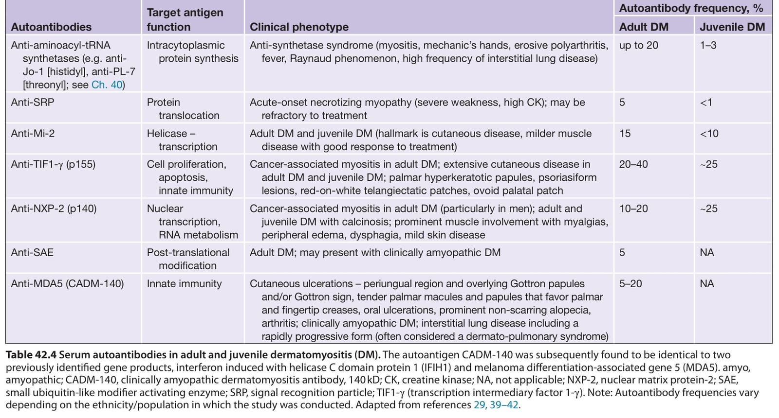
Table 42.4 Serum autoantibodies in adult and juvenile dermatomyositis (DM). The autoantigen CADM-140 was subsequently found to be identical to two previously identified gene products, interferon induced with helicase C domain protein 1 (IFIH1) and melanoma differentiation-associated gene 5 (MDA5). amyo, amyopathic; CADM-140, clinically amyopathic dermatomyositis antibody, 140 kD; CK, creatine kinase; NA, not applicable; NXP-2, nuclear matrix protein-2; SAE, small ubiquitin-like modifier activating enzyme; SRP, signal recognition particle; TIF1-γ (transcription intermediary factor 1-γ). Note: Autoantibody frequencies vary depending on the ethnicity/population in which the study was conducted. Adapted from references 29, 39–42.
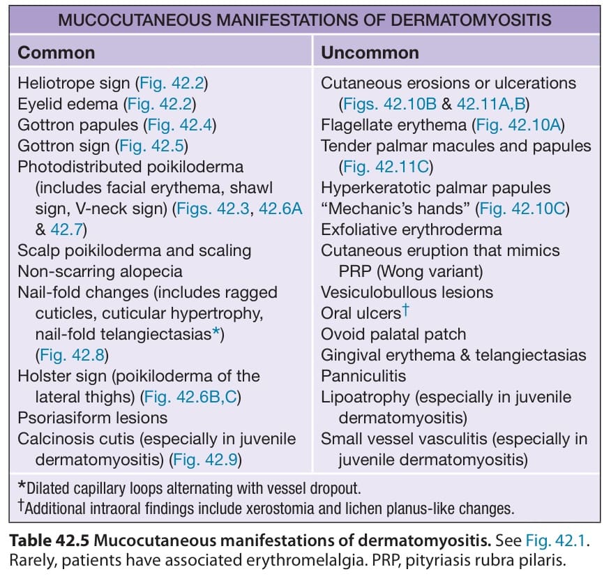
Table 42.5 Mucocutaneous manifestations of dermatomyositis. See Fig. 42.1. Rarely, patients have associated erythromelalgia. PRP, pityriasis rubra pilaris.
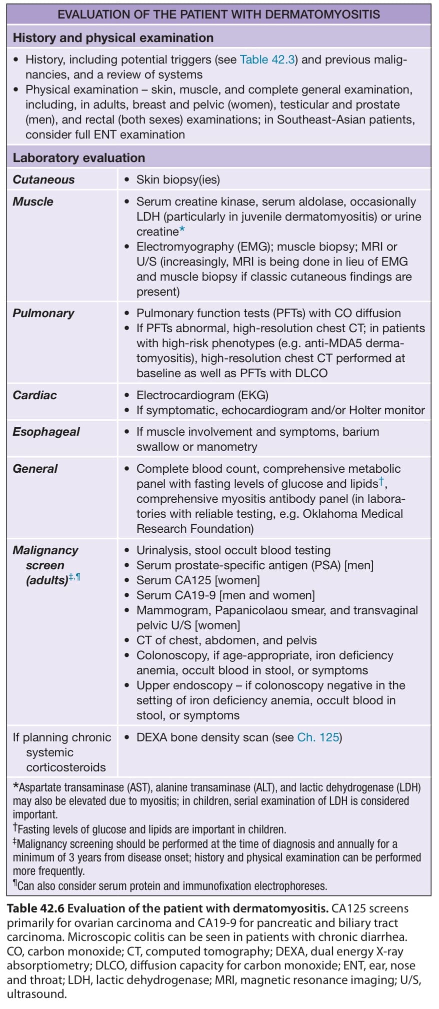
Table 42.6 Evaluation of the patient with dermatomyositis. CA125 screens primarily for ovarian carcinoma and CA19-9 for pancreatic and biliary tract carcinoma. Microscopic colitis can be seen in patients with chronic diarrhea. CO, carbon monoxide; CT, computed tomography; DEXA, dual energy X-ray absorptiometry; DLCO, diffusion capacity for carbon monoxide; ENT, ear, nose and throat; LDH, lactic dehydrogenase; MRI, magnetic resonance imaging; U/S, ultrasound.