PATHOLOGY
The characteristic changes seen in skin biopsy specimens from patients with dermatomyositis can be very subtle (Fig. 42.12) and include epidermal atrophy, signs of interface dermatitis with vacuolar alteration of basal keratinocytes and pigment incontinence, and dermal changes consisting of interstitial mucin deposition and a sparse lymphocytic infiltrate. In some cases, a colloidal iron stain may help to highlight
the presence of mucin. The histopathologic changes may be indistinguishable from those of cutaneous LE, in particular acute and subacute. Gottron papules show an interface dermatitis but have acanthosis and hyperkeratosis rather than epidermal atrophy. A lobular panniculitis can also occur in patients with dermatomyositis and can resemble lupus panniculitis.
Muscle biopsy specimens also show characteristic changes. The combination of type II muscle fiber atrophy, necrosis, regeneration, and hypertrophy with centralized sarcolemmal nuclei, plus lymphocytes in both a perifascicular and a perivascular distribution, is classic. It is best for the clinician to request that the surgeon sample triceps muscle rather than the usually biopsied deltoid muscle because the latter is often spared until late in the disease. Another option, especially for patients with mild involvement, is to choose the biopsy site based upon MRI findings (see below).
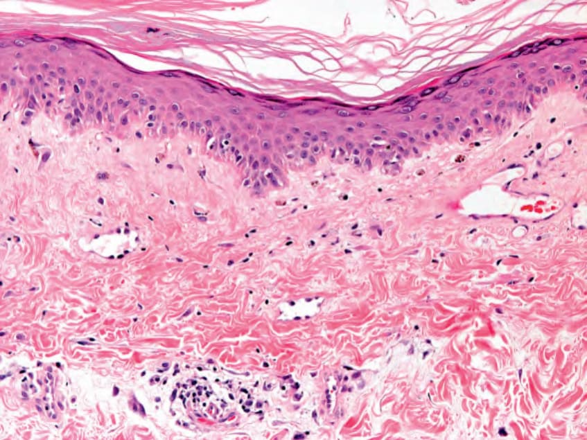
Fig. 42.12 Dermatomyositis – histopathologic features. The vacuolar changes within the basal layer can be subtle. In the dermis, dilated blood vessels and a sparse lymphocytic infiltrate are seen (poikilodermatous changes). Courtesy Lorenzo Cerroni, MD.