DIFFERENTIAL DIAGNOSIS AND EVALUATION
A detailed differential diagnosis of the patient who presents with proximal muscle weakness in the absence of skin lesions, i.e. the differential diagnosis of polymyositis, is beyond the scope of this dermatologic review. The patient with a characteristic cutaneous eruption should have a skin biopsy performed to confirm the clinical impression of dermatomyositis, keeping in mind that cutaneous LE can have similar histologic findings (Fig. 42.13). Table 42.7 summarizes the differential diagnosis of the dermatologic findings in dermatomyositis. Two validated instruments, the Dermatomyositis Skin Severity Index (DSSI) and the updated Cutaneous Dermatomyositis Disease Area and Severity Index (CDASI), can be used to determine the severity of cutaneous disease. These indices are helpful in assessing therapeutic responses in clinical trials.
Once the presumed diagnosis is confirmed histopathologically, a search for muscle and/or associated systemic disease must be under-taken (see Table 42.6), because it is their presence or absence that dictates therapeutic decisions. The evaluation for myositis can include: strength testing of proximal extensor muscles and other muscles including the neck flexors, serum levels of muscle enzymes, an electromyogram, and/or triceps muscle biopsy. However, increasingly, an MRI (Fig. 42.14) or ultrasound (Fig. 42.15) of proximal muscles is obtained prior to, or in lieu of, a muscle biopsy, especially in patients with classic cutaneous lesions. Individuals with normal muscle enzyme levels should be followed at regular intervals because these patients can have occult muscle disease, evolve into classic dermatomyositis, or remain as amyopathic dermatomyositis (see Fig. 42.13).
Additional baseline investigations for pulmonary disease (predicted in part by autoantibody profile; see above and Table 42.4), for cardiac
disease, for symptomatic esophageal disease (barium swallow or manometry), and for features of overlap autoimmune connective tissue disease should be performed at this stage, prior to the institution of systemic therapy unless immediate therapy is warranted to halt rapidly progressive disease. Adult patients should have screening for malignancy annually for at least 3 years from disease onset (see Table 42.6), or more frequently based upon symptoms. A detailed history, review of symptoms, and complete physical examination should be performed at regular intervals of 4–6 months. Adult patients with amyopathic dermatomyositis should have similar evaluations for malignancy and associated pulmonary disease.
The classic muscle enzymes that should be assessed at baseline and at regular intervals throughout the treatment period are creatine kinase (CK) and aldolase. Aspartate and alanine transaminases and lactate dehydrogenase are often elevated as well, since they also are released from damaged muscle tissue. It has been estimated that 95% of patients with classic dermatomyositis have an elevated CK at some point during their illness. Urine creatine is elevated in some patients who have a normal serum CK, but this test is not routinely performed.
Electromyography (EMG) is a sensitive but nonspecific test in patients with dermatomyositis. Over 90% of patients with classic dermatomyositis have abnormal EMG results. In the absence of classic cutaneous lesions, the muscle biopsy is a very important test for confirming the diagnosis and excluding other inflammatory myopathies. Surgeons will usually biopsy the deltoid muscle for convenience, but this muscle is relatively spared until late in the disease; therefore, the surgeon should be directed to perform a triceps muscle biopsy.
MRI (see Fig. 42.14) and ultrasound (see Fig. 42.15) of muscles are sensitive tests that can add greatly to patient evaluation. Both can be used to determine the appropriate site for a muscle biopsy. Ultrasound is less expensive and easier to perform sequentially. MRI may be the most sensitive test for muscle disease, but it is more expensive. In children with juvenile dermatomyositis, bilateral thigh MRI utilizing a myositis protocol is considered standard of care in many medical centers. MRI may also be useful in evaluating children for soft tissue changes that might precede calcinosis cutis.
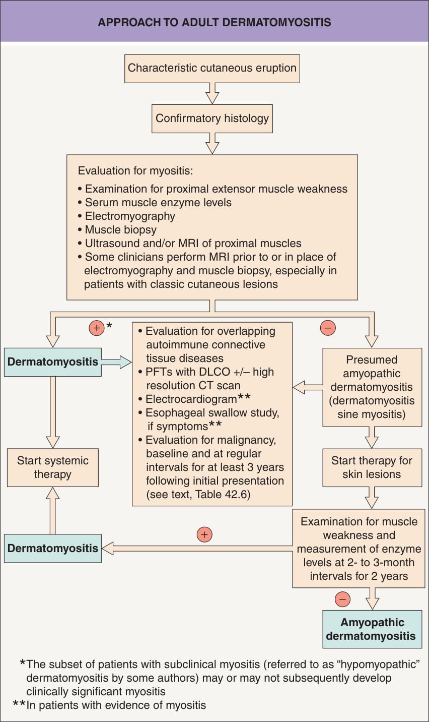
Fig. 42.13 Approach to adult dermatomyositis. The approach for children is similar but without the malignancy evaluation; in addition, pulmonary testing is not performed in children in the absence of symptoms given the low risk of interstitial lung disease. Table 42.1 outlines the definitions of “provisional” and “confirmed” amyopathic dermatomyositis but patients in the former group rarely go on to develop muscle disease. PFTs, pulmonary function tests.

Fig. 42.14 T2-weighted MR images of the proximal thigh in a patient with dermatomyositis. Note the increased signal intensity, primarily in the extensor muscles (white color, arrows). The increased signal correlates with inflammation.
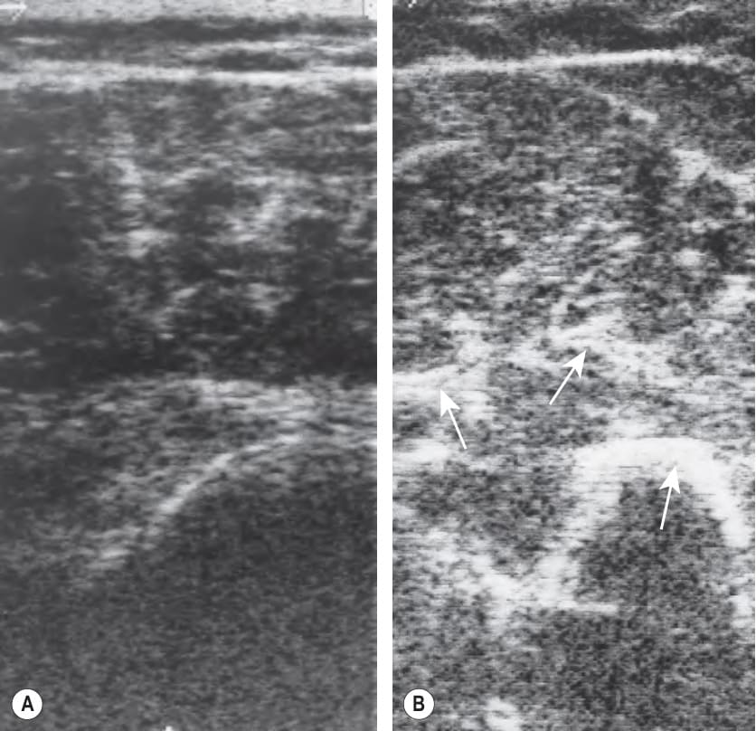
Fig. 42.15 Ultrasound images of the triceps muscle.A Cross-sectional ultrasound image from normal (control) triceps muscle. B Ultrasound image from affected triceps muscle in a patient with dermatomyositis. An increase in inter-stitial echoes is seen (arrows). This image highlights the triceps muscle, which is a higher yield biopsy site in dermatomyositis given that the deltoid muscle is often relatively spared until late in the disease.
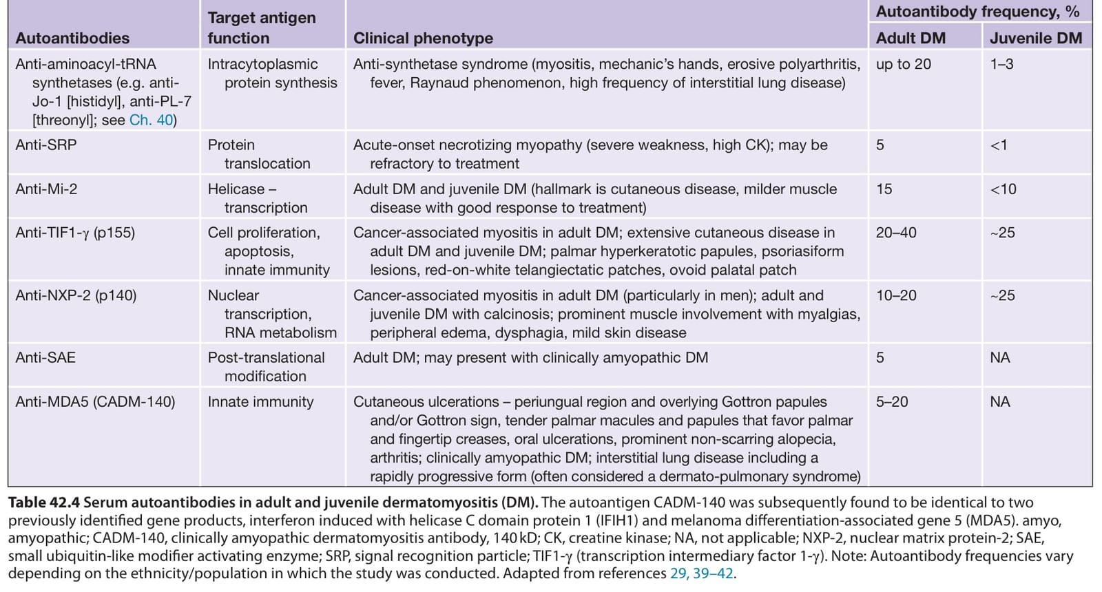
Table 42.4 Serum autoantibodies in adult and juvenile dermatomyositis (DM). The autoantigen CADM-140 was subsequently found to be identical to two previously identified gene products, interferon induced with helicase C domain protein 1 (IFIH1) and melanoma differentiation-associated gene 5 (MDA5). amyo, amyopathic; CADM-140, clinically amyopathic dermatomyositis antibody, 140 kD; CK, creatine kinase; NA, not applicable; NXP-2, nuclear matrix protein-2; SAE, small ubiquitin-like modifier activating enzyme; SRP, signal recognition particle; TIF1-γ (transcription intermediary factor 1-γ). Note: Autoantibody frequencies vary depending on the ethnicity/population in which the study was conducted. Adapted from references 29, 39–42.
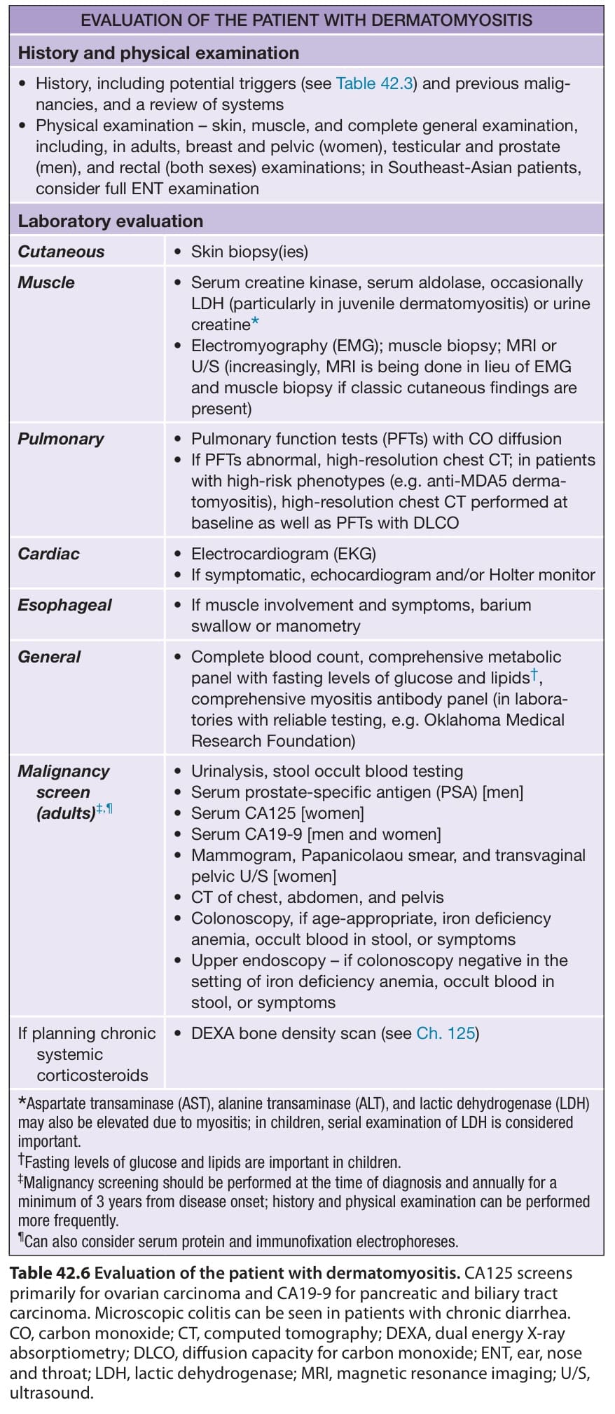
Table 42.6 Evaluation of the patient with dermatomyositis. CA125 screens primarily for ovarian carcinoma and CA19-9 for pancreatic and biliary tract carcinoma. Microscopic colitis can be seen in patients with chronic diarrhea. CO, carbon monoxide; CT, computed tomography; DEXA, dual energy X-ray absorptiometry; DLCO, diffusion capacity for carbon monoxide; ENT, ear, nose and throat; LDH, lactic dehydrogenase; MRI, magnetic resonance imaging; U/S, ultrasound.
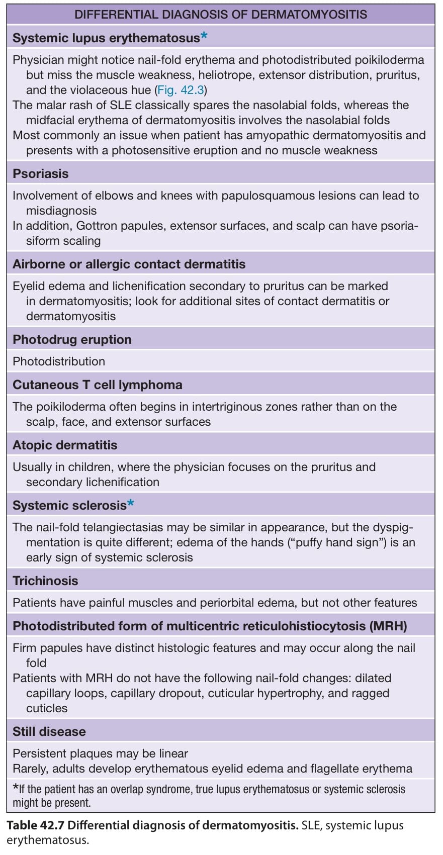
Table 42.7 Differential diagnosis of dermatomyositis. SLE, systemic lupus erythematosus.