TREATMENT
A therapeutic ladder for the treatment of patients with dermatomyositis is summarized in Table 42.8. As with many rare dermatologic diseases, there are few double-blind, placebo-controlled trials. However, a dermatology-based case series supported the belief that a number of patients (75%–85%) can become muscle disease-free, off treatment, after a period of 24–48 months, utilizing a slow corticosteroid taper. Long-term follow-up of pediatric patients also confirmed this conclusion. There is a clear discordance between response of the muscle disease to therapy and response of the skin disease. Available therapy for the skin disease is clearly inadequate, with many patients experiencing persistent cutaneous disease and pruritus, especially of the scalp, which can become major complaints once the muscle disease is under control. Aggressive topical therapy and systemic therapy (e.g. antimalarials and/or weekly methotrexate) added specifically for the skin disease often do not provide adequate relief. However, it is clear that, except in adult patients with an advanced malignancy, the prognosis for corticosteroid-treated dermatomyositis in children and adults is excellent, especially when compared with a mortality rate of up to 50% in the pre-corticosteroid era.
The treatment of dermatomyositis must take into account the discordance between the response of the muscle disease and that of the dermatologic disease to systemic therapy. If muscle disease is confirmed, then
systemic corticosteroid therapy is initiated in doses aimed at control of the myositis (i.e. generally prednisone 1 mg/kg/day). Patients with dermatologic manifestations alone can be treated as described below, with repeated clinical muscle examination and enzyme screening (generally CK and aldolase) at 2- to 3-month intervals.
If CK and aldolase serum levels are minimally elevated and corticosteroid therapy is initiated early, the disease generally comes under control rapidly over 2–4 weeks, with a taper to half the starting dose by 6 months. If the disease is advanced and enzyme levels are high (e.g. CK >1000 U/L), the disease can be much more difficult to control and often requires intravenous pulse corticosteroids plus early initiation of a steroid-sparing agent such as low-dose weekly methotrexate. Again, an attempt is made to taper the prednisone to 0.5 mg/kg/day in a single morning dose by 6–8 months.
Attention to general health maintenance principles and osteoporosis prevention is a key element of patient management (see Ch. 125). This can be accomplished by joint longitudinal care with an internist or pediatrician. Adults also need frequent (e.g. every 4–6 months for at least 3 years) complete physical examinations for malignancy.
Children with juvenile dermatomyositis should be followed at least twice yearly by a pediatrician comfortable with monitoring their developmental milestones while on immunosuppressive therapy. In addition, lipodystrophy and metabolic abnormalities, including hypertriglyceridemia and insulin resistance, have become increasingly recognized as potential complications of juvenile dermatomyositis.
When muscle disease is present, there is support for steroid-sparing therapies (especially in the setting of severe or recalcitrant disease), including IVIg, methotrexate, azathioprine, mycophenolate mofetil, rituximab, pulse cyclophosphamide, and cyclosporine (also for lung disease). Less often tacrolimus, sirolimus, infliximab, fludarabine, and hematopoietic stem cell transplantation are used. Caution should be exercised if TNF inhibitors are being considered as cases of drug-induced and drug-exacerbated dermatomyositis have been reported. Consequently, many experts recommend avoiding these agents in patients with dermatomyositis.
Of the limited number of randomized, controlled trials performed to date, two assessed IVIg, with both demonstrating benefit in both muscle and skin disease, and one examined rituximab. The rituximab trial failed to reach its primary and secondary endpoints for muscle disease, but this was potentially related to study design – muscle disease improved in 83% of patients and a steroid-sparing effect was noted. In this trial, none of the core measures of disease activity were skin-specific and no validated skin score was used; however, follow-up analysis utilizing a cutaneous visual analog scale demonstrated improvement of skin disease in both adult and juvenile dermatomyositis. In contrast, a pilot trial of rituximab that used a validated skin score found no improvement in cutaneous disease activity. A small, randomized pilot study that investigated abatacept for muscle disease in both dermatomyositis and polymyositis observed a reduction in disease activity in nearly 50% of treated patients. Notably, a placebo-controlled study showed no benefit from plasmapheresis or leukapheresis in dermatomyositis. In children with severe or refractory disease, IVIg is often administered.
Adjunctive physical therapy is also very important in improving muscle strength and endurance. Benefits of exercise and rehabilitation have been demonstrated, even in the course of active disease, without inducing flares of the myositis.
Dermatologic disease can be particularly challenging therapeutically, and not all patients clear their cutaneous manifestations when their muscle disease remits. Patients with dermatologic disease without active muscle disease are generally not treated with systemic corticosteroids as a mainstay of their therapy, and they often present a unique therapeutic challenge. As a result, the treatment of amyopathic dermatomyositis often differs significantly from the treatment of classic dermatomyositis in that systemic corticosteroids are not traditionally utilized.
For cutaneous disease, anecdotal reports, case series, retrospective reviews, and limited prospective data support the use of topical corticosteroids, topical tacrolimus, antimalarial therapy (either as monotherapy or in combination but with a less dramatic effect than in cutaneous LE), low-dose weekly methotrexate, mycophenolate mofetil, IVIg, dapsone, tofacitinib, and thalidomide, as well as additional agents listed in Table 42.8. Of note, there is an increased risk of a cutaneous drug reaction to hydroxychloroquine in patients with dermatomyositis as compared to those with LE. This medication is still frequently used as first-line therapy, but a pre-treatment warning is recommended. Some patients who develop a cutaneous reaction to hydroxychloroquine may tolerate chloroquine.
Clinical studies to support a specific treatment algorithm for cutaneous dermatomyositis are lacking. A common therapeutic approach includes a trial of antimalarials for mild cutaneous disease, whereas patients with moderate to severe skin disease are often treated with methotrexate, mycophenolate mofetil, and/or IVIg. As noted previously, antimalarials are considered less effective for cutaneous dermatomyositis as compared to cutaneous LE. In fact, one study demonstrated that only 11% of patients with clinically amyopathic dermatomyositis achieved adequate control of their skin disease with antimalarial therapy.
Calcinosis cutis may respond to diltiazem and/or surgical excision; other therapies include low-dose warfarin, bisphosphonates, probenecid, colchicine, TNF inhibitors, aluminum hydroxide, sodium thiosulfate,
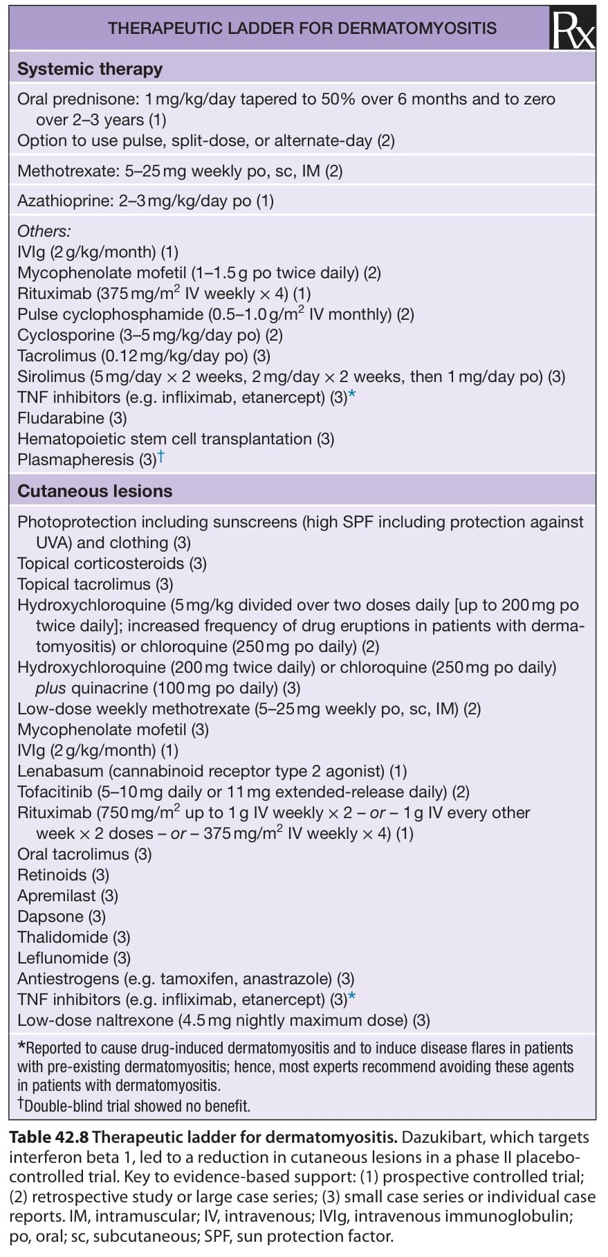
Table 42.8 Therapeutic ladder for dermatomyositis. Dazukibart, which targets interferon beta 1, led to a reduction in cutaneous lesions in a phase II placebocontrolled trial. Key to evidence-based support: (1) prospective controlled trial; (2) retrospective study or large case series; (3) small case series or individual case reports. IM, intramuscular; IV, intravenous; IVIg, intravenous immunoglobulin; po, oral; sc, subcutaneous; SPF, sun protection factor.
and electric shock wave lithotripsy (ESWL). Early and aggressive therapy of juvenile dermatomyositis does seem to reduce the risk of calcinosis cutis, which is often extremely challenging to treat.
Disorders of Skeletal Muscle. Philadelphia: Lippincott, Williams & Wilkins; 2000:268–269.23. Pachman LM, Litt DL, Rowley AH, et al. Lack of detection
cutaneous and systemic features associated with antitranscriptional intermediary factor-1gamma of cyclooxygenase-1, cyclooxygenase-2 and