PATHOGENESIS
Autoantibodies are usually as prevalent in morphea as in the general population, with two exceptions: (1) an increased prevalence of anti-single strand DNA (ssDNA), -topoisomerase IIα, -phospholipid, -fibrillin-1, and -histone antibodies (AHA) in patients with morphea; and (2) high titers of anti-nuclear antibodies (ANA) in juvenile patients with linear morphea and individuals with generalized morphea. In patients with linear morphea, the presence of ssDNA antibodies or AHA seems to be associated with increased risk of functional impairment.
Neither histology nor immunohistology of a single lesion will distinguish between morphea and SSc. In most clinicopathologic reviews, it is assumed that the two diseases are triggered by distinct events. However, the development of sclerosis following the initiating event seems to follow a common pathway. It is therefore assumed that pathogenic steps leading to SSc also contribute to the development of morphea, so they will be included in this discussion.
Currently, sclerosis of the skin is thought to involve three major, closely connected components: vascular damage, activated T cells, and altered connective tissue production by fibroblasts (Fig. 44.1).
Vascular Changes
A prominent feature of advanced sclerosis is a reduction in the number of capillaries. Studies performed in SSc suggest that hypoxia resulting from microvascular injury is a very early, or even the primary, event. Endothelial cells respond to various stimuli, including vascular endothelial growth factor (VEGF), platelet-derived growth factor (PDGF), and transforming growth factor-β (TGF-β) (see Ch. 102). In the sera of patients with SSc, markers indicative of endothelial cell activation (e.g. soluble adhesion molecules, VEGF) are elevated. Morphologic changes principally affect capillaries and small arterioles, and the initial changes in SSc include expression of adhesion molecules and endothelial swelling, followed by thickening of the basement membrane and intimal hyperplasia. An extrapolation is that similar changes may occur early on in morphea.
Control of Fibroblast Function by T Cell-Derived Cytokines
Pioneering work by E. Carwile Leroy showed that in vitro cultured fibroblasts isolated from sclerotic tissue produce increased amounts of collagen (types I, II, and III) and other extracellular matrix proteins. These fibroblasts can maintain this phenotype for weeks over several passages. This raised the question as to whether SSc results from an inborn or acquired error in collagen metabolism within fibroblasts. Today, most data favor the concept that abnormal collagen production is directed by surrounding cells. T cells in particular have the capacity to modify collagen synthesis by fibroblasts, and T cells are regularly present, at least early on, in a perivascular location and especially at the leading edge of developing sclerosis (see Fig. 44.1).
Enhanced production of collagen and other extracellular matrix proteins is induced by T cell-derived cytokines, especially interleukin (IL)-4, IL-13, IL-17A, and TGF-β. IL-4 is produced by CD4+ T helper type 2 (Th2) lymphocytes (see Ch. 4) and can directly enhance TGF-β production. Macrophages activated via the IL-4 receptor α (IL-4Rα) have been shown to influence collagen fibril assembly and promote fibrosis. In contrast, production of collagen and other extracellular matrix proteins can be significantly suppressed by interferons that are associated with Th1 lymphocytes. Th1 differentiation also implicates IL-12 signaling via the transcription factor STAT4. Of note, an association between STAT4 polymorphisms (e.g. rs7574865) and susceptibility to SSc, especially the limited subtype, has been reported. In a rare form of dominantly inherited pansclerotic morphea, gain-offunction variants in STAT4 were detected, with JAK inhibition leading to reduction of the hyperinflammatory fibroblast phenotype in vitro and clinical improvement11a. Additional susceptibility loci associated with SSc include immune-related genes (e.g. CD247, IRF5, HLA region) and the promoter region of the gene that encodes connective-tissue growth factor (CTGF).
Circulating levels of the chemokine ligand CXCL4 (secreted by plasmacytoid dendritic cells) have been found to be significantly elevated in SSc. Increased CXCL4 levels correlated with a higher prevalence of lung fibrosis and faster progression of skin fibrosis. Interestingly, CXCL4 promotes the Th2 cytokines IL-4 and IL-13 and has anti-angiogenic properties.
IL-4 is the most potent force in driving differentiation of Th2 lymphocytes (see Fig. 4.9). Because IL-4 enhances pathologic collagen production by fibroblasts and induces recruitment of eosinophils, it is believed that immune responses dominated by IL-4- as well as IL-13- and TGF-β-producing cells are critical in the initiation of skin sclerosis. This concept is supported by clinical and experimental data:
●In situ analysis of the advancing inflammatory margins of skin sclerosis revealed predominantly IL-4 expression.
●Th2-associated IL-13 and IL-33 have been shown to promote skin fibrosis in mice, and early treatment of scleroderma-developing mice with anti-IL-4 antibodies prevented scleroderma.
●Th2-like regulatory T (Treg) cells present in the skin of patients with SSc produce IL-4 and IL-13 and are responsive to IL-33.
●Systemic retinoids (which interact with TGF-β signaling) appear to reduce pathologic collagen synthesis by fibroblasts and may improve skin sclerosis in patients with chronic GVHD.
●Th2-promoting CXCL4 is so closely associated with SSc that it has been suggested as a biomarker for SSc. One theoretical therapeutic approach for morphea would be to redirect Th2 responses into Th1 responses. Unfortunately, placebocontrolled clinical trials of the Th1-inducing cytokines IFN-γ or IFN-α in patients with morphea produced negative results. The finding of elevated expression of IFN-related factors (e.g. CXCL9, CXCL10) in the skin and serum of cohorts with active morphea might provide an explanation, as it suggests that Th2 and Th1 cells may be activated simultaneously. Reports of the development of morphea and SSc during treatment with anti-PD-1 antibodies also point to the importance of activated T cells in their pathogenesis. It remains to be determined whether highly effective, broader inhibitors of the Th2 cytokines IL-4 and IL-13 or other morphea-associated T cell responses could help prevent the inflammatory phase of morphea and attendant skin sclerosis. More recently, antibodies directed against type 1 IFN-triggered dendritic cell-derived epiregulin, which blocked activation of EGFR on fibroblasts and subsequent paracrine NOTCH activation, were shown to reverse fibrosis in skin explants21a.
Lastly, when SSc fibroblasts are compared to control fibroblasts, there are differences in intracellular signaling proteins. Examples include activation of p38 mitogen-activated protein kinase and higher levels of Ha-Ras protein and reactive oxygen species. These findings plus data from animal studies demonstrating a link between skin fibrosis and pro-oxidative agents support hypoxia and oxidative stress as key factors in the pathogenesis of skin sclerosis.
Animal Model and Genetics
Currently, the best animal model for studying pathogenic events in SSc is the tight skin (TSK) mouse (Fig. 44.2). A partial duplication of the fibrillin-1 gene is thought to be responsible for the increased synthesis and accumulation of collagen in the skin and internal organs of TSK mice. TSK mice not only have increased collagen deposits, but also the collagen fibers are reduced in length and the quantity of hydroxyproline is increased in the dermis. Moreover, the mice have high antibody titers against topoisomerase I and fibrillin-1, similar to patients with SSc and morphea, respectively. One major difference is that blood vessels remain uninvolved in TSK mice, demonstrating that sclerosis of the skin can develop in the presence of apparently normal vessels. The phenotype can be transferred from diseased animals to healthy syngeneic mice via bone marrow cells (see Fig. 44.2).
Fibroblasts from TSK mice have elevated IL-4Rα expression and back-crossing the TSK mice onto a genetic background that either cannot respond to IL-4 or is deficient in producing TGF-β prevents skin sclerosis. Moreover, this back-cross normalizes collagen length, skin thickness, and hydroxyproline content and prevents antibody formation to topoisomerase I (see Fig. 44.2). In this murine model of SSc, inter-ventions that promoted Th1 responses or targeted PDGF receptor activity (e.g. imatinib) reduced dermal thickening and fibrosis. More recent strategies for improving experimental skin fibrosis have focused on the role of platelet-derived serotonin, β-integrins, TGF-β, and
Janus kinases. Of note, estrogen inhibition can exacerbate experimental dermal fibrosis driven by TGF-β.
In addition to the phenotypic similarities, the genetic locus associated with the TSK phenotype appears to be related to the risk of developing SSc. In humans, this susceptibility locus is on chromosome 15q in a region that contains the fibrillin-1 gene (FBN1); mutations in FBN1 have been detected in patients with stiff skin syndrome (see Ch. 43).
Triggering Events
Most immunologic and metabolic studies have been directed toward under-standing SSc, and the question remains as to whether they can also explain focal, asymmetric inflammation and sclerosis of the skin. The major discriminating factor between morphea and SSc might be the triggering event, with morphea being caused by a local trigger within the skin and SSc initiated by a systemic insult. The putative local triggering events for morphea include mechanical trauma, injections, vaccinations, and X-irradiation.
These questions initiated the search for potential triggers and studies of diseases that share clinical features with morphea and SSc. One intensively investigated area was the potential role of infection with Borrelia burgdorferi. The isolation of B. burgdorferi from the skin of some selected patients with morphea originally supported this speculation. However, larger studies failed to confirm this initial observation. Thus, this theory has been abandoned.
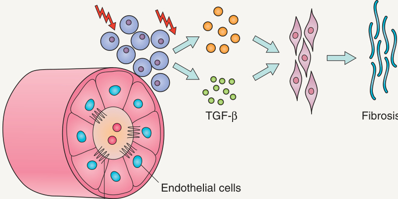
Fig. 44.1 Pathogenesis of sclerosis. Three components are involved during the formation of sclerosis: vascular damage, lymphocyte activation, and altered connective tissue production. IL, interleukin; TGF, transforming growth factor.

Fig. 44.2 Animal model for scleroderma. Transfer of scleroderma phenotype from diseased animals (TSK mice) to healthy syngeneic mice (C57BL/6) by bone marrow cells. Lack of tight skin and autoantibody production by back-crossing TSK mice with mice that cannot respond to IL-4 or mice that produce minimal amounts of TGF-β. IL, interleukin; TGF, transforming growth factor; TSK, tight skin strain of mice.