CLINICAL FEATURES
As outlined in Table 44.1, there are several classification schemes for morphea. Major forms include plaque-type (circumscribed), linear, generalized, and other less common variants (e.g. nodular or keloidal). There are also inflammatory syndromes leading to superficial or deep sclerosis of the skin that resemble morphea (i.e. morpheaform; see below). Patients with morphea do not have Raynaud phenomenon or involvement of internal organs, except for underlying fascia, muscle, and bone in some patients with linear morphea and the occasional patient with linear morphea of the head who has ocular or neurologic symptoms. In rare patients with presumptive morphea, clinically insignificant fibrosis of the lung or slightly reduced esophageal motility has been detected radiographically or by scintigraphy. Clinically relevant involvement of internal organs is observed in certain types of pseudoscleroderma (see Ch. 43).
Plaque-Type (Circumscribed) Morphea
Plaque-type (circumscribed) morphea, the most frequent variant, is characterized by the insidious onset of a slightly elevated, erythematous
or violaceous, slightly edematous plaque that undergoes centrifugal expansion (Fig. 44.3). It is generally asymptomatic and therefore frequently goes unnoticed by the patient. The central portion of the progressing lesion starts to transform into sclerotic, scar-like tissue. Depending on the depth of the sclerosis, the skin becomes progressively indurated. Centrally, it can acquire a shiny white color and peripherally, a violaceous or “lilac” ring (Fig. 44.4A). Postinflammatory hyperpigmentation often dominates over the white sclerosis as the lesions mature (Fig. 44.4B).
Skin structures such as hairs and sweat glands are frequently lost. Some patients complain of itch, perhaps due to associated xerosis. Once the “lilac” ring vanishes, the lesion’s progression also halts. Although this inflammatory margin may be difficult to appreciate, especially in linear sclerosis, it is an important indicator of clinical activity.
The plaques of morphea most commonly develop on the trunk and in general are between 2 and 15 cm in diameter. They are usually multiple and asymmetric, but marked variation exists. Individual lesions may enlarge significantly or remain stable in size.
The course of morphea is also variable. In most patients, morphea progresses over 3–5 years, then arrests and eventually resolves spontaneously. However, residual atrophy and dyspigmentation are commonly observed. Patients rarely have relapsing disease for more than 5 years, but may have sequelae such as calcinosis cutis (see Fig. 50.2).
Variants
Various manifestations of morphea have become associated with specific names. They do not really reflect unique entities, but rather different morphologic features, distribution patterns, or depth of involvement. In most patients, morphea seems to be randomly distributed. However, in some patients, the lesions are unilateral, and they may follow dermatomes or the lines of Blaschko.
●Guttate morphea presents primarily as multiple, rather superficial, nummular plaques. Even though relatively small, they may become deeply indurated.
●Atrophoderma of Pasini and Pierini is considered by some to be a very superficial variant of plaque-type morphea, while others consider it to be a separate entity in the differential diagnosis of “burnt-out” morphea (see Ch. 99). Hyperpigmented patches are seen most commonly on the posterior trunk; occasionally, lesions follow the lines of Blaschko (linear atrophoderma of Moulin).
●Deep morphea denotes inflammation and sclerosis that affects primarily the deep dermis and subcutaneous fat and may even involve underlying structures (e.g. the fascia) (Fig. 44.5A). Patients normally develop a small number of hard deep plaques that may impair motility of the skin; ultimately, lesions may calcify, leading to dystrophic calcinosis cutis. Because of the location of the sclerosis, individual lesions may resemble eosinophilic fasciitis (Fig. 44.5B and see Ch. 43).
●In nodular or keloidal morphea, inflammation within the dermis leads to thick, keloid-like nodules (Fig. 44.6) or bands. It can be clinically indistinguishable from indurated keloids.
●In rare patients, especially those where sclerosis of the skin is associated with diffuse, rapidly progressive edema, lymphoceles may result from stasis of lymphatic fluid. When the latter develop into bullae, this is referred to as bullous morphea. It is more frequently observed in generalized morphea and sclerodermoid or morpheaform GVHD and is rarely a feature of plaquetype morphea. Bullous morphea has to be distinguished from mechanical blisters that may occur centrally within plaques and are secondary to impaired mechanical stability of the dermal–epidermal junction.
Linear Morphea and Parry–Romberg Syndrome
Linear morphea is different from plaque-type morphea, with respect to age of onset, distribution, clinical outcome, and serologies. Morphea en coup de sabre (meaning stroke or blow with a single-edged sword) is a term used for linear morphea of the forehead and scalp. Hemifacial atrophy, or Parry–Romberg syndrome, is probably a very severe variant of linear morphea, but it may be a phenotype for more than one entity. There is a progressive loss of subcutaneous fat, but little or no sclerosis (Fig. 44.7). The entire distribution of the trigeminal nerve, including the eye and the tongue, may be affected.
Linear morphea may present initially as a linear, erythematous (inflammatory) streak, but more frequently begins as a harmlessappearing lesion of plaque-type morphea that extends longitudinally as a series of plaques that then join to form a scar-like band (Fig. 44.8). This band may severely impair the mobility of the affected limb. Linear morphea tends to involve the underlying fascia, muscle, and tendons. This leads not only to muscle weakness but also to shortening of the muscles and fascia which then impairs joint mobility. Linear morphea is especially problematic when it extends over joints, as this almost invariably results in a decrease in range of motion and functional impairment. In some patients, involvement is circular rather than linear and results in progressive atrophy of the limb similar to the Parry–Romberg variant of facial morphea. The linear variant can have prolonged periods of quiescence followed by reactivation.
The en coup de sabre type of morphea represents linear morphea of the head (Fig. 44.9). It is typically unilateral and extends from the forehead into the frontal scalp. It may start either as a linear streak, which may initially mimic a port-wine birthmark, or as a row of small plaques that coalesce. A paramedian location is most common. Like plaque-type morphea, it may initially be surrounded by a discrete lilac ring that extends longitudinally and may reach the eyebrows, nose, and even the cheeks. As inflammation wanes, a linear, hairless crevice develops that in some patients is sclerotic, while in others appears more atrophic.
En coup de sabre morphea can also involve the underlying muscles and osseous structures. Rarely, the inflammation and sclerosis progress to involve the meninges and even the brain, creating a potential focus for seizures. Ocular involvement has been reported in up to 15% of juvenile and 25% of adult patients.
Generalized Morphea
Generalized morphea normally begins insidiously on the trunk as plaque-type morphea. While individual lesions are indistinguishable in these two forms, in generalized morphea they do not stop expanding. Just as in the diffuse form of SSc, multiple plaques rapidly coalesce and can affect nearly the entire trunk, often only sparing the areolae and nipples (Fig. 44.10). The sclerosis may involve the extremities down to the hands (presenting initially as puffy edema). As the cutaneous sclerosis progresses, it may result in disabling constrictions that even cause difficulty in breathing due to impaired thorax mobility and inflammation of the intercostal muscles. Although an aggressive therapeutic approach is recommended, the disease is usually persistent given its often limited response to treatment.
On the forehead, the linear band(s) of sclerosis and atrophy are usually paramedian (rather than midline). A Both hyper- and hypopigmentation are present as well as subtle depression. B An obviously depressed, sclerotic, hypopigmented linear band. C Scarring alopecia develops when there is involvement of scalp; “lonely hairs” are also seen in other forms of scarring alopecia. D Note the prominent veins and loss of the medial eyebrow. Progression to Parry–Romberg syndrome would be a concern.
Morphea in Childhood
Approximately 20% of individuals with morphea are children and teenagers. The female-to-male ratio for plaque-type morphea in this age group is about 2 : 1, with a mean age at disease onset of 7 years. In two-thirds of patients with linear morphea, disease onset is prior to age 18 years, providing an explanation for the possibility of growth retardation of the affected limb. Thus, if left untreated, linear morphea may result not only in decreased range of motion of joints but also in permanent limb asymmetry from unilateral hypoplasia (Fig. 44.11).
Pansclerotic morphea, also referred to as disabling pansclerotic morphea of children, is similar to generalized morphea of the adult. It usually starts before 14 years of age and tends to cause lifelong severe disability due to persistent atrophy of the underlying muscles and contractures of the involved joints. The disease tends to extend from the trunk to the hands and feet. Periodontal atrophy may occur, but there is only slight involvement of the esophagus and lung. Distinction from SSc may prove challenging.
Importantly, in a large multicenter study of children with morphea, 22% had extracutaneous findings that were primarily articular (11%), neurologic (4%), and ocular (2%). The latter two findings were seen primarily in those with linear morphea of the head or Parry–Romberg syndrome, i.e., 85% of affected patients had these two variants.
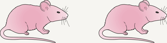
Fig. 44.2 Animal model for scleroderma. Transfer of scleroderma phenotype from diseased animals (TSK mice) to healthy syngeneic mice (C57BL/6) by bone marrow cells. Lack of tight skin and autoantibody production by back-crossing TSK mice with mice that cannot respond to IL-4 or mice that produce minimal amounts of TGF-β. IL, interleukin; TGF, transforming growth factor; TSK, tight skin strain of mice.
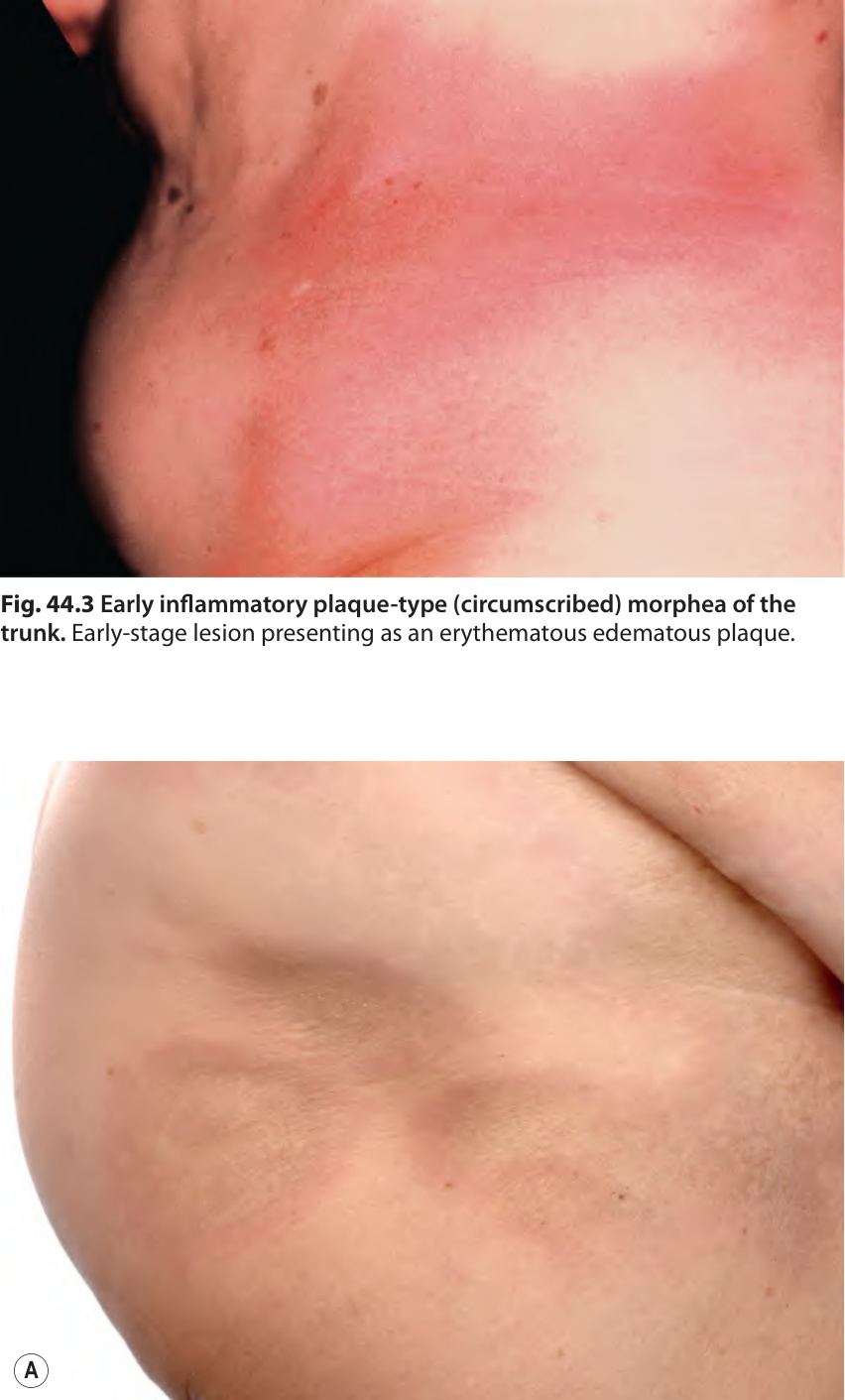
Fig. 44.3 Early inflammatory plaque-type (circumscribed) morphea of the trunk. Early-stage lesion presenting as an erythematous edematous plaque.
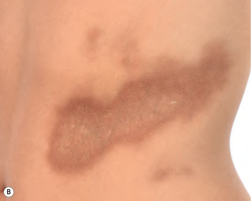
Fig. 44.4 Plaque-type (circumscribed) morphea.A Sclerotic plaques with the characteristic lilac-colored rim. Histologic findings will differ depending upon whether the biopsy specimen is obtained from the inflammatory edge or the central area of sclerosis (see Fig. 44.12). B The predominant lesion is a large, well-demarcated, hyperpigmented plaque that is depressed centrally; the trunk is a common location for plaque-type morphea.
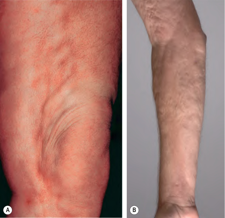
Fig. 44.5 Comparison of deep morphea and eosinophilic fasciitis.A Note the “pseudo-cellulite” appearance of the involved skin of the thigh in deep morphea. B In eosinophilic fasciitis, the level of fibrosis is also deep, resulting in a similar rippled appearance. This patient had the eosinophilic fasciitis-like form of chronic GVHD, also referred to as chronic GVHD-related fasciitis. B, Courtesy Edward Cowen, MD.
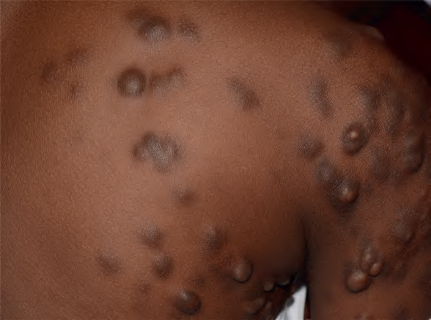
Fig. 44.6 Nodular or keloidal morphea. Multiple firm, hyperpigmented nodules mimicking keloids in a 10-year-old girl. Courtesy Julie V. Schaffer, MD.
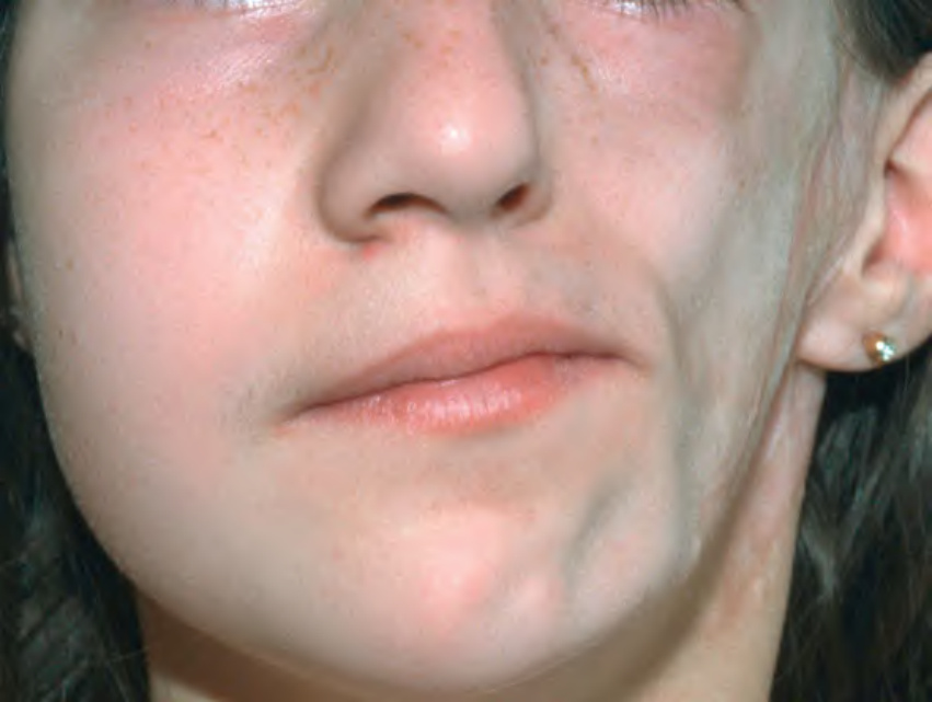
Fig. 44.7 Parry–Romberg syndrome. Unilateral sclerosis and loss of subcutaneous fat, leading to facial asymmetry. Courtesy Lorenzo Cerroni, MD.
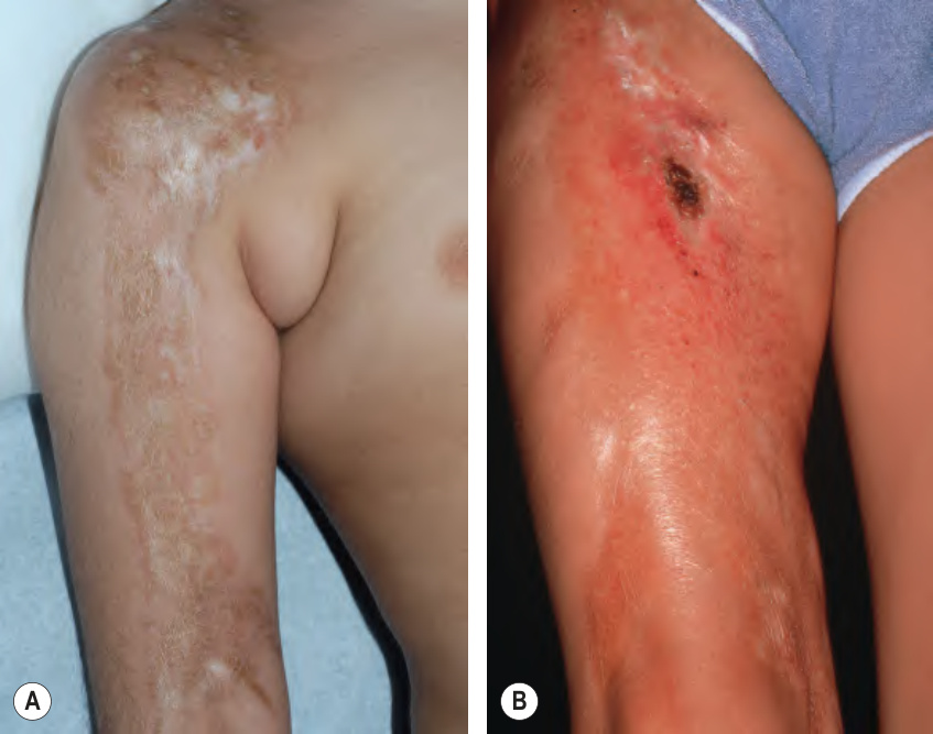
Fig. 44.8 Linear morphea of an extremity.A Linear sclerotic band of the arm with both hyper- and hypopigmentation. The majority of patients with linear morphea have unilateral involvement. B More inflammatory phase with ulceration in addition to induration. The differential diagnosis includes linear melorheostosis which is often associated with underlying candlewax-like linear hyperostosis. A, Courtesy Julie V. Schaffer, MD.
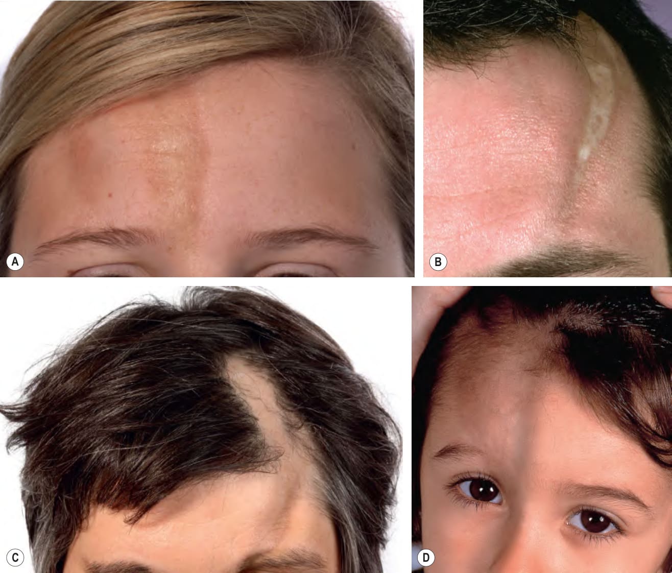
Fig. 44.9 Morphea en coup de sabre.
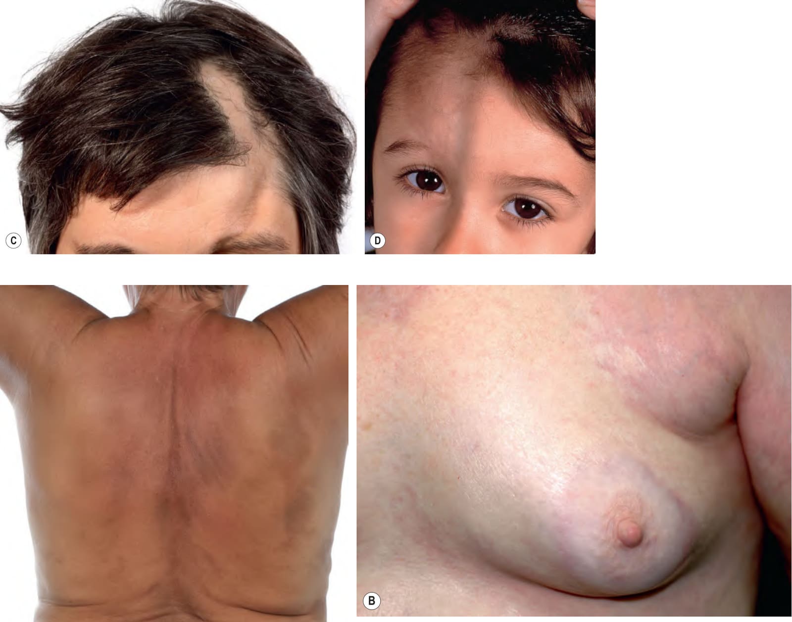
Fig. 44.10 Generalized morphea.A Multiple hyperpigmented plaques on the trunk that are becoming confluent. B Circumferential involvement with sparing of the central portion of the breast is a classic distribution pattern. B, Courtesy Luis Requena, MD.
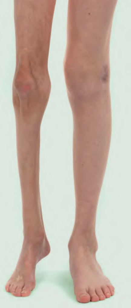
Fig. 44.11 Extensive linear morphea of the right leg leading to hypoplasia. In addition to marked hypoplasia, there is induration and a flexion contracture of the knee.
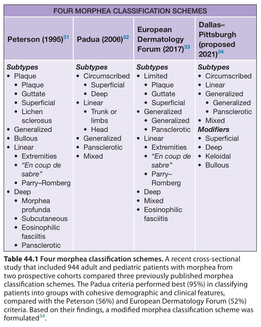
Table 44.1 Four morphea classification schemes. A recent cross-sectional study that included 944 adult and pediatric patients with morphea from two prospective cohorts compared three previously published morphea classification schemes. The Padua criteria performed best (95%) in classifying patients into groups with cohesive demographic and clinical features, compared with the Peterson (56%) and European Dermatology Forum (52%) criteria. Based on their findings, a modified morphea classification scheme was formulated.