TREATMENT
Various therapeutic modalities have been described for morphea, although quality evidence in support of many of these therapies has been limited. In addition, there has been a lack of standardized outcome measures for assessing the efficacy of various therapies. However, the localized scleroderma cutaneous assessment tool (LoSCAT) is gaining recognition as a valid and reproducible index for disease activity and damage in morphea. Radiologic imaging (e.g. ultrasound, MRI) may also provide useful information.
The major therapies for morphea include topical medications, photo therapy, systemic immunosuppressants, and physical/occupational therapy (Table 44.3). Several factors, including level of disease activity, subtype of morphea, depth of involvement, location and extent of disease, and degree of current or potential functional impairment or disfigurement, play a role in determining the most appropriate treatment.
Therapies are most effective when there is active disease (e.g. inflamed and expanding patches and plaques), whereas inactive disease (e.g. fixed sclerotic plaques) is less likely to respond. Plaque-type morphea
with limited body surface area involvement can often be managed with topical therapies while generalized or extensive disease responds best to phototherapy or systemic medications. Active deep morphea with involvement of the subcutaneous tissue, fascia, or muscles as well as morphea with the potential for functional impairment (e.g. linear morphea overlying joints) or significant disfigurement (e.g. facial involvement) should be managed aggressively with systemic immunosuppressants. In addition, patients at risk for functional impairment (e.g. joint contractures, loss of mobility) should be referred to physical and occupational therapists before the onset of such complications.
If persistent contractures do develop, reconstructive surgery may be required. Surgical revision can be offered for inactive linear or en coup de sabre morphea. Autologous fat transplantation has been used successfully for Parry–Romberg syndrome and injection of hyaluronic acid fillers, with or without fractional laser ablation, to correct facial morphea has been reported. Finally, patients with extracutaneous involvement may require additional evaluation and management, such as rheumatologic referral for arthritis or synovitis. Patients with morphea that involves the head are at risk for ocular and/or neurologic complications and, when indicated, referred to ophthalmology and neurology, respectively.
Phototherapy
Phototherapy for morphea was first described in 1994, and since then, efficacy has been confirmed by more than 30 publications and was recently supported by an open label controlled study. Various phototherapy modalities have been used, including UVA1, broadband UVA (BB-UVA), PUVA, and narrowband UVB (NB-UVB). When available, UVA1 has become the phototherapy of choice, followed by BB-UVA and PUVA. NB-UVB, because of its shallower depth of penetration, may be beneficial for superficial morphea but is less effective for deeper morphea. Treatment can be lesion-directed when there is a limited number of plaques or administered to the majority of the skin surface. The efficacy of phototherapy is thought to be due to immunomodulatory effects as well as upregulation of matrix metalloproteinases that stimulate collagen breakdown. Patients may also benefit from phototherapy plus low-dose systemic retinoids as this combination has led to improvement of sclerodermoid GVHD and plaque-type morphea.
Due to the nature of phototherapy, placebo-controlled studies have not been performed. Nonetheless, little doubt exists as to the efficacy of this therapy, as untreated morphea normally progresses over 3–5 years and then regresses very slowly over several years. More rapid spontaneous improvement only occurs in a small minority of patients. The clinical course is definitely different in patients receiving phototherapy. Following 30–36 treatments of bath PUVA therapy (at slightly suberythematous doses) or UVA1 (30–60 J/cm), morphea resolves or markedly improves in at least 60% of patients (Fig. 44.14). Further advantages of phototherapy, and especially UVA1, include clinical improvement in all skin types and sustained remissions. Recurrence of morphea plaques after successful phototherapy has been reported, but primarily in patients with a prolonged period of disease prior to initiation of phototherapy. The dose of UVA1 phototherapy (medium versus high) does not appear to correlate with risk of recurrence.
The ideal dose for UVA1 therapy has been debated. Most initial studies were performed with either low (20–30 J/cm) or medium doses (40–70 J/cm) of UVA1 and reported similar efficacy. A study that utilized 20 MHz ultrasound rather than clinical assessment of morphea lesions reported a better response with medium-dose (70 J/cm) than with low-dose (20 J/cm) UVA1. In agreement with these reports, we have observed that 36 treatments with either 30 J/cm or 50–60 J/cm
Disseminated morphea on the trunk, with sparing of the central portions of the breasts. Before (A) and after (B) PUVA bath photochemotherapy.
of UVA1 markedly improves morphea in the majority of patients. Importantly, in responding patients, lesions of morphea continue to improve beyond the end of the therapy. Thus, therapy can be terminated after 36 treatments, even if the sclerosis has not yet completely resolved. The only exception appears to be linear morphea, which may require up to 60 treatments.
Systemic Therapies
Indications for systemic therapies are outlined above and in Table 44.3. For decades, methotrexate, either alone or in combination with systemic corticosteroids, has represented a first-line treatment. Systemic corticosteroids are often added for severe involvement or when more rapid control of the disease is desired. Typically, the methotrexate is continued for at least 12 months and tapering is not begun until at least 6–12 months after the disease is inactive. Tapering of systemic corticosteroids (over several months) usually occurs sooner, once the disease is no longer active.
Recent data from retrospective cohort and longitudinal studies suggest that mycophenolate mofetil (MMF) is also beneficial and well tolerated in patients with severe morphea, even in those with a history of previously ineffective treatments. MMF is usually the next line of treatment should methotrexate +/− corticosteroids fail or be contraindicated. Efficacy data for other systemic agents are even more limited. In small case series, JAK inhibitors (e.g. tofacitinib, baricitinib) led to improvement in patients with recalcitrant morphea or eosinophilic fasciitis. Additional reported therapies include hydroxychloroquine and systemic retinoids (acitretin, isotretinoin). Of note, vitamin A derivatives have antifibrotic effects and can modulate collagen metabolism.
In case series and reports, improvement has been observed with tocilizumab, which blocks the IL-6R, including for pansclerotic morphea in children, as well as abatacept, a CTLA4 fusion protein that inhibits T cell activation. A placebo-controlled, multi-center trial with dupilumab, which blocks the interaction of the profibrotic cytokines IL-4 and IL-13 with IL-4Rα, is currently underway. Lastly, a novel approach based upon genetically modified autologous fibroblasts that express metalloproteinase-1 (MMP-1) has not progressed beyond phase I/II trial status.
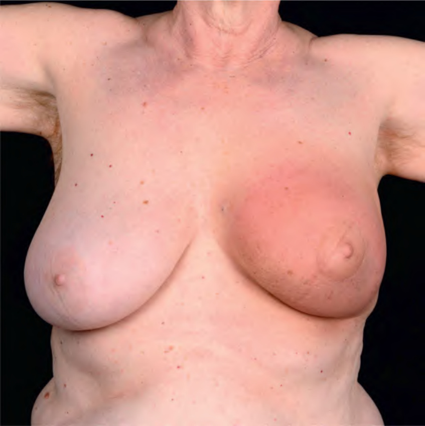
Fig 44.13 Radiation-induced morphea. Erythema and induration of the left breast. Courtesy Department of Dermatology, Medical University of Graz.
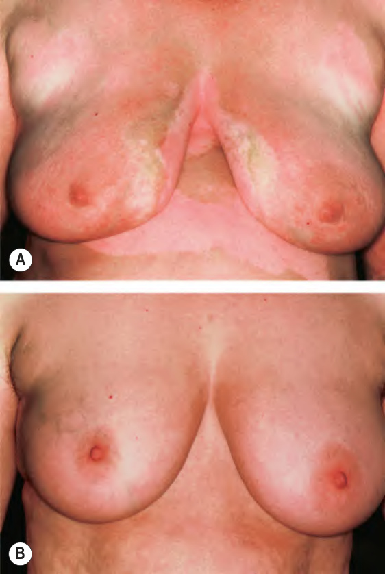
Fig. 44.14 Phototherapy for morphea.
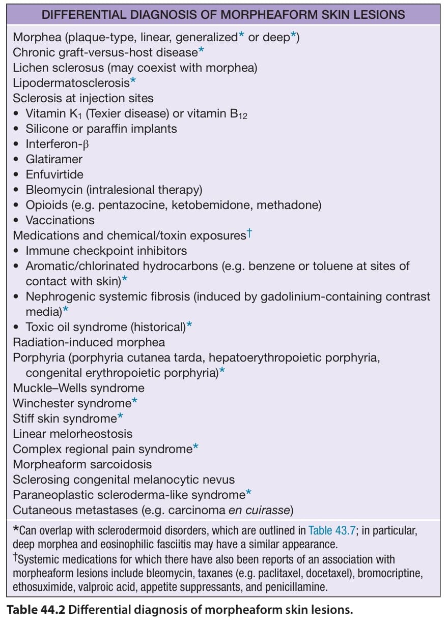
Table 44.2 Differential diagnosis of morpheaform skin lesions.
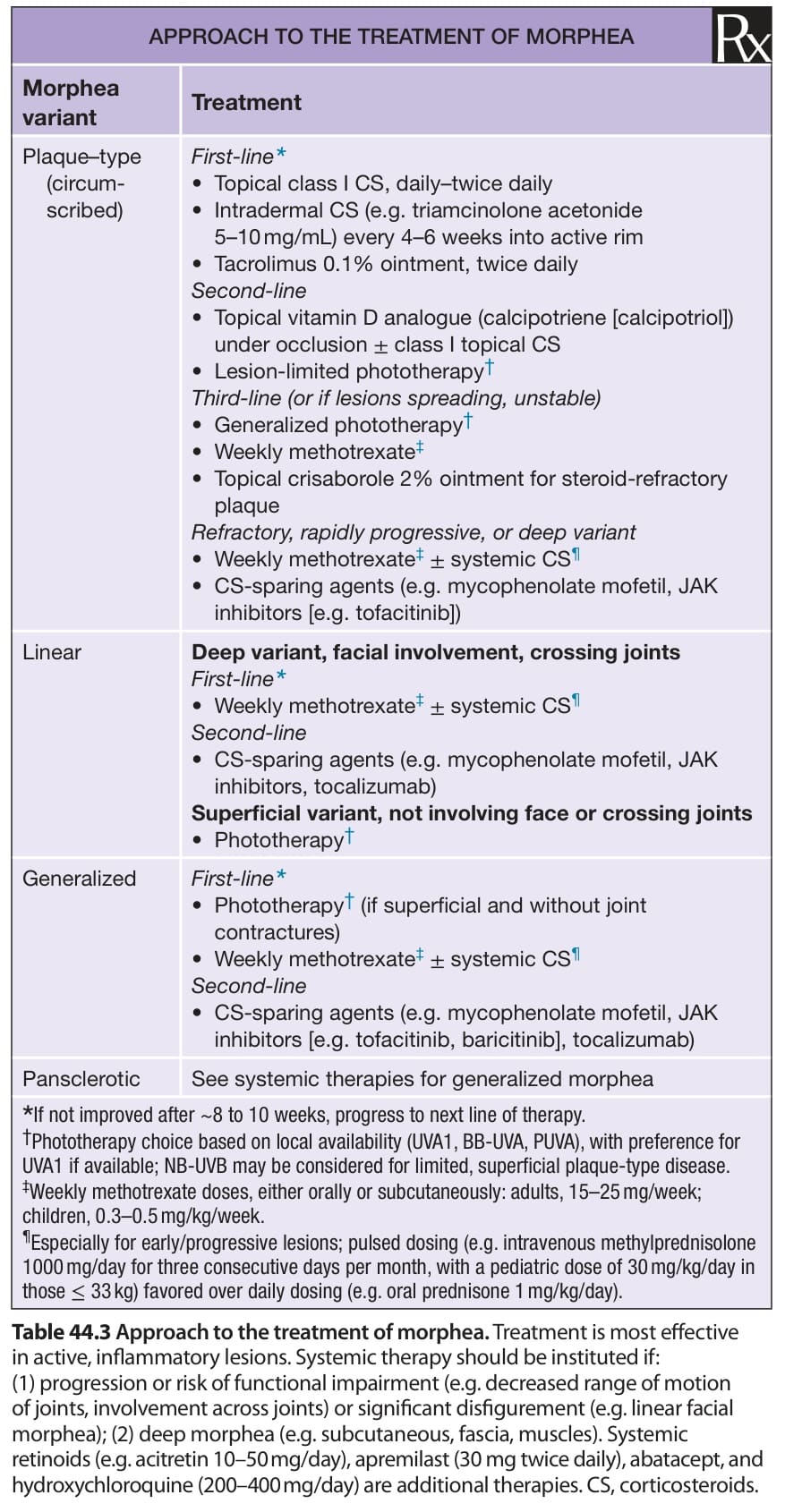
Table 44.3 Approach to the treatment of morphea. Treatment is most effective in active, inflammatory lesions. Systemic therapy should be instituted if: (1) progression or risk of functional impairment (e.g. decreased range of motion of joints, involvement across joints) or significant disfigurement (e.g. linear facial morphea); (2) deep morphea (e.g. subcutaneous, fascia, muscles). Systemic retinoids (e.g. acitretin 10–50 mg/day), apremilast (30 mg twice daily), abatacept, and hydroxychloroquine (200–400 mg/day) are additional therapies. CS, corticosteroids.