CLINICAL FEATURES
Lichen sclerosus most frequently affects the anogenital region, and less often extragenital skin. Lichen sclerosus of the oral cavity, palms, or soles is rare. Intraorally, bluish-white papules up to 5 mm in diameter may occur on the buccal mucosa or under the tongue. They can lead to superficial scar-like atrophy or erosions.
Extragenital LS normally does not cause symptoms except for dryness and associated pruritus. Lesions favor the trunk (Fig. 44.15) and the proximal extremities. More specific predilection sites include the neck, shoulders, flexor surfaces of the wrists, and sites of physical trauma or continuous pressure (e.g. the shoulder or hip). The periorbital area and scalp are rarely affected.
It is unusual to see extragenital disease during its early phases when single lesions begin as polygonal, bluish-white, shiny, slightly elevated, interfollicular papules. Over time, these papules often enlarge or coalesce into larger plaques. Most patients present with slightly sclerotic, scar-like papules and plaques that are usually ivory in color and have a shiny and/or wrinkled surface. Occasionally, lesions have a pink or light violet hue. At sites of continuous pressure, the surface may acquire a parchment-like appearance.
In more advanced stages, telangiectasias or follicular plugging can be seen (Fig. 44.16A). The flattened interface between the epidermis and dermis results in fragility of the dermal–epidermal junction. As a result, LS is occasionally complicated by the development of bullae that tend to become hemorrhagic (Fig. 44.16B).
In women, the vulva and the perianal region are commonly involved, often in a figure-of-eight configuration (see Ch. 73). Although the disease may be symptom-free, it frequently causes severe pruritus and soreness. These symptoms may be considerable and lead to dysuria, dyspareunia, or pain upon defecation (often manifesting as constipation in children). As a result, quality of life can be adversely affected, including in children and adolescents.
Vulvar LS often begins as a slightly elevated, sharply demarcated area of erythema, which can be slightly eroded. Over time, the involved skin becomes shiny, hypopigmented, and sclerotic, and fissures may develop, especially in the perineum (Fig. 44.17). When progressive, associated scarring can lead to burying of the clitoris and fusion of the
labia minora to the labia majora, and when untreated, an increased risk of malignancy. With severe disease, loss of the labia minora and significant narrowing of the introitus can occur. Sexual intercourse may become impossible. In general, LS does not worsen during pregnancy and when treated successfully, vaginal delivery is possible without an increased incidence of perineal tears and episiotomies (compared with healthy control populations).
Areas of purpura often develop within lesions of anogenital LS and can actually aid in the clinical diagnosis (see Fig. 73.3). However, the hemorrhage combined with erosions may be misdiagnosed as sexual abuse (see Ch. 90). A careful history and clinical examination, along with histologic confirmation when needed, can assist in establishing the diagnosis.
In boys and men, acquired phimosis or recurrent balanitis are the primary presenting features, and perianal involvement is rare. Itching and soreness are common. On the glans and the inner aspect of the foreskin, LS starts as sharply demarcated, sometimes slightly blue–red lesions, occasionally with erosions. This inflammation tends to evolve into an atrophic white sclerotic scar (Fig. 44.18). The constriction may cause pain on erection and, at advanced stages, dysuria and urinary obstruction. If the foreskin is affected in uncircumcised male patients, LS invariably leads to phimosis (difficulty in retracting the foreskin to uncover the glans), with the additional risk of paraphimosis (when a foreskin is retracted behind the glans and cannot be returned to its original position); the latter represents a urologic emergency. Many boys and men first present when the phimosis impairs repositioning of the foreskin;
in these situations, diagnosis depends on histology. With progression, poorly healing ulcers of the glans can develop. While circumcision is clearly a first-line therapy, LS may recur at the site of a circumcision.
A continuing debate is whether genital LS is a precancerous condition. Interpretation and comparison of the data are complicated by at least two factors: some of the patients reported as developing cancer had previously received X-ray therapy for the disease or had previous dysplasia due to human papillomavirus (HPV) infection. It is therefore very difficult to perform a useful meta-analysis based on existing data. Some data suggest that genital LS is not intrinsically precancerous, but a recent prospective cohort study of 507 women with vulvar LS noted the development of SCC or VIN in 5% of patients partially compliant with topical corticosteroid treatment and in none of the compliant patients, with a mean follow-up of 4.7 years. Whether the microRNA signature will assist in identifying those patients at increased risk of SCC remains to be determined80a.
Lentigines and melanocytic nevi can arise within areas of LS, primarily genital LS; histologically they may resemble persistent (recurrent) melanocytic nevi and therefore be confused with melanoma.
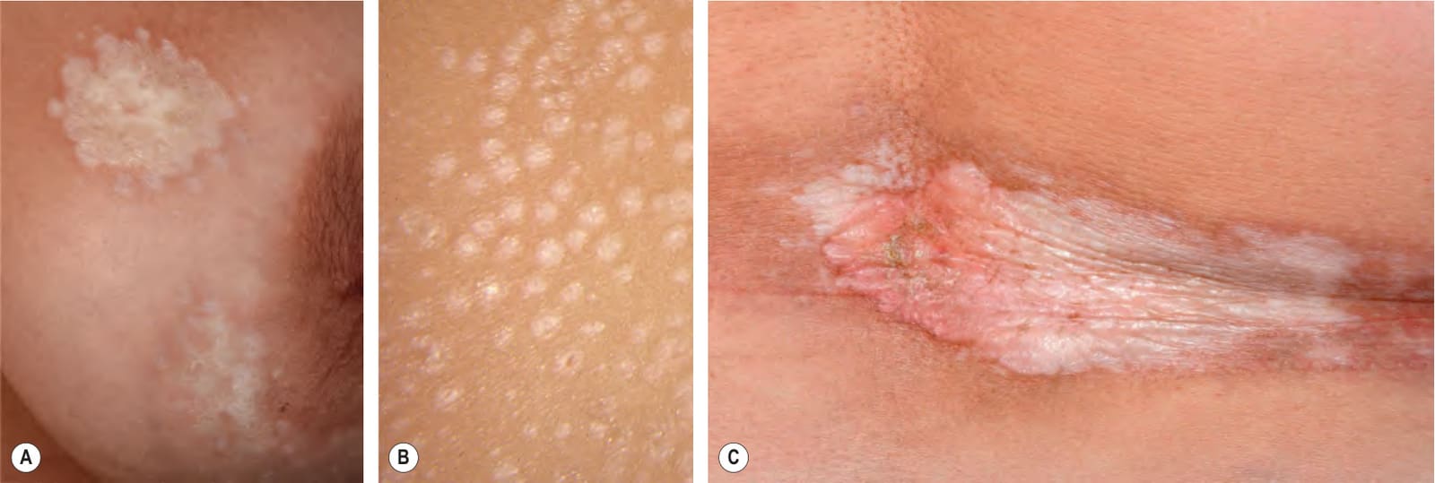
Fig. 44.15 Lichen sclerosus.A Shiny, ivory-colored plaques on the breast with several guttate satellite lesions. B In some patients, only guttate lesions are present. C Large ivory-colored, shiny plaque on the waistline, a site of persistent pressure. B, Courtesy Louis A. Fragola, Jr, MD.
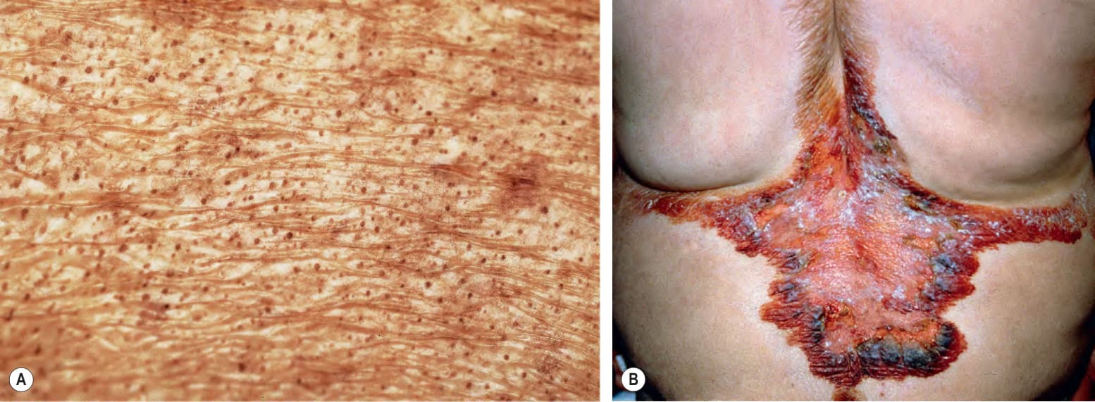
Fig. 44.16 Lichen sclerosus.A Follicular plugging in a plaque of lichen sclerosus on the back of a patient with chronic GVHD. B Hemorrhagic bullae primarily at the periphery of a large plaque on the back. A, Courtesy Jean L. Bolognia, MD; B, Courtesy Luis Requena, MD.
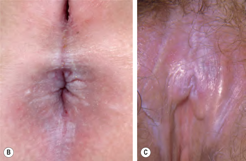
Fig. 44.17 Anogenital lichen sclerosus.A Characteristic vulvar, perineal, and perianal involvement in a child. Symmetric ivory-colored, shiny plaques with a thin pink peripheral rim are seen; a perianal pyramidal protrusion is also present. B Perianal lichen sclerosus in which the hypopigmented plaques are accompanied by a linear erosion (skin tear) at 6 o’clock and central fissuring of the perineum. C Involvement of the clitoral hood and clitoris as well as fusion of the left labia minora and labia majora. A, Courtesy A. Hernández-Martín, MD; C, Courtesy Louis A. Fragola, Jr, MD.
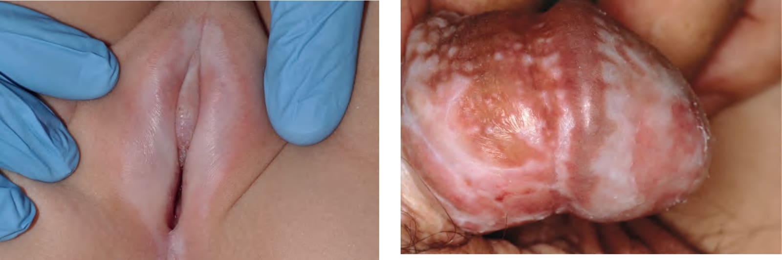
Fig. 44.18 Lichen sclerosus of the penis (balanitis xerotica obliterans). Note the ivory color, erosion, and scarring.