PATHOLOGY
Lichen sclerosus has a specific histologic pattern that is seen in all age groups. Initially, superficial dermal edema is associated with a band-like lymphocytic infiltrate (Fig. 44.19A). The epidermis is thinned,
with orthohyperkeratosis and vacuolar degeneration of the basal layer. Hyperkeratosis is especially pronounced at follicular openings and may lead to plugging. Vacuolar degeneration of the basal layer and flattening of the rete ridges predispose to the development of blisters, which may become hemorrhagic. The most important changes are found in the super-ficial dermis, where the pale staining reflects edema initially and then homogenized dermal collagen (Fig. 44.19B). Loss of elastic fibers is typical for LS and is not observed in morphea. Subepidermal clefting and hemorrhage within the homogenized papillary dermis is frequently observed.
During the early phases, the inflammatory infiltrate is especially pronounced along the zone of hyalinization and consists of lymphocytes (CD3+, CD4+, CD8+), macrophages, and mast cells. In older lesions, the mononuclear infiltrate is reduced and sparse, and patchy islands of mononuclear cells are dispersed within the hyalinized dermis. Ultrastructural studies reveal shortened collagen fibers.
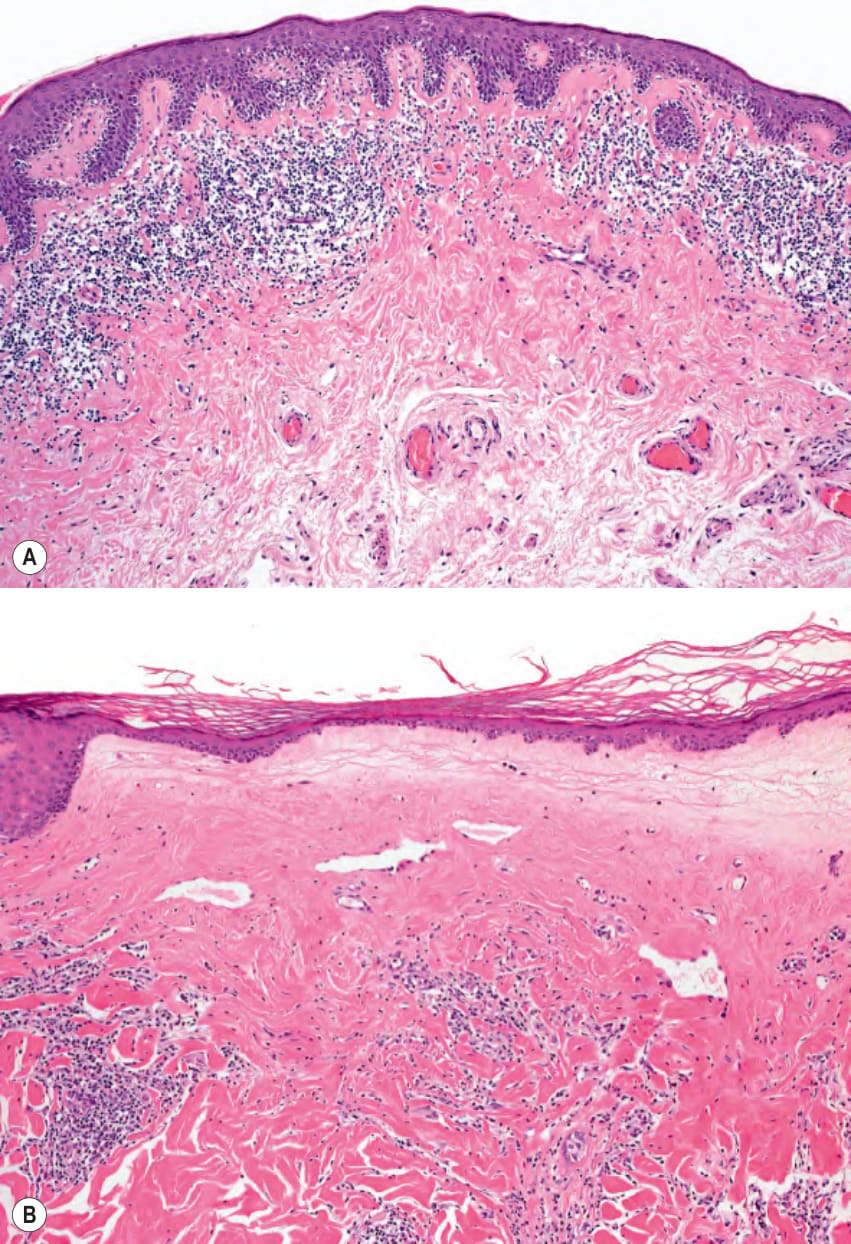
Fig. 44.19 Lichen sclerosus – histopathologic features.A Early lesion with band-like infiltrate of lymphocytes in the upper dermis and vacuolar change at the dermal–epidermal junction. Mild homogenization of the papillary dermis is present. B Late lesion with orthohyperkeratosis, thinning of the epidermis, and homogenization of the upper dermis. There is also hyalinization and sclerosis of the papillary dermis, telangiectasias, and an interstitial lymphocytic infiltrate. Courtesy Lorenzo Cerroni, MD.