INTRODUCTION
The cutaneous mucinoses are a heterogeneous group of disorders in which an abnormal amount of mucin accumulates in the skin, either diffusely or focally.
Mucin is a component of the dermal extracellular matrix and is normally produced in small amounts by fibroblasts. It is a jelly-like, amorphous mixture of acid glycosaminoglycans (formerly referred to as acid mucopolysaccharides), which are complex carbohydrates composed of multiple repeating polysaccharide units (see Ch. 95). The acid glycosaminoglycans may be attached to both sides of a protein core (proteoglycan monomer), as in the case of dermatan sulfate and chondroitin sulfate, or they may be free, as in the case of hyaluronic acid, which is the most important component of dermal mucin.
Mucin is capable of absorbing 1000 times its own weight in water, playing a major role in maintaining the salt and water balance of the dermis. In routinely stained sections, the presence of either a blue-staining material between separated collagen bundles or empty spaces within the dermis are good clues that there is mucin deposition. For confirmation, special stains can be used, such as Alcian blue, colloidal iron, or toluidine blue (Table 46.1). Furthermore, dermal mucin is PAS-negative and, if composed of hyaluronic acid, hyaluronidasesensitive. Fixation of biopsy specimens in absolute alcohol, rather than formalin, may improve dermal mucin detection. In addition, monoclonal antibodies have been used to detect sulfated acid glycosaminoglycans. An alternative approach is to utilize biotinylated hyaluronan (HA)-binding proteins coupled to an avidin–peroxidase reaction.
Why mucin abnormally accumulates within the skin of some individuals is unclear. Certain “serum factors”, e.g. immunoglobulins and/or cytokines, could promote the upregulation of glycosaminoglycan synthesis. For example, elevated serum immunoglobulin levels (monoclonal or polyclonal) and circulating autoantibodies can be found in association with cutaneous mucinoses such as scleromyxedema, Graves disease-associated pretibial myxedema, and papulonodular mucinosis of lupus erythematosus. However, the serum from these patients stimulates mucin production even after the elution of either the IgG paraprotein in those with scleromyxedema or the autoantibodies in those with Graves disease-associated pretibial myxedema. Circulating cytokines such
Franco Rongioletti Mucinoses 46
(mucopolysaccharides). GAG, glycosaminoglycan.
as interleukin (IL)-1, tumor necrosis factor (TNF), and TGF-β, which are known to stimulate glycosaminoglycan synthesis within the skin, could play a role in excess mucin deposition. A reduction in the normal catabolic degradation of mucin could also be a factor.
This chapter focuses on the disorders characterized by abnormal dermal deposits of mucin, composed primarily of hyaluronic acid (Fig. 46.1). The mucopolysaccharidoses, in which the predominant dermal mucin is dermatan sulfate or heparan sulfate (e.g. Hunter syndrome), are discussed in Chapter 48.
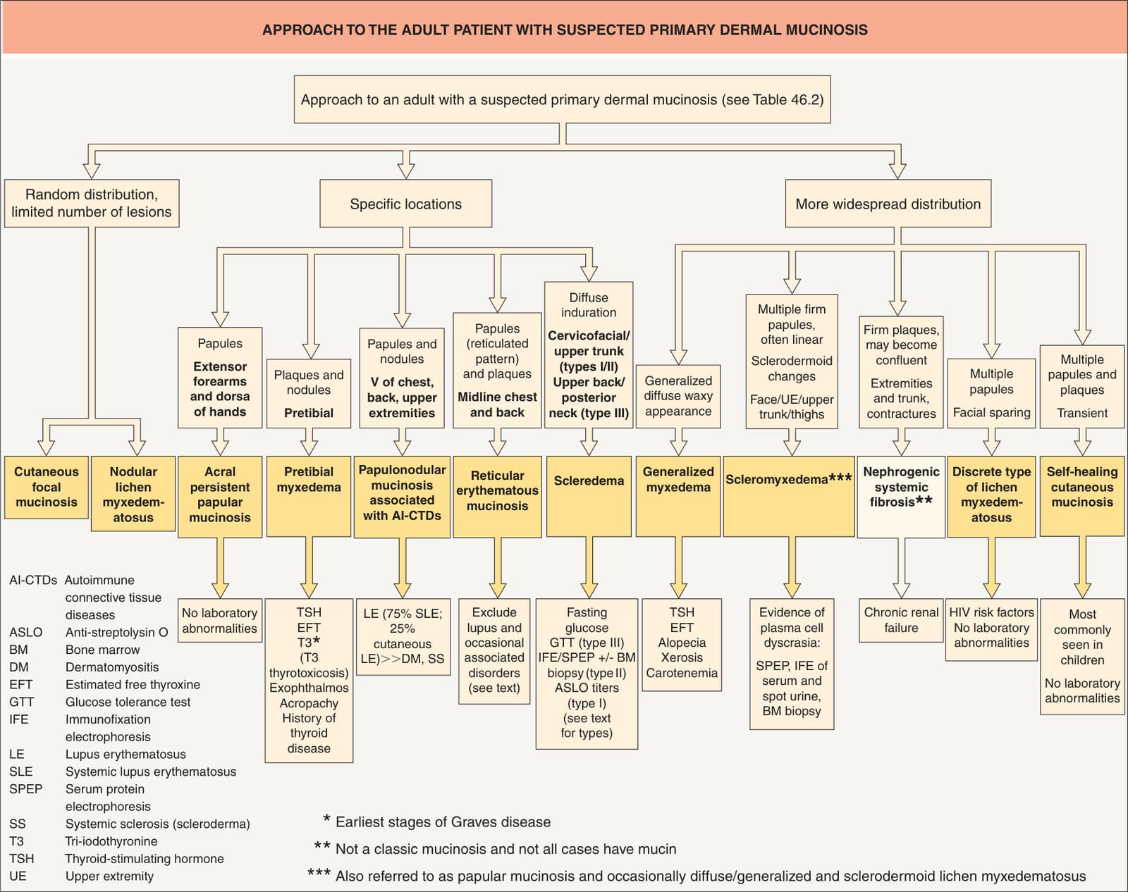
Fig. 46.1 Approach to the patient with suspected primary dermal mucinosis. Pretibial myxedema must be distinguished from obesity-associated lymphedematous mucinosis.
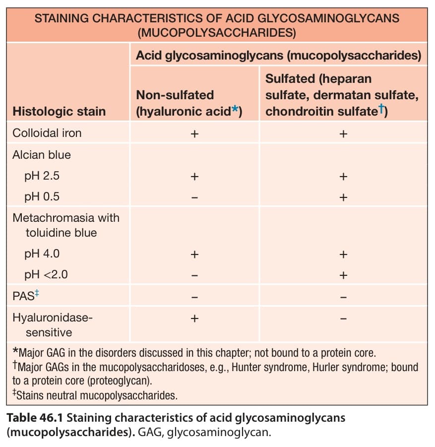
Table 46.1 Staining characteristics of acid glycosaminoglycans