PRIMARY DEGENERATIVE–INFLAMMATORY MUCINOSES
Dermal Mucinoses
Scleromyxedema
Synonyms: Papular mucinosis Diffuse/generalized and sclerodermoid lichen myxedematosus Arndt–Gottron syndrome
Introduction and definition Scleromyxedema is a chronic idiopathic disorder characterized by numerous firm papules and areas of induration that are due to dermal mucin deposition in association with an increase in dermal collagen. Nearly all patients also have a monoclonal gammopathy, and some may have systemic, even lethal, manifestations. This entity must be distinguished from localized variants of lichen myxedematosus where the skin is the sole site of involvement (Table 46.4). There are, however, occasional patients who have an atypical constellation of findings and fall between scleromyxedema and localized lichen myxedematosus (see “atypical forms” below).
History Although the first descriptions of scleromyxedema were attributed to Dubreuilh in 1906 and Reitman in 1908, it was not until the review of Montgomery and Underwood in 1953 that the disease was distinguished from scleroderma and generalized myxedema. A year later, Gottron and his colleagues gave the name “scleromyxedema” to the generalized and sclerotic form. The association of a monoclonal gammopathy with scleromyxedema was first described in 1963.
Epidemiology and pathogenesis Scleromyxedema is a fairly uncommon disease, affecting middle-aged adults of both sexes equally. The pathogenesis of scleromyxedema is unknown. The role of the associated monoclonal gammopathy remains a matter of debate. While most patients have a plasma cell dyscrasia, paraprotein levels do not correlate with either the extent or the progression of the disease. In addition, sera from patients with scleromyxedema enhanced fibroblast proliferation in vitro, but an immunoglobulin purified from paraprotein-containing sera failed to do so, suggesting a pathogenetic role for circulating factors other than the paraprotein. Clinical remission of scleromyxedema, along with a reduction in the M-protein, that follows autologous hematopoietic stem cell transplantation (HSCT) points to the bone marrow as a source of these circulating factors.
In addition to circulating cytokines, such as IL-1 and TNF, which are known to stimulate glycosaminoglycan synthesis and fibroblast proliferation in the skin, analysis of RNA in involved skin tissue also confirmed a promoting role for TGF-β. Abnormally high secretion of IL-4, a profibrotic cytokine, was recently detected in the serum of scleromyxedema patients, suggesting a chronic Th2-skewed response against an unknown target antigen. Lastly, the development of scleromyxedema following a cutaneous granulomatous reaction to intra-dermal injections of hyaluronic gel or silicone breast implants suggests autoimmune phenomena.
Clinical features In scleromyxedema, numerous, 2–3 mm, firm, waxy, closely aligned papules develop in a relatively widespread symmetrical pattern. The most common sites of involvement are the head and neck region, upper trunk,
(secondary mucinoses). A limited amount of dermal mucin can occasionally be observed in virtually all cutaneous disorders. CLL, chronic lymphocytic leukemia; DFSP, dermatofibrosarcoma protuberans; GVHD, graft-versus-host disease.
hands, forearms, and thighs (Fig. 46.2). Papules are often arranged in a strikingly linear array. The surrounding skin is shiny and indurated, i.e. sclerodermoid in appearance, and the glabella is typically involved with deep longitudinal furrowing. Severe involvement of the face can result in
a leonine facies (Fig. 46.3A). Deep furrowing can also occur on the trunk and extremities and is referred to as the “Shar-Pei sign” (Fig. 46.3B).
Erythema, edema, and a brownish discoloration may also be seen in the involved areas, and pruritus is not uncommon. The mucous membranes and scalp are spared. As the condition progresses, erythematous and infiltrated plaques may appear with skin stiffening, sclerodactyly, and decreased motility of the mouth and joints. Overlying the proximal interphalangeal joints, a central depression surrounded by an elevated rim (due to skin thickening) can be seen and is referred to as
the “doughnut sign”. Mat and cuticular telangiectasias and calcinosis, as seen in systemic sclerosis, are absent.
Scleromyxedema is almost always associated with paraproteinemia. The monoclonal gammopathy is usually IgG and the light chains are more commonly lambda. Although a mild plasmacytosis may be observed in bone marrow biopsies, <10% of patients with scleromyxedema progress to symptomatic myeloma. Patients with scleromyxedema can have a number of internal manifestations, in particular muscular, neurologic, rheumatologic, pulmonary, renal, and cardiovascular. Dysphagia, proximal muscle weakness due to myositis, disturbances of the CNS leading to unexplained coma, peripheral neuropathy, arthropathies, carpal tunnel syndrome, restrictive or obstructive lung disease, and scleroderma-like renal disease may accompany or follow the cutaneous manifestations. While the predominantly sensory peripheral neuropathy typically affects older men and has an insidious onset, the dermato-neuro syndrome is a potentially life-threatening encephalopathy. This syndrome begins abruptly with a worsening of skin lesions, a flu-like prodrome, fever, and seizures, and it can eventuate in an unexplained coma. Possible triggers include viral infections and vaccines (e.g. SARS-CoV-2)7a.
Pathology Scleromyxedema is characterized by a triad of microscopic features:
●a diffuse deposit of mucin in the upper and mid reticular dermis
●an increase in collagen deposition
●a marked proliferation of irregularly arranged fibroblasts (Fig. 46.4).
The epidermis may be normal or thinned by the pressure of the underlying mucin and fibrosis; the hair follicles may be atrophic. A slight superficial, perivascular, lymphoplasmacytic infiltrate is often present. The elastic fibers are fragmented and decreased in number. More recently, an interstitial granuloma annulare-like pattern was described. Mucin may fill the walls of myocardial blood vessels as well as the parenchyma of the kidney, pancreas, adrenal glands, nerves, and lymph nodes. In the dermato-neuro syndrome, autopsy findings have not proved helpful in elucidating an underlying pathogenesis.
Differential diagnosis The primary differential diagnosis for scleromyxedema is systemic sclerosis (scleroderma) and scleredema. The presence of papules, especially in linear arrays, is a very helpful clinical sign in distinguishing scleromyxedema. In the occasional patient with systemic sclerosis who develops primary cutaneous amyloidosis (see Fig. 47.6), histologic findings allow distinction. Additional entities in the sclerodermoid differential diagnosis should be excluded (see Ch. 43). For example, nephrogenic systemic fibrosis, which develops in individuals with renal impairment exposed to gadolinium-containing contrast media, may show mucin in biopsy specimens, but patients lack both facial involvement (commonly seen in scleromyxedema) and paraproteinemia. Criteria for diagnosing scleromyxedema versus localized variants of lichen myxedematosus are summarized in Table 46.4, and the dermatologic disorders in which leonine facies can develop are outlined in Table 46.5.
Treatment Recommendations are still based on case reports and open-label small case series. In the past, monthly courses of melphalan were often the therapy of choice, targeting the plasma cell dyscrasia. However, while this alkylating agent can result in some clinical improvement, it has also been implicated in 30% of the deaths secondary to its induction of hematologic malignancies and septic complications. Other chemotherapeutic agents (e.g. cyclophosphamide, methotrexate, chlorambucil, 2-chlorodeoxyadenosine) have been tried, but with no better results and the risk of significant side effects.
IVIg, alone or in combination with systemic medications (see below), has gained widespread acceptance as first-line therapy for both cutaneous involvement and associated systemic manifestations, including the dermato-neuro syndrome. Although long-lasting remissions after cessation of IVIg therapy have been reported, the response is usually not permanent and maintenance infusions are required. Either thalidomide or lenalidomide or a combination of lenalidomide, a proteasome inhibitor (e.g. bortezomib) and dexamethasone, standard therapy for multiple myeloma, is considered second-line treatment and is often
administered in combination with IVIg. This multi-drug regimen may represent first-line therapy for individuals with severe disease including CNS or cardiac involvement. Autologous HSCT represents third-line therapy, especially for individuals with disabling or potentially life-threatening disease. Due to the excellent response observed in patients with myeloma, the anti-CD38 monoclonal antibodies daratumumab and isatuximab may represent future therapies.
Additional therapies, including PUVA, UVA1, systemic retinoids, cyclosporine, electron beam radiation, plasmapheresis, and extracorporeal photochemotherapy, have all produced variable results. Dysarthria and a flu-like illness may herald life-threatening coma, and the patient should be promptly admitted to the hospital for close observation and consideration of treatment with a multi-drug regimen (see above) plus IVIg and plasmapheresis.
Lichen myxedematosus (localized variants)
Introduction In the localized variants of lichen myxedematosus, patients develop small, firm, waxy papules (or nodules and plaques produced by the confluence of papules) that are limited to only a few sites – usually the upper and lower extremities and/or trunk. The skin is the only site of involvement and these variants, in contrast to scleromyxedema, are not associated with sclerosis, paraproteinemia, or systemic involvement nor are they associated with thyroid disease (see Fig. 46.1). While most dermatologists equate lichen myxedematosus with localized, skinlimited disease, a source of potential confusion is the uncommon and more historical use of the term “diffuse/generalized and sclerodermoid lichen myxedematosus” to describe scleromyxedema.
The localized variants of lichen myxedematosus are subdivided into four subtypes:
●a discrete papular form
●acral persistent papular mucinosis
●cutaneous mucinosis of infancy
●a pure nodular form. Localized variants of lichen myxedematosus may be observed in association with HIV infection, exposure to toxic oil or L-tryptophan (historical), and hepatitis C viral (HCV) infection.
Epidemiology Exact incidence and prevalence rates for the localized variants of lichen myxedematosus are not known.
Clinical features Discrete papular lichen myxedematosus is characterized by 2–5 mm papules, numbering from just a few to hundreds and involving the limbs and trunk in a symmetrical pattern (Fig. 46.5). The affected skin is not indurated and the face is spared. Lesions progress slowly without
systemic involvement. However, they rarely resolve spontaneously. To date, progression to scleromyxedema has never been definitively proven.
In acral persistent papular mucinosis, first described in 1986 by Rongioletti et al., multiple ivory to skin-colored papules develop exclusively on the dorsal aspect of the hands and extensor surface of the distal forearms (Fig. 46.6). A female : male ratio of 3 : 1 has been noted. The lesions persist, but without systemic manifestations.
In cutaneous mucinosis of infancy (syn. papular mucinosis of infancy), first described by Lum in 1980, firm opalescent papules appear on the neck (Fig. 46.7), upper arms (especially the elbows), and trunk. In contrast to self-healing mucinosis, neither systemic symptoms nor spontaneous resolution are observed. Of the handful of patients described to date, at least two have had a congenital linear variant, which might be better categorized as an example of a mucinous nevus.
Nodular lichen myxedematosus is characterized by multiple nodules on the limbs and trunk, with a mild or absent papular component. Atypical tuberous myxedema (Jadassohn–Dosseker) probably represents a pure nodular variant of lichen myxedematosus1,17a.
Localized lichen myxedematosus in HIV-infected patients. The majority of patients developed either the discrete papular form or the nodular variant of lichen myxedematosus, with lesions on the limbs and trunk. All became infected with HIV before the onset of the lichen myxedematosus. Most had hypergammaglobulinemia, a well-recognized manifestation of HIV infection, and to date only two had paraproteinemia. Of note, up to 10% of HIV-infected individuals may have monoclonal gammopathy of undetermined significance (MGUS) and how this relates to the mucinosis remains to be determined. None of the patients had visceral involvement due to mucin deposition.
Localized lichen myxedematosus in “toxic” syndromes. Multiple papules due to mucin deposition with a clinical appearance similar to the discrete form of lichen myxedematosus have also been described
in the setting of toxic oil syndrome and L-tryptophan-associated eosinophilia–myalgia syndrome. Although unrelated epidemiologically and both of historical interest, toxic oil syndrome (due to the ingestion of adulterated rapeseed oil in Spain in the early 1980s) and L-tryptophan-associated eosinophilia–myalgia (related to a contaminant in L-tryptophan-containing products used as sedatives during the late 1980s) share several clinical features, including constitutional symptoms, peripheral eosinophilia, hyperpigmentation, and a sclerodermoid appearance, in addition to this mucinous papular eruption. Usually 1–5 months after the onset of the illness, 1–5 mm, white or skin-colored papules appeared, primarily on the limbs. The lesions resolved slowly after exposure to the toxic substance ceased.
Localized lichen myxedematosus and HCV infection. An association of lichen myxedematosus with chronic hepatitis due to HCV infection has been reported, especially from Japan, but this relationship needs to be confirmed by more extensive studies.
Cutaneous mucinosis consistent with localized lichen myxedematosus has been associated with several medications including targeted immunomodulators (“biologics”), inhibitors of CSF-1R (colonystimulating factor-1 receptor), and interferons (including at injection sites) in addition to trauma (e.g. mechanical, following total knee replacement).
Atypical forms of lichen myxedematosus. Occasionally patients with lichen myxedematosus have atypical features or features inter-mediate between scleromyxedema and localized lichen myxedematosus. This group includes patients with scleromyxedema who lack a monoclonal gammopathy as well as individuals with localized forms of lichen myxedematosus who also have a monoclonal gammopathy and/ or systemic symptoms. One possible explanation for the former group is performance of just a serum protein electrophoresis (SPEP), rather than an SPEP plus an immunofixation electrophoresis (IFE) of serum and urine, as IFE is a more sensitive assay.
Pathology In the localized forms of lichen myxedematosus, the histologic changes are less characteristic than in scleromyxedema. Mucin accumulates in the upper and mid reticular dermis and fibroblast proliferation is variable; fibrosis is not marked and may even be absent. In acral persistent papular mucinosis, mucin accumulates focally in the upper reticular dermis (sparing a subepidermal zone) and fibroblasts are not increased in number. In cutaneous mucinosis of infancy, the mucin may be so superficial as to look as if it were “enclosed” by epidermis, although mucin may also deposit in the reticular dermis.
Differential diagnosis Histologic examination of the skin helps to distinguish localized variants of lichen myxedematosus from several papular eruptions that can have a similar appearance, such as papular granuloma annulare, amyloidosis, colloid milium, molluscum contagiosum, papular elastorrhexis, and
eruptive collagenomas. It is also important to differentiate these entities from scleromyxedema and the cutaneous mucinosis that can occur in the setting of autoimmune connective tissue diseases (see below).
Treatment Localized lichen myxedematosus does not require therapy, and a waitand-see approach is recommended. Topical application of corticosteroids, pimecrolimus, or tacrolimus may be of some benefit. One patient with associated HIV infection had a complete remission after treatment with oral isotretinoin. Spontaneous resolution may occur, even in the setting of HIV infection.
Self-healing cutaneous mucinosis
Although previously considered a subtype of localized lichen myxedematosus, self-healing cutaneous mucinosis is best regarded as a distinct rare form of primary dermal mucinosis. It was initially described in children, with ages of reported patients ranging from 1 to 15 years. The same disorder was subsequently observed in a few adults, but in the author’s opinion, some of the adult patients were actually examples of localized lichen myxedematosus with spontaneous resolution.
Self-healing cutaneous mucinosis is characterized by the following clinical presentations: (1) an acute eruption of multiple papules, sometimes coalescing into linear infiltrated plaques, on the face, neck, scalp, abdomen, and thighs; and (2) mucinous subcutaneous nodules in periarticular areas and on the face, with periorbital swelling (Fig. 46.8). In addition, systemic symptoms (e.g. fever, arthralgias, and muscle tenderness) may accompany the cutaneous lesions, but paraproteinemia, bone marrow plasmacytosis, and thyroid dysfunction are not observed. Spontaneous resolution over a period of 1–8 months is characteristic, hence the name. Histologically, papular lesions show dermal mucin deposition with mild inflammation and a small increase in the number of fibroblasts, whereas nodules have deep mucinous deposits associated with bands of fibrosis and a prominent proliferation of fibroblast-like cells and gangliocyte-like cells mimicking proliferative fasciitis. While spontaneous complete regression is the expectation, longitudinal evaluation is recommended due to the possible development of dermato-rheumatologic disorders such as fibroblastic rheumatism.
Scleredema
Synonyms: Scleredema adultorum of Buschke Scleredema diabeticorum
Introduction Scleredema is a symmetrical diffuse induration of the upper part of the body due to a thickened dermis and deposition of mucin.
History Although Buschke has been credited with first describing scleredema, the original description dates back to Pitford in 1876. A relationship with diabetes mellitus was only established in 1970.
Epidemiology and pathogenesis Scleredema is an unusual disease that affects all races. The form that is associated with diabetes mellitus is more prevalent in men, while other forms are seen more commonly in women. Irreversible glycosylation of collagen and resistance to degradation by collagenase may lead to an accumulation of collagen. Alternatively, enhanced stimulation by insulin, microvascular damage, and hypoxia may increase the synthesis of collagen and mucin. Streptococcal hypersensitivity (type I), injury to lymphatics, and paraproteinemia (type II) may also play a role.
Clinical features Classically, there are three types of scleredema, although a simpler division into those with and those without diabetes has been suggested. The first type affects primarily children and middleaged women. It is preceded by fever, malaise, and an infection (usually streptococcal) of the upper or lower respiratory tract. The skin of the cervicofacial region suddenly hardens with extension to the trunk and proximal upper limbs. The face is expressionless and opening of the mouth and swallowing are difficult because of the involvement of the tongue and pharynx. This type usually resolves spontaneously.
The second type shares the same clinical features as the first but has a more subtle onset, without a preceding illness. It persists for years. This type is more frequently associated with a monoclonal gammopathy.
The third type occurs primarily in obese middle-aged men with insulin-dependent diabetes (scleredema diabeticorum). The onset is subtle and the involvement persistent. Erythema and induration of the posterior neck and the back are commonly observed (Fig. 46.9), as is a peau d’orange appearance of the skin. In those patients who also have cheiroarthropathy, the skin of the distal extremities may appear waxy, presumably due to increased amounts of glycosylated collagen.
Occasionally, patients with limited site involvement (e.g. periorbital skin) have also been reported. In all three forms, systemic manifestations such as serositis, myositis, dysarthria, dysphagia, parotitis, and ocular and cardiac abnormalities (e.g. ophthalmoplegia, congestive heart failure) may occur, but are unusual. Scleredema may be idiopathic and anecdotal associations with autoimmune diseases (e.g. rheumatoid arthritis, Sjögren syndrome, primary biliary cholangitis, dermatomyositis, lichen sclerosus), internal neoplasms (malignant insulinoma, gallbladder carcinoma, carcinoid, pituitary–adrenocortical neoplasm), and HIV infection have been described. Except for limitation of movement, scleredema usually causes little morbidity. While postinfectious scleredema may clear within 6–24 months, scleredema associated
with diabetes or a monoclonal gammopathy has a chronic unremitting course. Rarely, death may occur when internal organs are involved.
Pathology The principal alteration in scleredema is the thickening of the reticular dermis, with large collagen bundles separated from one another by clear spaces filled with mucin, resulting in fenestration of the dermis (Fig. 46.10). There is no increase in the number of fibroblasts, but the elastic fibers are reduced in number. At times, the mucin deposition can be so slight that multiple biopsies or staining of unfixed frozen sections are required to detect it. There is sometimes a sparse perivascular lymphocytic infiltrate. Direct immunofluorescence is usually negative, but deposits of IgG and C3 have been observed at the dermal–epidermal junction. Mucin also accumulates within skeletal muscle and the heart.
Differential diagnosis Scleredema may be confused with systemic sclerosis, but the absence of Raynaud phenomenon and cuticular and mat telangiectasias points to scleredema. Histologically, mucin deposition may be observed in systemic sclerosis, but adnexal structures are atrophic or absent and the dermis is diffusely sclerotic, as opposed to the “fenestration” observed in scleredema. Patients with scleromyxedema also have firm papules (often in a linear array) in addition to dermal induration as well as a proliferation of fibroblasts histologically. The other types of cutaneous mucinoses are usually distinguished on the basis of clinical findings. Additional entities in the sclerodermoid differential diagnosis should be excluded (see Ch. 43). Occasionally, because of associated erythema, diabetes-associated scleredema is sometimes misdiagnosed as cellulitis (usually by non-dermatologists).
Treatment Therapy is unnecessary for scleredema associated with streptococcal infections because it is self-limited. Management of scleredema associated with diabetes or a monoclonal gammopathy is more difficult and no specific treatment is available. Phototherapy, in particular UVA1 and narrowband UVB, is considered first-line therapy as it can stimulate fibroblasts to synthesize collagenase, thereby enhancing degradation of sclerotic tissue. For scleredema associated with a plasma cell dyscrasia, systemic medications used to treat myeloma (see above), may be effective. Some, but not all, patients with diabetes-associated scleredema improve with strict glycemic control.
Systemic and intralesional corticosteroids, intralesional hyaluronidase, antibiotics, methotrexate, cyclosporine, pulse therapy with cyclophosphamide plus oral corticosteroids, tamoxifen, and allopurinol have all been tried, with variable results. Electron beam radiotherapy, IVIg, and extracorporeal photopheresis are additional reported therapies. In patients with limited mobility or respiratory difficulty, ultrasonic massage and physical therapy can be employed. Aggressive therapies should be limited to individuals with disabling disease or systemic manifestations.
Mucinoses associated with altered thyroid function
Definition
Localized or pretibial myxedema is characterized by cutaneous induration of the shins due to mucin deposition. It is often associated with hyperthyroidism (most commonly due to Graves disease), but may appear following treatment of the thyroid disease.
Epidemiology and pathogenesis Graves disease is seven times more common in women than in men and onset is usually during the third or fourth decade. Pretibial myxedema is one sign of Graves disease, along with goiter, exophthalmos, thyroid acropachy, and thyroid-stimulating immunoglobulins that recognize the thyroid-stimulating hormone (TSH) receptor. Overall, pretibial myxedema is found in 1%–5% of patients with Graves disease, but in up to 25% of those with exophthalmos. It can also appear in the setting of hypothyroidism following the treatment of Graves disease. Rarely, localized myxedema occurs in the setting of Hashimoto thyroiditis without thyrotoxicosis, and even in euthyroid patients.
A serum factor (unrelated to thyroid-stimulating immunoglobulins) may incite fibroblasts to produce mucin. Fibroblasts from the dermis of the lower extremities have been found to be more sensitive to such a factor than are fibroblasts from other areas of the body. An insulin-like growth factor, trauma, and lymphatic obstruction due to mucin may also play a role.
Clinical features Localized myxedema presents as erythematous to skin-colored, but sometimes purple–brown or yellowish, waxy indurated nodules or plaques. A peau d’orange appearance is a characteristic finding (Fig. 46.11). The lesions are usually located on the anterolateral aspect of the lower legs or the feet. Localized myxedema can also present as a diffuse non-pitting edema of the shins or feet that evolves into elephantiasis. Even more rarely, localized myxedema affects the face, shoulders, upper extremities, lower abdomen, scars, or donor graft sites. Large plaques are often painful and pruritic. When present, hypertrichosis and hyperhidrosis are confined to pretibial myxedematous skin.
Apart from appearance and possible pain, associated morbidity is usually minimal. Entrapment of peroneal nerves by mucinous connective tissue may cause foot drop or faulty dorsiflexion.
Pathology Large quantities of mucin are deposited within the reticular dermis, causing collagen bundles to separate and the dermis to thicken. A subepidermal grenz zone of normal collagen is also observed. A perivascular and periadnexal lymphocytic infiltrate with mast cells is seen, as well as large stellate fibroblasts. Elastic fibers are reduced in number. There is often hyperkeratosis in addition to papillomatosis and hyperplasia of the epidermis.
Differential diagnosis In addition to lichen simplex chronicus and hypertrophic lichen planus, pretibial myxedema should be distinguished from lymphedema, lipedema, and obesity-associated lymphedematous mucinosis. In the latter disorder, semi-translucent papules, plaques with woody induration, and occasionally vesicles develop on the shins of euthyroid, morbidly obese individuals with significant lower extremity edema. Histologically, mucin deposition is found within the superficial papillary dermis and around vessels, in addition to epidermal atrophy (with effacement of the rete ridge pattern), vertically oriented capillaries, and deposits of hemosiderin.
Treatment Corticosteroids, either applied under occlusive dressings or delivered by intralesional injection (e.g. triamcinolone 10–40 mg/cc), are considered first-line therapy. Intralesional hyaluronidase, alone or in combination with intralesional corticosteroids, may also be tried. More recently, teprotumumab, an IGF-1R inhibitor that is FDA-approved for the treatment of thyroid-associated ophthalmopathy, was reported to lead to improvement33a. For the severe elephantiasis form, medical treatments, including IVIg, rituximab, plasmapheresis and octreotide, have been of benefit and are sometimes combined with surgery. Unfortunately, skin grafting is usually followed by recurrence. Compression stockings and gradient pneumatic compression are useful for the associated lymphedema. Addressing risk factors such as obesity and smoking is recommended. Therapy for hyperthyroidism does not improve the cutaneous lesions and, often, localized myxedema develops after treatment has been instituted or completed. Localized myxedema may clear spontaneously after an average of 3.5 years.
Definition Generalized myxedema is a manifestation of severe hypothyroidism in which mucin is deposited in the dermis, leading to waxiness of the skin.
Pathogenesis Generalized myxedema is due to a quantitative or functional deficiency of thyroxine. Impaired degradation of mucin, rather than increased synthesis, has been suggested as the cause.
Clinical features Hypothyroidism may be congenital, with sequelae formerly referred to as cretinism, or of juvenile or adult onset.
Congenital hypothyroidism affects 1 in 5000 neonates and may cause a distinctive syndrome of dwarfism, intellectual disability, and systemic and cutaneous findings. Somnolence, constipation, poor feeding, poor muscle tone, persistence of jaundice, and respiratory distress all suggest the diagnosis. However, more than a third of infants have no symptoms. Periorbital tissues and the tongue, lips, hands, and genitals are puffy. The skin is dry, cold, and pale. Nails and hairs are dry and brittle, and there can be patchy alopecia. The presence of a clavicular pad is diagnostic.
Juvenile hypothyroidism develops in a previously euthyroid child. Clinical features include short stature, abnormal physical and mental development, retardation of sexual maturity, and poor performance at school. Hypertrichosis may develop on the shoulders and upper back.
Adult hypothyroidism is the most common form of the disease. It is seen primarily in women 40–60 years of age, often as a result of autoimmune disease (usually Hashimoto thyroiditis), therapy of hyperthyroidism (usually Graves disease) or, rarely, pituitary or hypothalamic failure. Rarely, the ingestion of large quantities of foodstuffs that contain inhibitors of iodine uptake by the thyroid gland, e.g. raw bok choy, can lead to myxedema.
The initial symptoms are subtle and include mental and physical sluggishness, weight gain, constipation, leg cramps, loss of appetite, and cold intolerance. The face has a dull expression. The eyelids, lips, tongue, and hands are puffy, the nose is broad (Fig. 46.12), and the speech becomes hoarse and slurred. The skin is pale, cool, waxy, and dry; an absence of sweating can lead to acquired ichthyosis or eczema craquelé. A yellowish discoloration of the palms and soles due to carotenoderma may appear. Hair and nails are dry and brittle, and a diffuse non-scarring alopecia is common; alopecia of the lateral third of the eyebrows can also be seen. Purpura involving the extremities, blue telangiectatic fingertips, delayed wound healing, and xanthomas may be observed. Systemic manifestations include cardiomegaly, megacolon or bowel obstruction, psychiatric symptoms mimicking Alzheimer disease, serositis, carpal tunnel syndrome, and seventh nerve paralysis.
Pathology Mucin deposits, mainly perivascular and perifollicular, splay collagen bundles and may extend into the subcutaneous fat and nerves. Fibroblasts are not increased in number, but elastic fibers are reduced. Mucin deposits in the brain probably cause the psychiatric symptoms.
Diagnosis and differential diagnosis The diagnosis is suspected clinically. Low levels of circulating free T confirm the diagnosis. Serum TSH level is high in primary hypothyroidism and low in secondary hypothyroidism. Myxedema does not seem to occur in secondary hypothyroidism.
Treatment Early treatment is crucial for proper mental development of neonates with hypothyroidism, but it is also critical for the juvenile and adult forms. Measurement of serum levels of T and/or TSH is performed between 2 and 4 days of age in newborn screening panels, and treatment should be initiated within the first 2 weeks of life. Usually, symptoms subside with thyroxine administration and recur if it is discontinued.
Even areas of hair loss regrow with proper treatment. If untreated, patients can die as a result of “myxedema coma”.
Reticular erythematous mucinosis
Synonyms: Plaque-like cutaneous mucinosis Reticular erythematous mucinosis syndrome Midline mucinosis
Introduction and definition Reticular erythematous mucinosis (REM) is a disorder in which persistent, erythematous papules or plaques develop in the midline of the back or chest. The papules often have a reticular or net-like configuration. This disorder has some overlap with lupus erythematosus (LE) tumidus.
History Steigleder and colleagues coined the term “reticular erythematous mucinosis” in 1974. However, in 1960, Perry and colleagues described three similar patients with plaque-like cutaneous mucinosis. Some authors regard these two entities as identical, while others view them as being on a continuum.
Epidemiology and pathogenesis REM is a rare disorder that occurs most often in middle-aged women, although men and children are not spared. It is seen worldwide and can be provoked or exacerbated by a wide range of factors, from heat and perspiration to oral contraceptives and pregnancy to radiation therapy. There is debate as to whether REM is a photoaggravated disorder. Familial cases suggest a genetic predisposition.
Within endothelial cells and pericytes of lesional skin, tubuloreticular inclusions have been detected. Although these inclusions occur in viral infections, they can also be produced by high levels of interferon and are also found within endothelial cells in LE. Lastly, the fibroblasts of REM patients exhibit an abnormal overstimulation when exposed to exogenous IL-1.
Clinical features Plaque-like cutaneous mucinosis and REM are probably different clinical presentations of the same rare syndrome. On the mid back or chest, pink to red macules and papules merge into reticulated and annular patterns (Fig. 46.13) or plaque-like lesions. They may be slightly pruritic and sometimes lesions spread to the abdomen. While sun exposure may worsen the eruption, it has also been reported to be beneficial. Provocative phototests (UVA and/or UVB) can sometimes reproduce REM lesions. In general, REM is not associated with systemic diseases or abnormal laboratory tests. However, there are reports of autoimmune diseases (e.g. SLE), hypothyroidism, and more common internal malignancies in patients with REM.
Pathology The epidermis is normal. Interstitial deposits of small amounts of mucin are seen in the upper dermis, along with a perivascular and, at times, perifollicular T cell infiltrate. Vascular dilation is present. Usually, direct immunofluorescence is negative, but, rarely, granular deposits of IgM, IgA, and C3 have been seen at the dermal–epidermal junction.
Differential diagnosis In biopsy specimens of discoid lesions of LE there is involvement of the epidermis and deposits of IgG and C3 are found at the dermal–epidermal junction, and in lymphocytic infiltrate of Jessner, there is minimal, if any, dermal mucin. LE tumidus is very difficult to distinguish microscopically from REM. Both diseases have perivascular dermal infiltrates of T lymphocytes, primarily CD4+, plus increased dermal mucin. In REM, the mucin and the lymphocytes tend to be more superficial and the lymphocytes more scattered. In LE tumidus, plasmacytoid dendritic cells were found to be clustered whereas in REM they were fewer in number and appeared as single cells. These findings aside, some authors view REM and LE tumidus as variants of the same disease and along a continuum with cutaneous LE. It would be unusual for polymorphous light eruption to be restricted to the chest and histologically there is less abundant mucin and sometimes pronounced edema of the super-ficial dermis. Seborrheic dermatitis and tinea versicolor may involve the central chest, but they have associated scale and can be diagnosed clinically, as can confluent and reticulated papillomatosis.
Treatment Hydroxychloroquine or chloroquine (+/− quinacrine) are usually effective in clearing the lesions within 4–8 weeks. Results from other treatments such as topical and systemic corticosteroids, topical calcineurin inhibitors, oral antihistamines, tetracyclines, cyclosporine, UVB irradiation, and pulsed dye laser are quite variable. Despite the potential for an exacerbation, improvement following UVA1 or UVB irradiation has been reported. The lesions may clear spontaneously, even after 15 years.
Papulonodular mucinosis associated with autoimmune
Synonyms (when associated with LE): Cutaneous lupus mucinosis Papulonodular mucinosis in (systemic) lupus erythematosus Papular and nodular mucinosis of Gold
Papules, nodules, and plaques secondary to mucin deposition may accompany or even antedate an autoimmune connective tissue disease, most commonly LE, and rarely dermatomyositis or systemic sclerosis. Cutaneous lupus mucinosis occurs in ~1%–2% of patients with LE. Only in Japan has a male predominance been observed. The lesions present as asymptomatic, skin-colored, at times erythematous, 0.5–2 cm papules and nodules (Fig. 46.14); rarely, they merge into large plaques. Tangential illumination can be used to enhance detection of the lesions. A central depression and pigmentation may be additional features. The back, V of the chest, and the upper extremities are the
Skin-colored papules and nodules leading to a “lumpy” appearance of the chest.
most common sites of involvement. In only a few reported patients was there a flare of the papulonodules after sun exposure.
Cutaneous lupus mucinosis can antedate LE or begin concurrently. The clinical course may correlate, in some patients, with underlying disease activity. Approximately 75% of patients with LE who develop papular and nodular mucinosis have systemic involvement, primarily renal and articular. The minority have only skin involvement, usually discoid or subacute cutaneous LE lesions.
Histologically, mucin abounds in the upper and mid dermis but may involve the subcutaneous fat as well. Sometimes, it is accompanied by a slight perivascular lymphocytic infiltrate. The epidermal changes of LE are absent, but linear or granular deposits of immunoglobulin and/or C3 are detected at the dermal–epidermal junction.
Cutaneous lesions of dermatomyositis often have dermal mucin histologically; rarely, patients develop erythematous nodules and plaques due to mucin deposition. The latter appear primarily on the trunk and usually follow the myositis. Papular and nodular mucinosis can also occur in association with systemic sclerosis.
Treatment Therapies for papular and nodular mucinosis are similar to those used for LE and dermatomyositis, i.e. sunscreens, topical corticosteroids, and oral antimalarials. For unresponsive cases, systemic corticosteroids can be prescribed. Intralesional corticosteroids may prove useful for reducing the size of large nodules and plaques.
Cutaneous focal mucinosis
Cutaneous focal mucinosis presents as an asymptomatic, skin-colored papule or nodule, <1 cm in diameter. The lesion can occur anywhere on the body or in the oral cavity. Cutaneous focal mucinosis favors adults and only rarely has it been linked to thyroid disorders (without myxedema), REM, or scleromyxedema. Trauma can be an inciting factor. The diagnosis is based upon histologic findings.
Histologically, mucin is dispersed throughout the upper and mid dermis, sparing the subcutaneous fat. Cleft-like spaces, but no cysts, are seen. Spindle-shaped or stellate, vimentin-positive fibroblasts are present. There is also a minor population of dermal dendrocytes that are partially factor XIIIa-positive and partially CD34-positive. The elastic and reticulum fibers are absent, but capillaries are normal in number.
Cutaneous focal mucinosis results from a “muciparous” reaction of the connective tissue to nonspecific stimuli and should be distinguished from digital mucous cyst or from angiomyxoma, which represents a true neoplasm. Cutaneous focal mucinosis can be surgically excised, and relapses are uncommon.
Digital mucous cyst (myxoid cyst) is discussed in Chapter 110.
Primary Follicular Mucinoses
Mucin accumulates within the follicular epithelium in two distinctive primary disorders: Pinkus follicular mucinosis and urticaria-like follicular mucinosis. Otherwise, follicular mucinosis is considered a histologic epiphenomenon (i.e. secondary mucinosis), most often associated with cutaneous T cell lymphoma, in particular mycosis fungoides and Sézary syndrome, or other skin diseases (e.g. atopic dermatitis; see Table 46.3).
Follicular mucinosis
Synonyms: Alopecia mucinosa Mucinosis follicularis Pinkus follicular mucinosis – benign primary form
History, epidemiology, and pathogenesis This uncommon inflammatory disorder, described in 1957 by Pinkus, has a predilection for children and for adults in the third and fourth decades of life. It is unclear why dermal-type mucin is deposited selectively within an epithelial structure. Although follicular keratinocytes have been considered to be the source of the mucin, an etiologic role for cell-mediated immune mechanisms has been proposed, including a reaction to persistent antigens such as Staphylococcus aureus.
Clinical features Primary follicular mucinosis is an idiopathic benign form of the disease, apparently not linked to lymphoma. Clinically, it presents as an acute or subacute eruption in children and young adults and is characterized by one or several pink plaques, often composed of grouped follicular papules. There may be associated scale, and lesions are limited to the face (Fig. 46.15A) and scalp and are associated with alopecia. Papulonodules (Fig. 46.15B), annular plaques, folliculitis, follicular spines, and acneiform eruptions have also been described. A second type of follicular mucinosis, characterized by: (1) a more generalized distribution (extremities, trunk and face; Fig. 46.15C); (2) larger and more numerous plaques; (3) a chronic clinical course; and (4) occurrence in a slightly older age group, is probably best regarded as a secondary follicular mucinosis associated with atopic dermatitis or cutaneous T cell lymphoma, rather than a primary condition. Follicular mucinosis has occasionally been observed in patients with hematologic malignancies other than mycosis fungoides.
Pathology Mucin accumulates within the follicular epithelium and sebaceous glands, causing keratinocytes to disconnect (Fig. 46.15D). In more advanced lesions, the follicles are converted into cystic spaces containing mucin, inflammatory cells, and altered keratinocytes. A perifollicular and intra-follicular infiltrate of lymphocytes, histiocytes, and eosinophils is seen.
Differential diagnosis The differentiation between primary follicular mucinosis and mycosis fungoides-associated follicular mucinosis is very difficult, and there is no single reliable criterion. Although the existence of a primary form of follicular mucinosis has been questioned by some authors (who consider it as an “indolent” localized form of cutaneous T cell lymphoma), features in favor of a primary form are the young age of the patient, a solitary plaque or limited number of lesions in the head and neck region, spontaneous resolution, and the absence histologically of epidermotropism and atypical lymphocytes. Detection of clonal T cell gene rearrangements does not seem to help differentiate the two types42,42a.
Treatment There is no specific treatment. A wait-and-see approach is recommended for primary follicular mucinosis as many cases resolve spontaneously within 2–24 months. Topical, intralesional, and systemic corticosteroids, antimalarials, phototherapy (PUVA, UVA1), minocycline, dapsone, indomethacin, oral isotretinoin, interferon-α-2b, and orthovoltage irradiation have been reported to be beneficial. For secondary forms, treatment of the underlying disease is indicated. Longitudinal evaluation and assessment for cutaneous T cell lymphoma is recommended for patients with presumed primary follicular mucinosis that is persistent or becomes more extensive.
Urticaria-like follicular mucinosis
Urticaria-like follicular mucinosis is a very rare disorder that occurs primarily in middle-aged men. Pruritic urticarial papules or plaques appear on the head and neck within an erythematous “seborrheic” background. As lesions resolve, red macules persist for a few weeks. Hair-bearing regions may be involved, but neither follicular plugging nor alopecia is seen. Urticaria-like follicular mucinosis waxes and wanes irregularly over a period that can vary from a few months to 15 years. There are no associated systemic diseases. Response to natural sunlight has been inconsistent, but it has been beneficial in a small number of cases.
As in primary follicular mucinosis, mucin-filled cystic spaces occupy hair follicles. In the upper dermis, lymphocytes and eosinophils are seen around blood vessels and hair follicles. In only a single patient to date were vascular C3 deposits seen by direct immunofluorescence. The prognosis is good, and based upon a limited number of case reports, antimalarials and dapsone were reportedly beneficial.
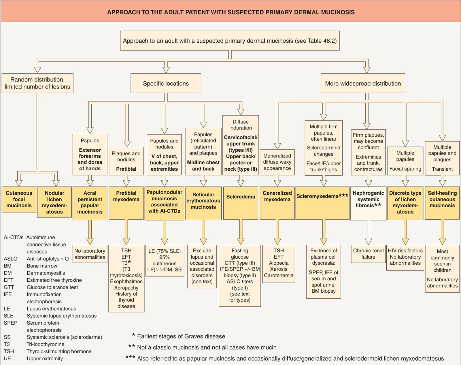
Fig. 46.1 Approach to the patient with suspected primary dermal mucinosis. Pretibial myxedema must be distinguished from obesity-associated lymphedematous mucinosis.
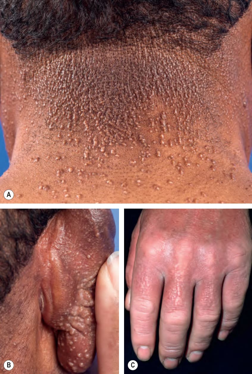
Fig. 46.2 Scleromyxedema.A, B Numerous monomorphic, firm, skin-colored to tan papules which can have a strikingly linear arrangement. This is most obvious on the posterior neck. C The skin can also become shiny and indurated, leading to a sclerodermoid appearance, but the small firm papules in a linear array are a clue to the diagnosis. A, B, Courtesy Edward Cowen, MD; C, Courtesy Lorenzo Cerroni, MD.
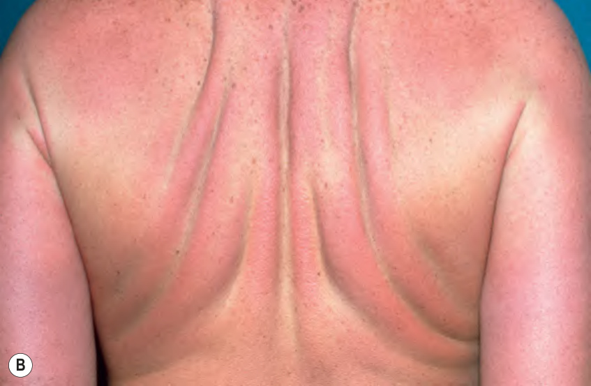
Fig. 46.3 Scleromyxedema. Thickening of the skin of the forehead (A) and trunk (B), leading to deep furrows and folds. Scleromyxedema is one of the causes of leonine facies. A, Courtesy Joyce Rico, MD; B, Courtesy Lorenzo Cerroni, MD.
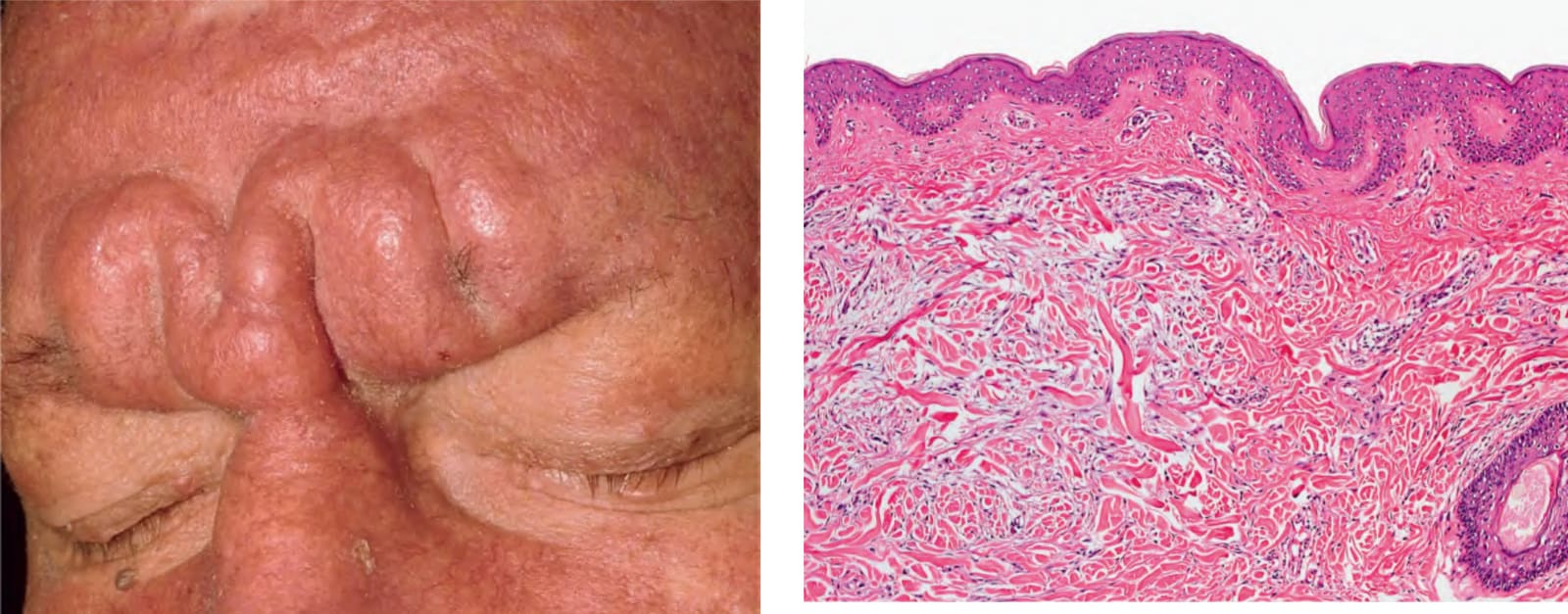
Fig. 46.4 Scleromyxedema – histopathologic features. Typical triad of fibrosis, an increased number of irregularly arranged fibroblasts, and interstitial deposits of mucin in the upper and mid reticular dermis. Courtesy Lorenzo Cerroni, MD.
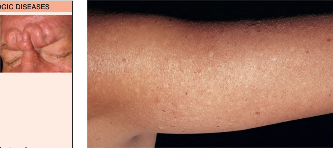
Fig. 46.5 Localized lichen myxedematosus – discrete papular type. Persistent whitish papules on the arms.
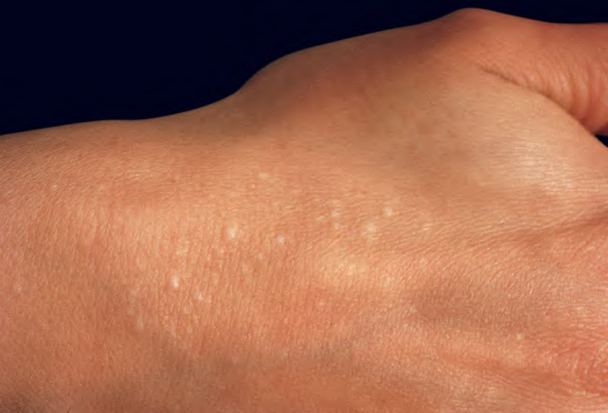
Fig. 46.6 Acral persistent papular mucinosis. Skin-colored papules on the dorsal aspect of the hands.
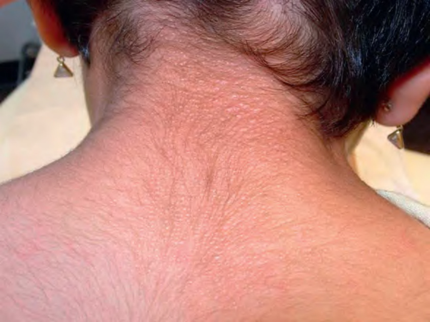
Fig. 46.7 Cutaneous mucinosis of infancy. Skin-colored papules on the back of the neck in a young girl.
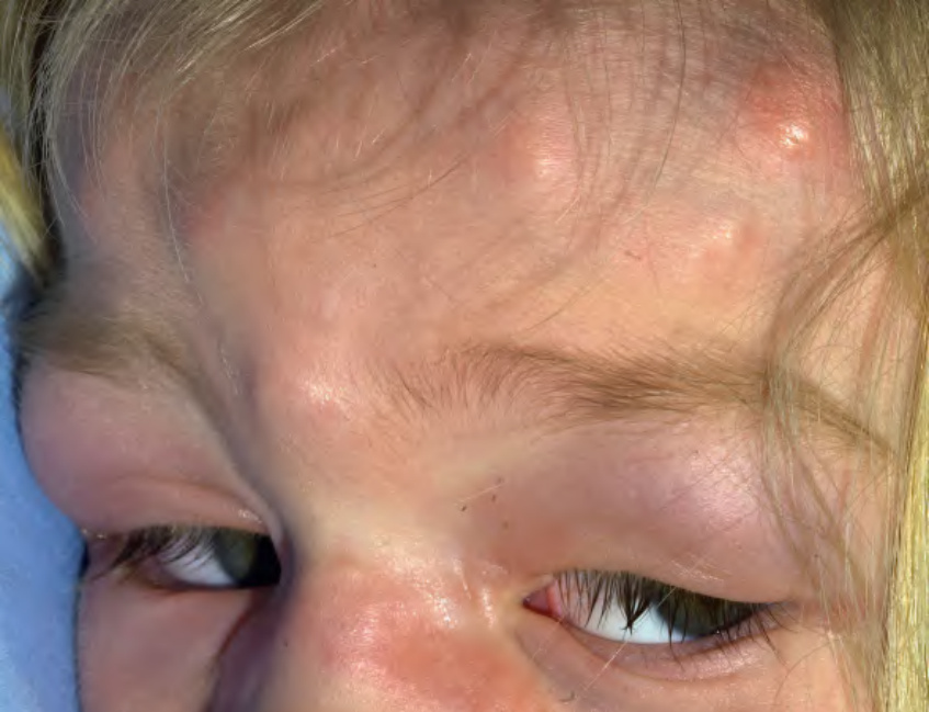
Fig. 46.8 Self-healing cutaneous mucinosis. Pink to skin-colored dermal and subcutaneous nodules as well as periorbital swelling. Courtesy Drs Hansgeorg Müller and Heinz Kofler.
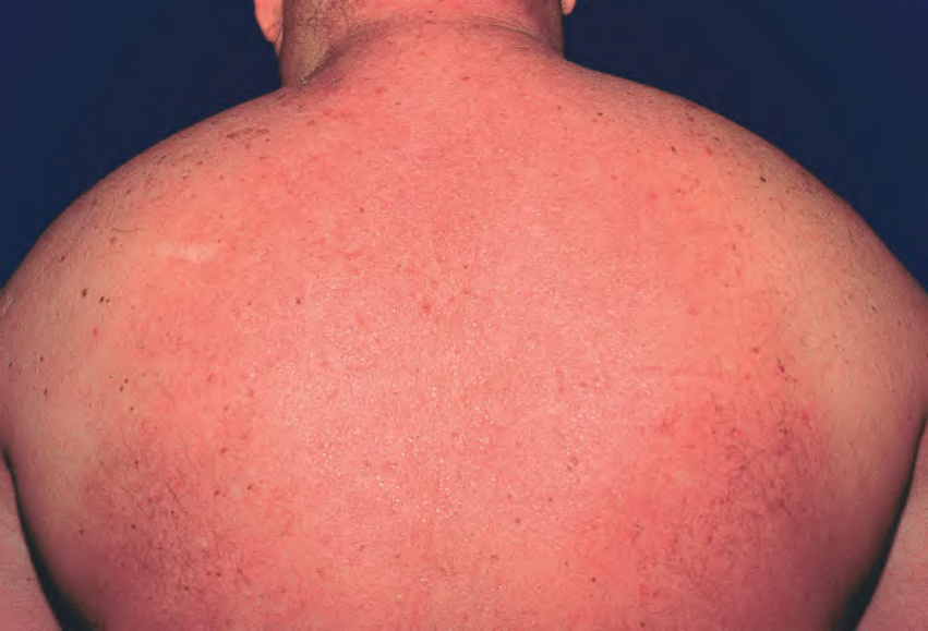
Fig. 46.9 Scleredema in association with diabetes mellitus. Diffuse induration of the upper part of the body with overlying erythema.
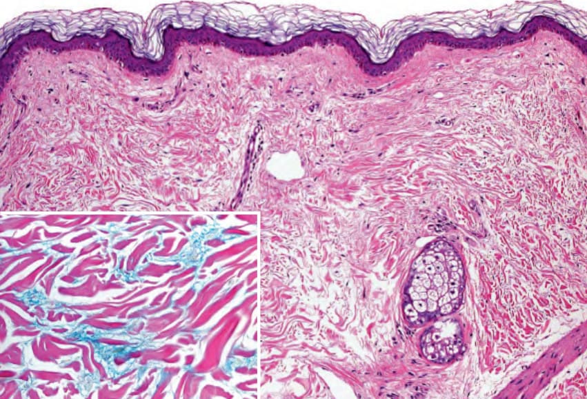
Fig. 46.10 Scleredema – histopathologic features. Slightly separated collagen bundles and normal dermal cellularity. Inset: Mucin deposits between collagen bundles. Courtesy Lorenzo Cerroni, MD.
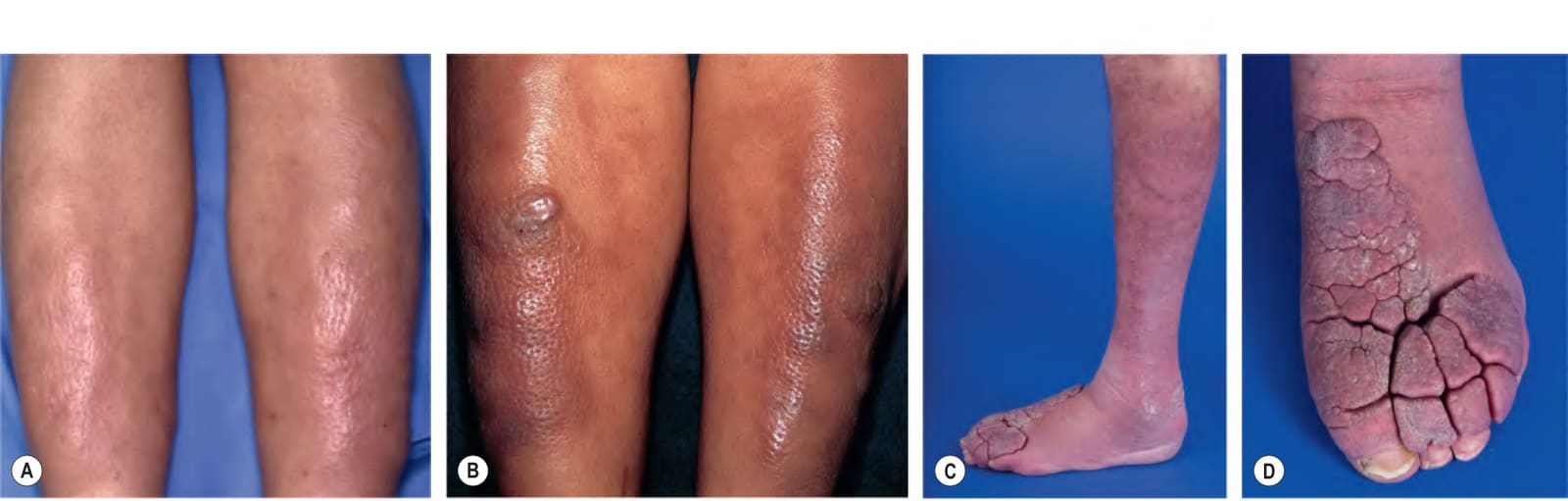
Fig. 46.11 Pretibial myxedema in patients with Graves disease.A Skin-colored to pink thick plaques on the shins. B Purple–brown nodules and plaques on the shins of a patient with a darker skin phototype. A peau d’orange appearance is present in both patients. C, D Induration and violaceous erythema of the legs with disfiguring soft tissue overgrowth of the dorsal feet and toes. C, D, Courtesy Edward Cowen, MD.
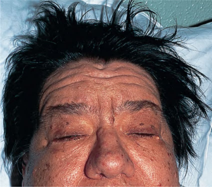
Fig. 46.12 Generalized myxedema in an adult with hypothyroidism. The face is puffy with a dull expression and the hair is dry.
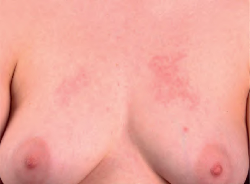
Fig. 46.13 Reticular erythematous mucinosis. Grouped pink papules and plaques on the chest. No surface changes are present and there is a reticulated configuration. Courtesy Lorenzo Cerroni, MD.
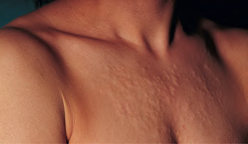
Fig. 46.14 Papulonodular mucinosis in a patient with lupus erythematosus.
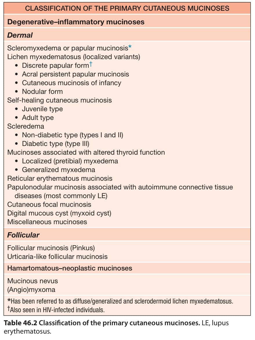
Table 46.2 Classification of the primary cutaneous mucinoses. LE, lupus erythematosus.
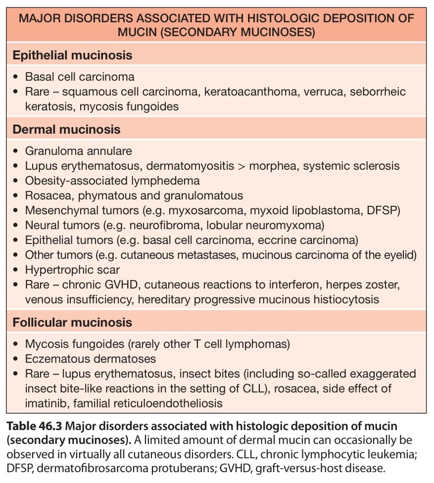
Table 46.3 Major disorders associated with histologic deposition of mucin
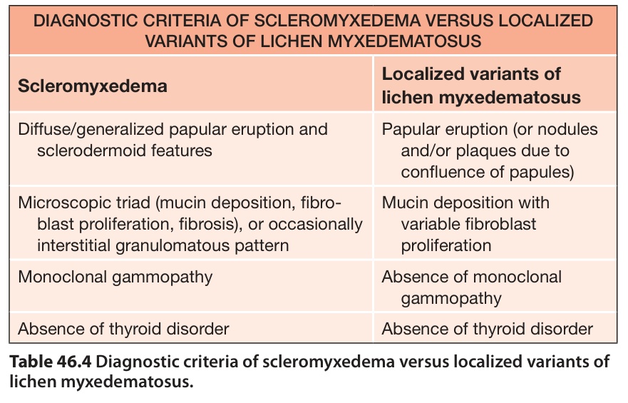
Table 46.4 Diagnostic criteria of scleromyxedema versus localized variants of lichen myxedematosus.
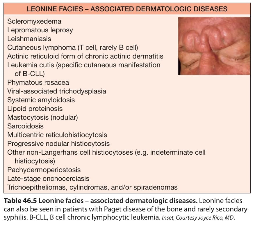
Table 46.5 Leonine facies – associated dermatologic diseases. Leonine facies can also be seen in patients with Paget disease of the bone and rarely secondary syphilis. B-CLL, B cell chronic lymphocytic leukemia. Inset, Courtesy Joyce Rico, MD.