PRIMARY HAMARTOMATOUS–NEOPLASTIC MUCINOSES
Although stromal mucin deposition may be found in a variety of cutaneous tumors as an additional finding (e.g. basal cell carcinomas, neurofibromas), only in mucinous nevus and (angio)myxoma is the mucin deposition a distinctive feature.
Mucinous Nevus
A mucinous nevus (nevus mucinosus) is a benign hamartoma that can be congenital or acquired. It consists of a plaque that usually has a unilateral linear nevoid pattern. Histologically, a diffuse deposit of mucin is seen in the upper dermis, and collagen and elastic fibers are absent within the mucinous area. The epidermis can be normal or it may be acanthotic with elongation of the rete ridges and hyperkeratosis, as in an epidermal nevus. The latter set of findings points to a combined hamartoma, in which features of an epidermal nevus are associated with those of a connective nevus of the proteoglycan type. The cellular component of a mucinous nevus consists of CD34+ fibroblasts and scarce factor XIIIa-positive dendritic cells.
A Pink plaque containing multiple follicular papules in a young boy. B Skin-colored papules on the cheek of a child. C Grouped follicular papules on the leg of an older adult in association with erythematous plaques. Note the associated alopecia. The hemorrhagic crust is the site of a previous biopsy. D Histology of follicular mucinosis (patient in A). Mucin deposition (wispy) within the follicular epithelia. A, D, Courtesy Lorenzo Cerroni, MD; B, Courtesy Julie V. Schaffer, MD.
Superficial (Angio)Myxoma
Superficial (angio)myxoma, also known as cutaneous myxoma, is an acquired benign neoplasm that is usually nodular and measures 1–5 cm in diameter. It favors the trunk, head and neck, and genital region; rarely, cutaneous myxomas have an acral location. These tumors can be solitary, without any systemic abnormalities, or multiple. When multiple and involving the external ears, eyelids, or nipples, they may be a manifestation of Carney complex (cutaneous myxomas, cardiac myxomas, numerous lentigines, multiple blue nevi, endocrinopathies).
Histologically, a cutaneous myxoma is a lobulated, well-defined lesion characterized by a mucinous matrix within the dermis and the subcutis, with variably shaped fibroblasts, mast cells, and a few collagen and reticulin fibers. Bizarre multinucleated cells and regular mitotic figures can be seen. The stromal cells are positive for vimentin and smooth muscle α-actin (SMA), with variable positivity for CD34, S-100, factor XIIIa, and CD68.
Myxomas must be distinguished from cutaneous focal mucinosis, a reactive lesion which is usually <1 cm and ill-defined histologically; the latter does not have an epithelial component and the mucin deposits are in the dermis but not the subcutis. This differentiation is important as
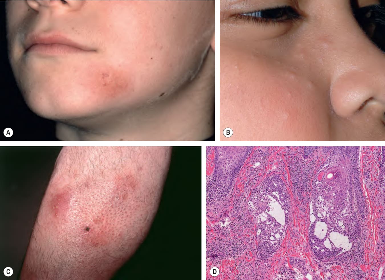
Fig. 46.15 Follicular mucinosis.