INTRODUCTION
Amyloidosis is not a single disease; rather, the term is used to refer to several diseases that share the common feature of abnormal extracellular deposition of amyloid, a fibrillar proteinaceous material, within tissues. Deposits of amyloid can be seen in a range of clinical disorders, from plasma cell dyscrasias and Alzheimer disease to familial polyneuropathies and primary cutaneous lichen amyloidosis.
Amyloid itself is not a single chemically distinct substance, and several types of amyloid have been described. However, regardless of the source, pathogenetic mechanisms, or underlying disease, amyloid material shares certain common tinctorial and physico-chemical properties, e.g. the cross-β-pleated sheet configuration.
The two clinical settings in which dermatologists are likely to encounter amyloidoses are: (1) the more common primary cutaneous forms of amyloidosis; and (2) the less common systemic amyloidosis with cutaneous manifestations (Fig. 47.1).
History
Virchow, in 1854, introduced the term “amyloid”. He believed that the substance resembled starch or cellulose because, like starch, it turned blue when stained with iodine followed by dilute sulfuric acid. In 1928, Gutmann first described a patient with clinical features of lichen amyloidosis, while Freudenthal, in 1930, introduced the term “lichen amyloidosus”.
Epidemiology
The precise epidemiology of systemic amyloidosis is not known, as the disease is often underdiagnosed or misdiagnosed. In the US, the incidence of primary systemic amyloidosis is estimated to be ~1300 to 3200 new cases per year; an increase in detection will likely accompany more widespread use of the assay for serum free light chains. Since 1980, there has been a marked decline in the incidence of symptomatic rheumatoid arthritis-associated secondary systemic amyloidosis due to better control of the inflammatory response.
Richard W. Groves Amyloidosis 47
Primary cutaneous amyloidosis is commonly observed in Southeast Asian countries, including Singapore, Taiwan, and Thailand. Lichen amyloidosis in particular appears to be more prevalent in those of Chinese descent. Macular amyloidosis is frequently seen in Central and South American countries, especially those close to the equator. In general, macular and lichen amyloidosis occur more often in individuals with skin phototypes III and IV.
Classification
Amyloidosis can be classified clinically into systemic (generalized) forms, with involvement of several organ systems, and organ-limited (localized) forms, in which deposits are limited to a single organ such as the skin (Table 47.1). In the localized forms, amyloid deposition occurs at or near the site of synthesis, while in the systemic forms the precursors are secreted into the circulation, followed by amyloid deposition at distant sites. Amyloidosis can also be classified according to its constituent precursor protein (Table 47.2; see next section).
Pathogenesis
The major component of amyloid is the fibril protein; the minor components are amyloid P component, glycosaminoglycans, and apoE lipoprotein. Amyloid P is a glycoprotein derived from serum amyloid P protein (SAP) and it has a specific calcium-dependent binding affinity for amyloid. Over 40 distinct forms of amyloid fibril proteins and their precursors have been identified9a, including: AL (amyloid light chains), containing immunoglobulin light chains; AA (amyloid-associated), composed of an acute phase protein synthesized by the liver; Aβ amyloid, found in cerebral lesions of Alzheimer disease; and ATTR (transthyretin-associated), found in some forms of familial amyloidosis (mutated) and senile systemic amyloidosis (wild-type). Each condition is associated with a specific precursor protein (see Table 47.2); these amyloid precursors are initially soluble proteins that undergo changes leading to aggregation, polymerization, fibril formation, and, finally, extracellular tissue deposition as insoluble amyloid.
The process by which this transformation takes place differs amongst the various types of amyloidoses. In primary systemic amyloidosis, substitution of amino acids at specific positions within the variable region of the immunoglobulin light chain potentially destabilizes these chains thereby increasing the likelihood of their conversion to amyloid fibrils. Similarly, mutations in the transthyretin gene have been shown to alter the stability of the transthyretin protein and increase its baseline mild amyloidogenicity. The accumulation of these relatively inert amyloid fibrils within vital organs leads to functional impairment.
The precise pathogenesis of primary cutaneous amyloidosis is not yet fully understood. Prolonged friction, genetic predisposition (e.g. OSMR, IL31RA), and environmental factors have all been implicated as possible etiologies. The precursor protein involved has not been fully characterized; however, for the macular and lichenoid variants of primary cutaneous amyloidosis it is thought to be keratinocyte-derived. This has been supported by ultrastructural studies demonstrating transitional forms between viable keratinocytes and amyloid, as well as by positive reactions with monoclonal antibodies directed against basal layer keratins. The fibrillar theory proposes that keratinocyte tonofilaments undergo degeneration and pass into the dermis, where they are presumably modified by histiocytes and fibroblasts into amyloid material. An alternative theory suggests that the material is produced at the epidermo-dermal interface, with precursor proteins being secreted by basal keratinocytes. This hypothesis has been supported by ultrastructural findings and demonstration of basement membrane antigens such as type IV collagen and laminin within amyloid deposits. Although these cutaneous amyloid deposits stain positively with anti-human antibodies directed against IgG, IgM and IgA, this staining is thought to be the result of nonspecific immunoglobulin absorption as opposed to an immunoglobulin being the putative precursor protein. Apolipoprotein E4, galectin-7, and actin have also been shown to be associated with primary cutaneous amyloid deposits and may be synthesized locally by keratinocytes. Small fiber neuropathy has been observed in primary cutaneous amyloidosis and may be related to the associated pruritus.
In nodular amyloidosis there is no specific staining with antikeratin antibodies, but rather the amyloid deposits are composed of immunoglobulin light chains, suggesting plasma cell derivation akin to the cutaneous lesions of primary systemic amyloidosis. Thus, its origin is very different from that of either macular or lichen amyloidosis. Presumably, there is local cutaneous production of light chains in nodular amyloidosis, whereas in primary systemic amyloidosis with skin involvement, the light chains are derived from the systemic circulation.
Amyloid Properties
In H&E-stained sections, amyloid appears as amorphous, eosinophilic fissured masses (Figs. 47.2, 47.3A & Table 47.3). With a Congo red stain, amyloid has an orange–red color on routine light microscopy, whereas under polarized light it exhibits green birefringence (Fig. 47.3B). Other special stains that can be used to detect amyloid deposits include crystal violet, methyl violet, periodic acid Schiff (PAS), Sirius red, pagoda red, Dylon stain, and Thioflavin T (Fig. 47.3C). AA (but not AL) amyloid loses its affinity for Congo red after exposure to potassium permanganate.
By electron microscopy, amyloid appears as 7 to 10 nm wide, non-branching, non-anastomosing fibrils (see Fig. 47.2). X-ray crystallography and infrared spectroscopy reveal a characteristic cross-β- pleated sheet conformation. These findings are identical in all types of amyloid, regardless of clinical setting or chemical composition. The cross-β-pleated sheet structure of amyloid is believed to be responsible for its staining and birefringence with Congo red stain.
Immunohistochemical stains utilizing antibodies directed against the amyloid fibril proteins (e.g. immunoglobulin γ light chain,
transthyretin, keratin, AA protein) can help to differentiate between the types of amyloid (Fig. 47.3D). However, these immunohistochemical stains have limitations. For precise protein identification, including AL, ATTR and Alect2, liquid chromatography–tandem mass spectrometry (LC-MS/MS) is performed on formalin-fixed, paraffin-embedded sections or subcutaneous fat fine needle aspirates (see Table 47.2). Laser capture technology can be used to obtain the sample for testing.
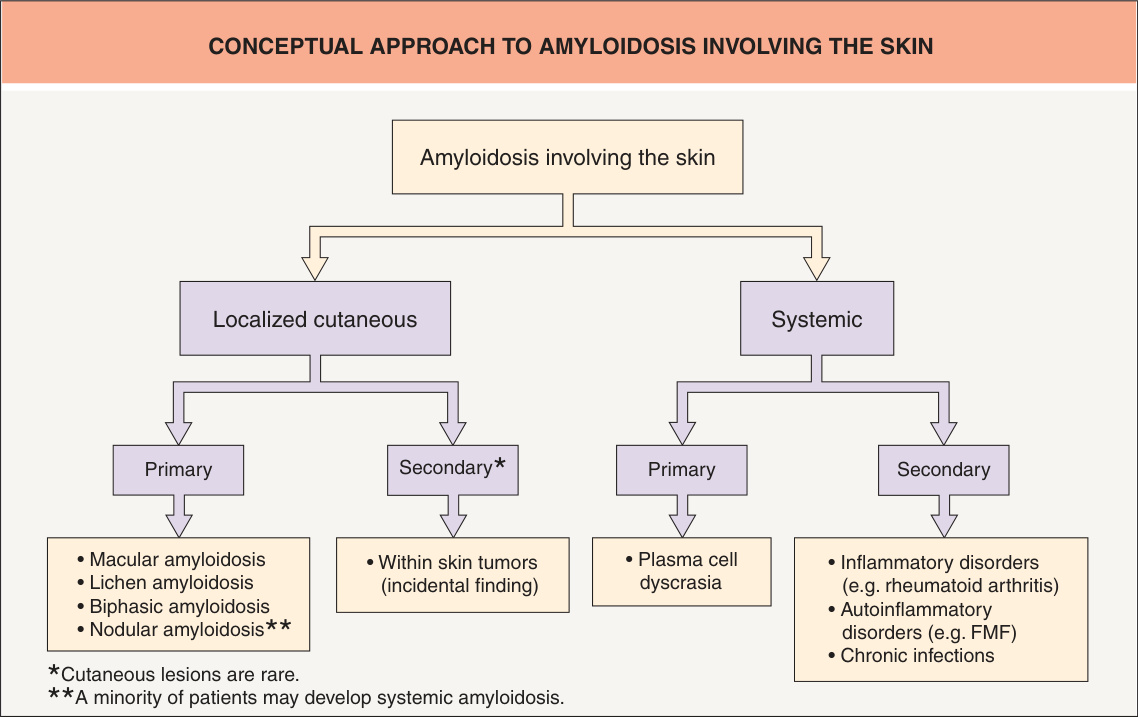
Fig. 47.1 Conceptual approach to amyloidosis involving the skin. FMF, familial Mediterranean fever.
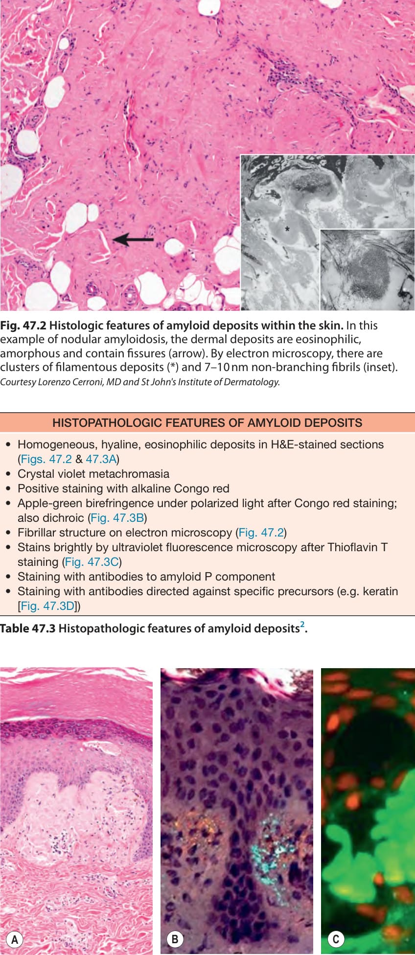
Fig. 47.2 Histologic features of amyloid deposits within the skin. In this example of nodular amyloidosis, the dermal deposits are eosinophilic, amorphous and contain fissures (arrow). By electron microscopy, there are clusters of filamentous deposits (*) and 7–10 nm non-branching fibrils (inset). Courtesy Lorenzo Cerroni, MD and St John’s Institute of Dermatology.
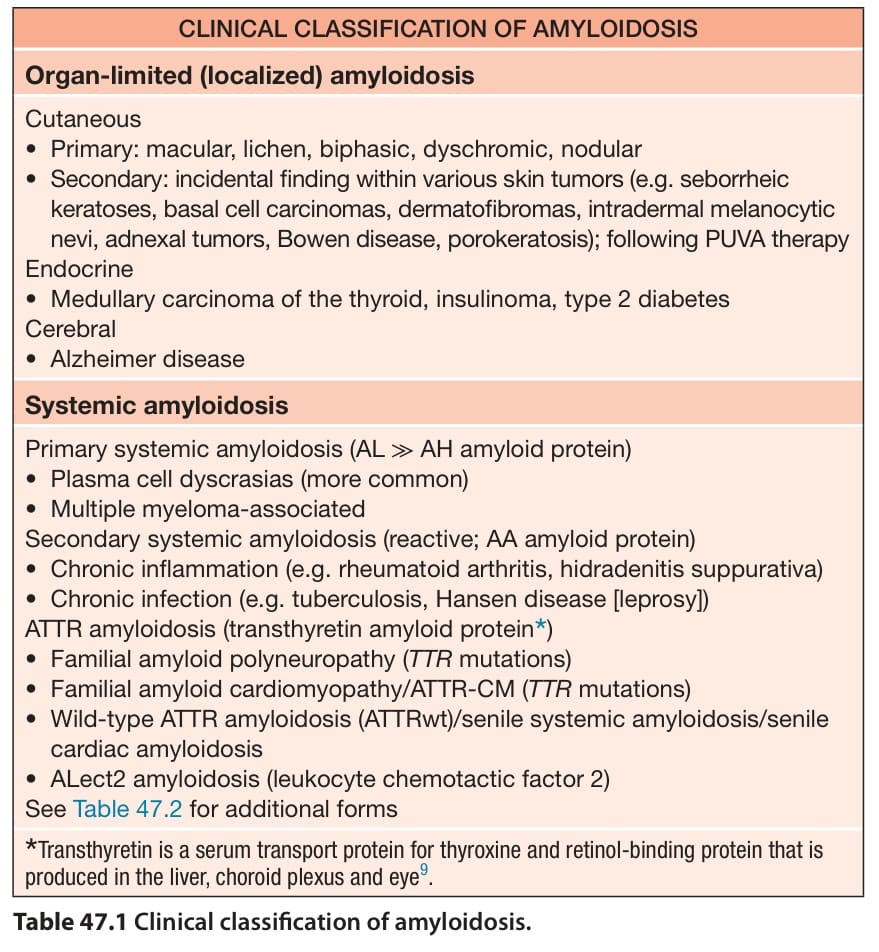
Table 47.1 Clinical classification of amyloidosis.
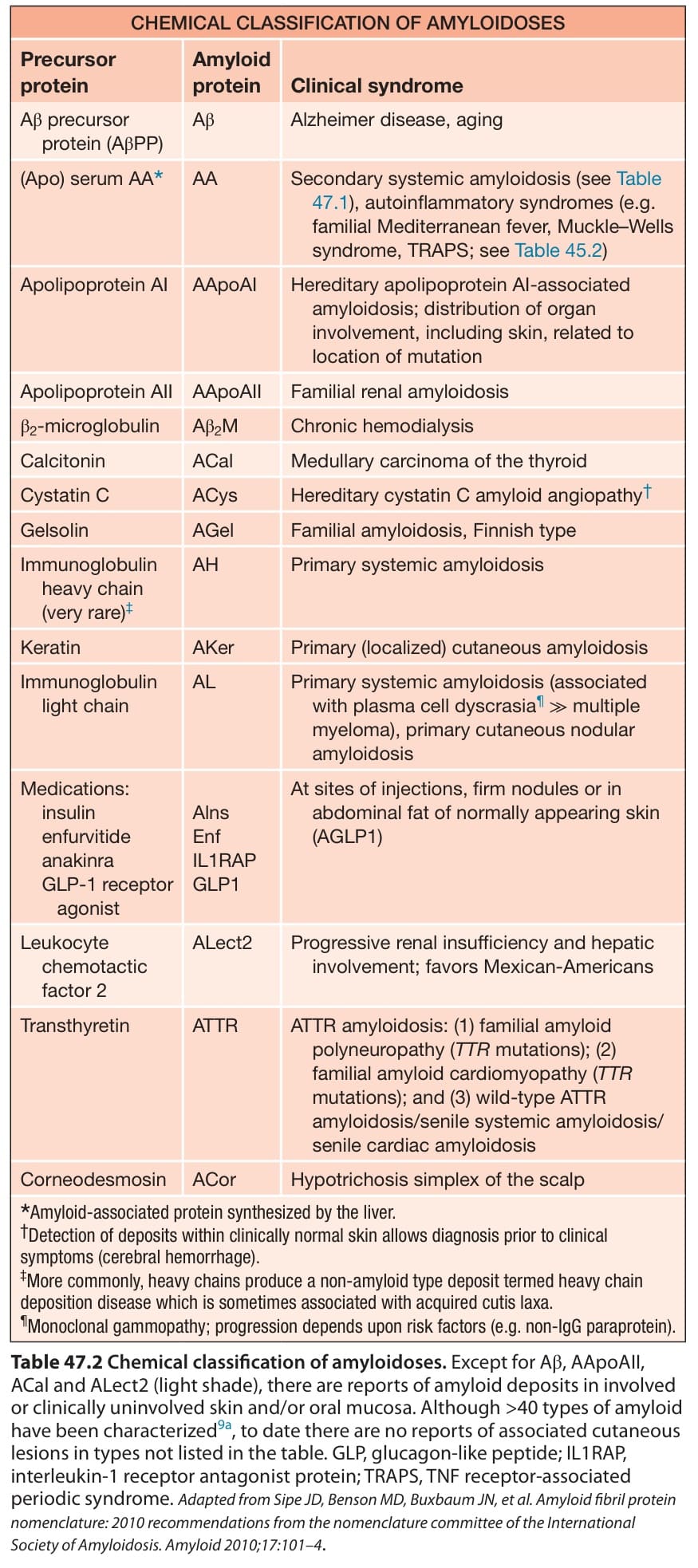
Table 47.2 Chemical classification of amyloidoses. Except for Aβ, AApoAII, ACal and ALect2 (light shade), there are reports of amyloid deposits in involved or clinically uninvolved skin and/or oral mucosa. Although >40 types of amyloid have been characterized9a, to date there are no reports of associated cutaneous lesions in types not listed in the table. GLP, glucagon-like peptide; IL1RAP, interleukin-1 receptor antagonist protein; TRAPS, TNF receptor-associated periodic syndrome. Adapted from Sipe JD, Benson MD, Buxbaum JN, et al. Amyloid fibril protein nomenclature: 2010 recommendations from the nomenclature committee of the International Society of Amyloidosis. Amyloid 2010;17:101–4.
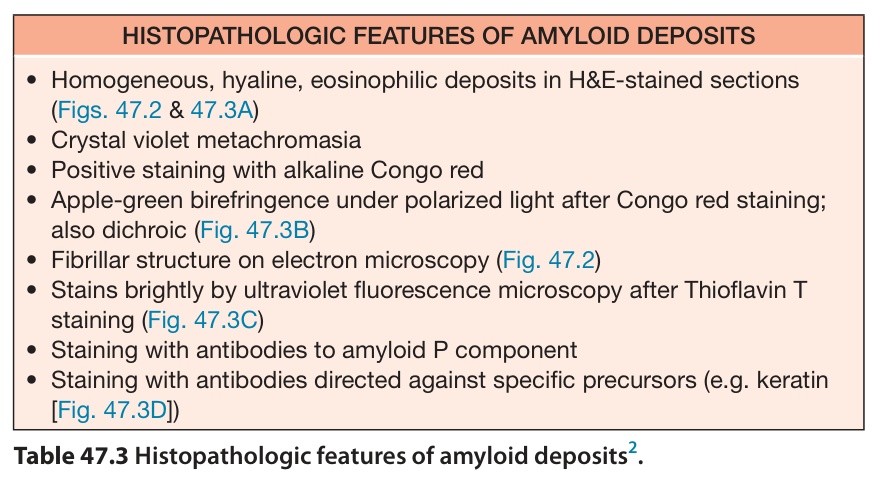
Table 47.3 Histopathologic features of amyloid deposits.