PRIMARY LOCALIZED CUTANEOUS AMYLOIDOSIS
Primary cutaneous amyloidosis is associated with the deposition of amyloid in the skin but not internal organs (see Table 47.1). The most common variants are macular amyloidosis, lichen amyloidosis, and biphasic amyloidosis.
Clinical Features – Macular, Lichenoid, Biphasic, and Dyschromic Amyloidosis
Although primary cutaneous amyloidosis is classically divided into macular (macular amyloidosis), papular (lichen amyloidosis), and nodular (nodular amyloidosis) forms, the first two entities actually represent the ends of a clinical spectrum. In some patients and even some lesions both macular and papular forms can be present, termed “biphasic amyloidosis”. Primary cutaneous amyloidosis can have a profound effect on quality of life due to the associated pruritus and visual appearance of the skin lesions.
Macular amyloidosis is often pruritic but it may be asymptomatic. The lesions of macular amyloidosis are hyperpigmented, either in a confluent or rippled pattern (Fig. 47.4A). The latter can be best appreciated by stretching the skin. The most common site of involvement is the upper back, especially over the scapular area, followed by the extensor surfaces of the extremities. A linear or even nevoid pattern may be seen (Fig. 47.4B). Macular amyloidosis usually presents in early adulthood, and women may be affected more often than men. In biphasic amyloidosis, fine papular lesions are superimposed upon a background of hyperpigmentation.
The term “friction amyloidosis” has been used to describe a subgroup of patients in whom local friction from nylon brushes, towels, and other rough materials contributes to the production of macular and lichenoid lesions. There is also significant clinical overlap between macular amyloidosis and the pigmented form of notalgia paresthetica, where pruritus of the scapular areas leads to rubbing and scratching (see Ch. 6).
Lichen amyloidosis is the most common form of primary cutaneous amyloidosis and usually presents as persistent, pruritic plaques on the shins or other extensor surfaces, e.g. anterior thighs or forearms (Fig. 47.5). Initial lesions are discrete, firm, scaly, skin-colored or hyperpigmented papules, which later coalesce into plaques that often have a rippled or ridged pattern. At the onset, lesions are usually unilateral, but over time a bilateral symmetric distribution pattern can develop.
Primary cutaneous amyloidosis manifesting as pigmentation and lichenification of the anal and sacral regions has been described as an ano-sacral variant, but these patients often have lichen amyloidosis
or biphasic amyloidosis elsewhere. Bullous lesions have also been described in the setting of lichen amyloidosis, but they are more commonly associated with systemic amyloidosis.
Macular and/or lichen amyloidosis has also been described in association with autoimmune connective tissue disorders (e.g. systemic sclerosis [Fig. 47.6], systemic lupus erythematosus, dermatomyositis), and primary biliary cirrhosis. One patient was observed to have sparing of lichen amyloidosis lesions in areas with higher
Note the numerous papules, some of which are in a grid-like array.
cutaneous temperatures (such as along the course of large superficial veins), suggesting that amyloid fibril formation may be a temperaturedependent process in vivo.
Familial primary cutaneous amyloidosis syndromes
A number of genetic disorders have been identified that predispose to the development of primary localized cutaneous amyloidosis (PLCA).
Several families with primary localized cutaneous amyloidosis have been shown to have pathogenic variants in the genes that encode either the β subunit of the oncostatin M receptor (OSMR; PLCA1) or the α subunit of the interleukin (IL)-31 receptor (IL-31RA; PLCA2). However, the protein product of OSMR serves as the β subunit of two receptors – not just OSMR but also IL-31R; this subunit then combines with IL-31RA to form the IL-31 receptor. Of note, IL-31 has been shown to be of importance in pruritic skin conditions (e.g. dermatitis, prurigo nodularis), and nemolizumab, an anti-IL-31RA antibody, reduced pruritus in patients with atopic dermatitis. In these families with primary localized cutaneous amyloidosis, IL-31 signalling may also be influenced by monocyte chemoattractant protein 1 (MCP-1).
Dyschromic amyloidosis, also referred to as primary localized cutaneous amyloidosis 3 (PLCA3), is an unusual variant in which a guttate leukoderma is superimposed upon a background of hyperpigmentation and admixed with characteristic lesions of macular and lichen amyloidosis (see Fig. 67.27). Homozygous and compound heterozygous mutations in GPNMB, which encodes glycoprotein nonmetastatic melanoma protein b, have been detected in patients with dyschromic amyloidosis.
Sipple syndrome is an autosomal dominant disorder with the triad of medullary carcinoma of the thyroid, pheochromocytomas, and hyperparathyroidism (see Ch. 63). Increased serum levels of calcitonin are a reflection of hyperplasia or carcinoma of the parafollicular calcitoninproducing C cells of the thyroid. Multiple endocrine neoplasia (MEN) type 2 A is associated with pruritic areas of hyperpigmentation on the upper back. The lesions have been described as notalgia paresthetica, macular amyloidosis, and lichen amyloidosis. The unusual aspect is the early age of onset, often before the age of 10 years. Recognition of this skin sign of systemic disease can lead to prophylactic thyroidectomy once the diagnosis is established. A study of 10 families with MEN 2 A found an incidence of lichen amyloidosis of 36%; moreover, all those with cutaneous amyloidosis had a specific pathogenic variant in codon 634 of the RET proto-oncogene.
Partington and colleagues described a family with an X-linked disease in which adult women had linear streaks of hyperpigmentation and male patients manifested reticulated, mottled brown skin pigmentation, which on biopsy demonstrated dermal deposits of amyloid (in adults but not children). In women, involvement was limited to the skin, but in male patients, recurrent respiratory infections, corneal dystrophy, and photophobia were seen (see Ch. 67). As in macular amyloidosis, the amyloid deposits are keratin-positive and absent in other tissues.
Pathogenic variants in POLA1 on chromosome Xp22, which encodes the catalytic subunit of DNA polymerase α1, have been detected in several families. This was associated with an activation of proinflammatory genes (e.g. NF-κB) and an increased type I interferon profile.
Other associated disorders
Primary cutaneous amyloidosis has also been reported in association with pachyonychia congenita, dyskeratosis congenita, and familial palmoplantar keratoderma. At sites of injection of several medications, e.g. insulin, cutaneous amyloid deposits can develop (see Table 47.2)
Clinical Features – Nodular Amyloidosis
Nodular amyloidosis is rare, and presents as single or multiple waxy nodules or infiltrated plaques, most commonly on the trunk or extremities (Fig. 47.7A,B). Immunoglobulin γ light chains as well as β-microglobulin have been demonstrated within the deposits, and both are thought to be produced by plasma cells in the vicinity (Fig. 47.7D). In several patients with nodular amyloidosis, the cutaneous plasma cells had monoclonal restriction of their immunoglobulin light chains as determined by immunohistochemistry or in situ hybridization; examination of the bone marrow in one patient showed no evidence of a clonal plasma cell proliferation. Of note, there may be an association between Sjögren syndrome and nodular amyloidosis of the skin or lung. It has been suggested that at least some cases of cutaneous nodular amyloidosis could represent a variant of primary cutaneous marginal zone lympho proliferative disorder (lymphoma) with unusual histopathologic features, but a similar excellent prognosis (see Ch. 119).
There are reports of the nodular variant progressing to systemic involvement, hence longitudinal evaluation is appropriate. However, recent observations suggest that this risk is ~7%, much less than the previously quoted 50%. Although 40% of patients in this long-term study had a paraproteinemia at presentation, the gammopathy remained static during the follow-up period and progression to systemic involvement was
Firm waxy plaques and nodules on the back and chin. The color can vary from skin-colored to pink–yellow to violaceous. C Histologically, there are extensive amyloid deposits within the dermis and around blood vessels. D An infiltrate of plasma cells can be seen in association with the amyloid deposits. D, Courtesy Lorenzo Cerroni, MD.
observed in only 1 of 15 patients, 23 years after the initial presentation. A similar study of 16 patients with nodular amyloidosis found a near equal sex distribution, with a tendency for an acral distribution. There was one patient who subsequently died from systemic amyloidosis.
Pathology
In macular and lichen amyloidosis, amyloid deposits are restricted to the upper dermis, particularly the papillary dermis (Fig. 47.4C). In lichen amyloidosis, the deposits may expand the papillae and displace the elongated rete ridges laterally. The overlying epidermis often has features of chronic rubbing, e.g. acanthosis, compact orthohyperkeratosis (see Fig. 47.3A). Melanophages and a sparse perivascular lymphohistiocytic infiltrate can be seen in both forms. Although antikeratin antibodies (e.g. MNF 116) may be useful in confirming the keratin origin of the amyloid deposits in these two forms of primary cutaneous amyloidosis, they are seldom required (see Fig. 47.3D).
In contrast, in the nodular variant, the dermis, subcutis, and blood vessel walls are diffusely infiltrated with amyloid (Fig. 47.7C). A perivascular infiltrate of plasma cells is nearly universally present, but in most cases is inconspicuous (see Fig. 47.7D). Immunostaining for light chain deposition can aid in the diagnosis of nodular amyloidosis. As noted previously, these plasma cells are usually monoclonal, and it has been recently suggested that a number of cases of primary cutaneous nodular amyloidosis (“nodular amyloidoma”) may represent a peculiar variant of cutaneous marginal zone lymphoproliferative disorder (lymphoma).
Differential Diagnosis
The diagnosis of primary cutaneous amyloidosis is based on clinical morphology and histologic demonstration of amyloid deposits within the skin (see Table 47.3).
There is significant overlap in the appearance of macular amyloidosis and notalgia paresthetica. The latter is also seen on the upper back and can have rippled hyperpigmentation. Histologically, the early stages of notalgia paresthetica contain scattered necrotic keratinocytes and a few melanophages but no amyloid deposits. However, later on, even within the same lesion, deposits can appear. When macular amyloidosis is more diffuse or confluent, it must be distinguished from postinflammatory hyperpigmentation. Other entities in the differential diagnosis may include pityriasis versicolor, atrophic lichen planus (LP), erythema dyschromicum perstans (ashy dermatosis), and drug-induced pigmentation.
The primary differential diagnosis for lichen amyloidosis includes lichen simplex chronicus (LSC) and hypertrophic LP. Both are characterized by chronic pruritic plaques, often on the shins and, histologically, exhibit hyperkeratosis, acanthosis, and a mild to moderate lymphohistiocytic inflammatory infiltrate but no amyloid deposits. In LSC, lichenification is often prominent, while hypertrophic LP lesions may have a violaceous hue and, on histologic examination, a lichenoid infiltrate with vacuolar degeneration. Other entities that may be considered in the clinical differential diagnosis include papular mucinosis, pretibial myxedema, prurigo simplex/nodularis, pemphigoid nodularis, lichen ruber moniliformis, and epidermolysis bullosa pruriginosa.
Nodular amyloidosis may resemble the cutaneous lesions of primary systemic amyloidosis and the latter should be excluded. The clinical differential diagnosis also includes other nodular lesions (e.g. cutaneous lymphoid hyperplasia, sarcoidosis, colloid milium), all of which have rather distinctive histopathologic features and therefore can be easily differentiated.
Treatment
No treatment modalities described to date have proven to be curative or uniformly effective in patients with macular or lichen amyloidosis (Table 47.4). Treatment must be directed at breaking the itch–scratch– itch cycle usually present in these patients. Chronic friction, scratching, and rubbing can serve as possible precipitating or aggravating factors and patients should be advised of this. Potent topical corticosteroids are useful to some extent, especially in mild cases, and application under occlusion or in combination with a mild keratolytic agent such as salicylic acid (particularly in lichen amyloidosis) may have added benefit. The involved sites can be protected from scratching by applying occlusive dressings such as hydrocolloids or gauze wraps impregnated
with zinc oxide for a period of weeks to months, but recurrence rates may be high. Based upon case reports, topical calcineurin inhibitors may play an adjunctive role, and more recently, dupilumab was observed to be an effective treatment for lichen amyloidosis. Whether nemolizumab, the anti-IL-31RA antibody, will lead to improvement remains to be determined.
In a study that compared the efficacy of topical corticosteroids versus either UVB or topical PUVA phototherapy for the treatment of lichen amyloidosis, there was greater improvement in the pruritus and roughness scores on the sides treated with either form of phototherapy, with PUVA being marginally better in reducing pruritus. Dermabrasion was shown to be beneficial in lichen amyloidosis involving the limbs with the effects lasting at least five years: the epidermis and part of the papillary dermis along with some of the amyloid was removed, permitting re-epithelialization to occur from the adnexal structures. CO or erbium:YAG laser therapy may also be of benefit in some patients with macular and lichen amyloidosis.
Retinoids including acitretin and alitretinoin have been reported to improve pruritus and result in flattening of lesions, with some clearance of the associated hyperpigmentation. In a preliminary study, low-dose cyclophosphamide (50 mg daily) was effective in reducing pruritus and papules in lichen amyloidosis. However, the side effects of these systemic medications (see Chs. 126 & 130) must be weighed against the possible benefits.
Surgical excision, cryotherapy, electrodesiccation, and CO laser can be used to treat nodular amyloidosis, but local recurrences are common. Rarely, the local recurrence lacks amyloid deposits and has the histopathologic features of a conventional primary cutaneous marginal zone lymphoma. There are scattered reports of the use of cyclophosphamide (see above).
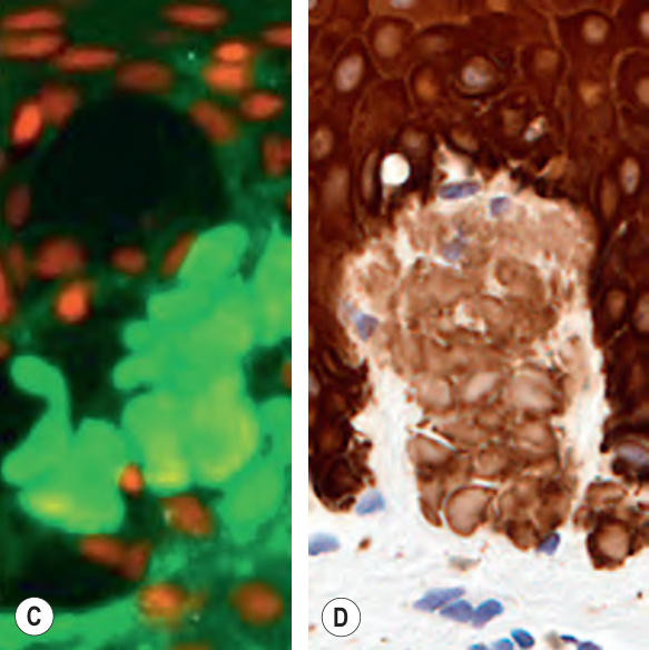
Fig. 47.3 Use of special stains to identify amyloid deposits within the papillary dermis in lichen amyloidosis.A In hematoxylin & eosin-stained sections, amyloid deposits, admixed with melanophages, are seen within the papillary dermis in addition to compact orthohyperkeratosis and acanthosis. B With Congo red stain, there is characteristic green birefringence with polarized light; the color can vary from yellow–green to blue–green. C With Thioflavin T stain, amyloid deposits stain brightly with ultraviolet fluorescence microscopy. D Positive immunohistochemical stain for keratin (MNF 116, broad-spectrum anti-keratin antibody). A, Courtesy Lorenzo Cerroni, MD; B,C, Courtesy St John’s Institute of Dermatology; D, Courtesy Jennifer McNiff, MD.
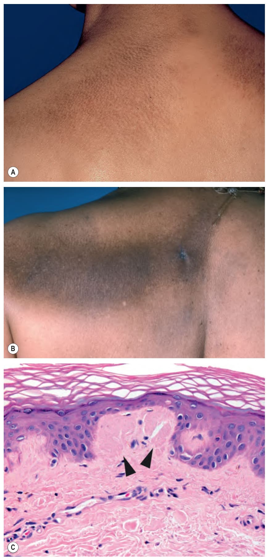
Fig. 47.4 Macular amyloidosis.A Characteristic rippled hyperpigmentation of macular amyloidosis. B Occasionally a linear accentuation is seen. Scattered papules can also be seen in both patients. C Histologically, deposits of amyloid within the dermal papillae (arrowheads) as well as melanophages within the papillary dermis; note the characteristic fissure in the amyloid deposit. These deposits may be subtle. C, Courtesy Lorenzo Cerroni, MD.
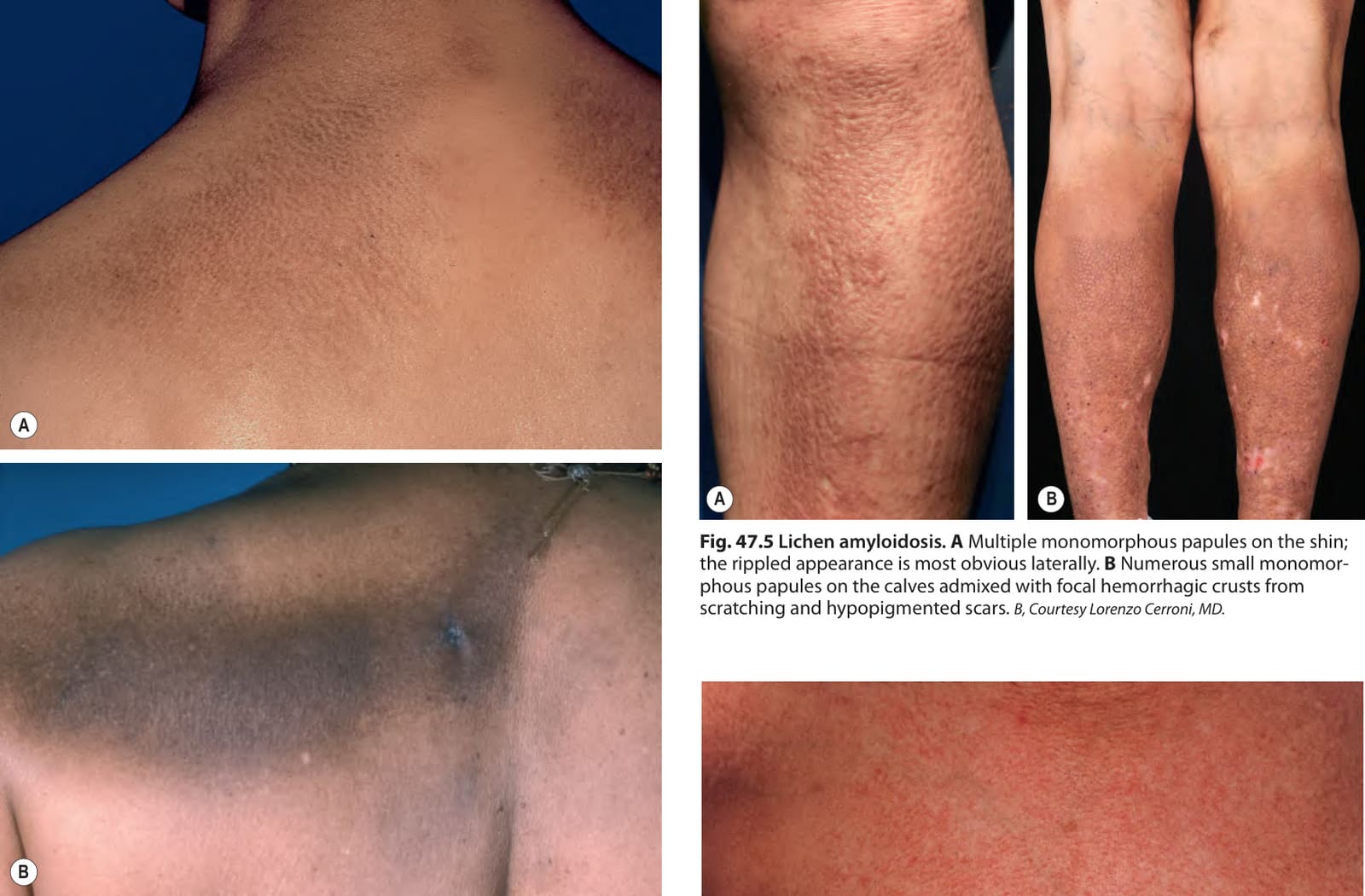
Fig. 47.5 Lichen amyloidosis.A Multiple monomorphous papules on the shin; the rippled appearance is most obvious laterally. B Numerous small monomorphous papules on the calves admixed with focal hemorrhagic crusts from scratching and hypopigmented scars. B, Courtesy Lorenzo Cerroni, MD.
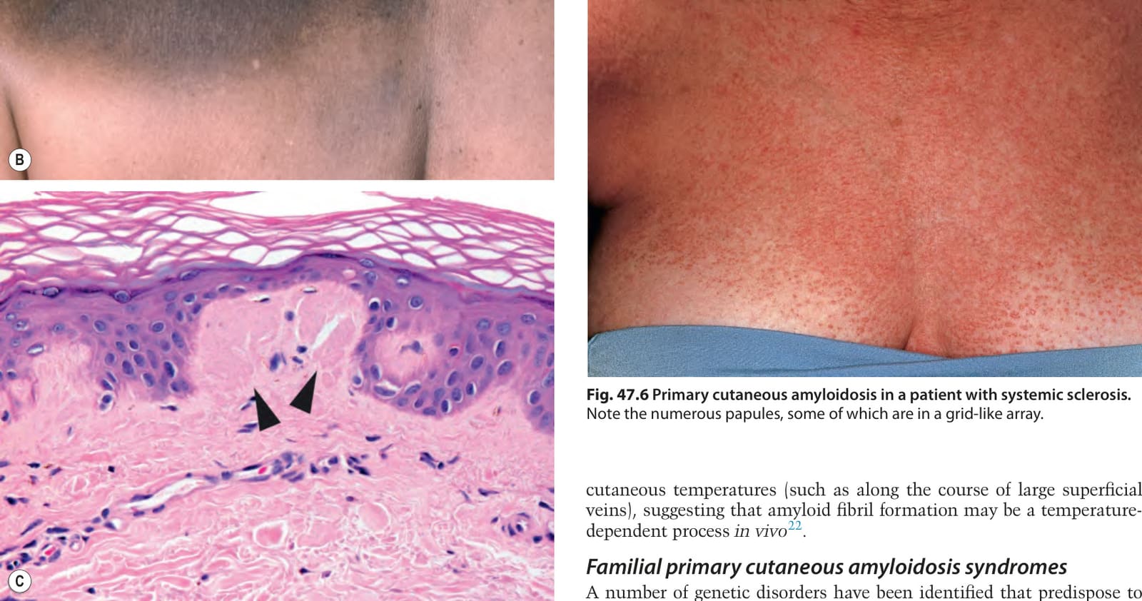
Fig. 47.6 Primary cutaneous amyloidosis in a patient with systemic sclerosis.
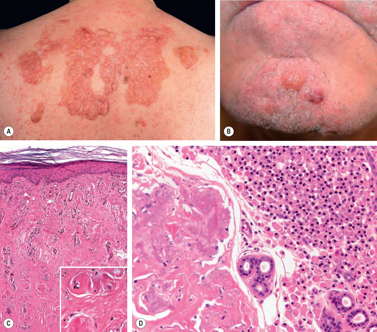
Fig. 47.7 Nodular amyloidosis.A, B
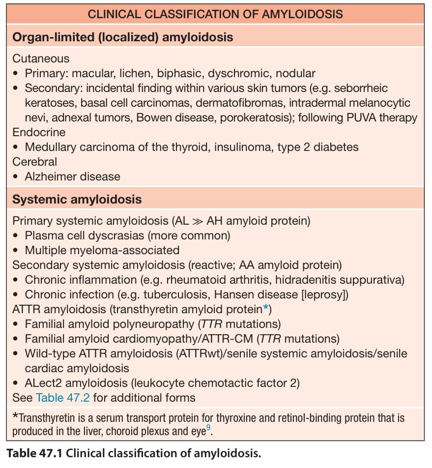
Table 47.1 Clinical classification of amyloidosis.
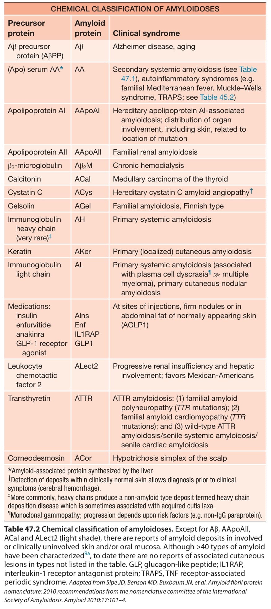
Table 47.2 Chemical classification of amyloidoses. Except for Aβ, AApoAII, ACal and ALect2 (light shade), there are reports of amyloid deposits in involved or clinically uninvolved skin and/or oral mucosa. Although >40 types of amyloid have been characterized9a, to date there are no reports of associated cutaneous lesions in types not listed in the table. GLP, glucagon-like peptide; IL1RAP, interleukin-1 receptor antagonist protein; TRAPS, TNF receptor-associated periodic syndrome. Adapted from Sipe JD, Benson MD, Buxbaum JN, et al. Amyloid fibril protein nomenclature: 2010 recommendations from the nomenclature committee of the International Society of Amyloidosis. Amyloid 2010;17:101–4.
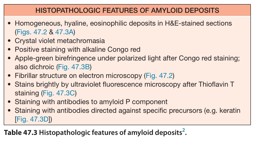
Table 47.3 Histopathologic features of amyloid deposits.
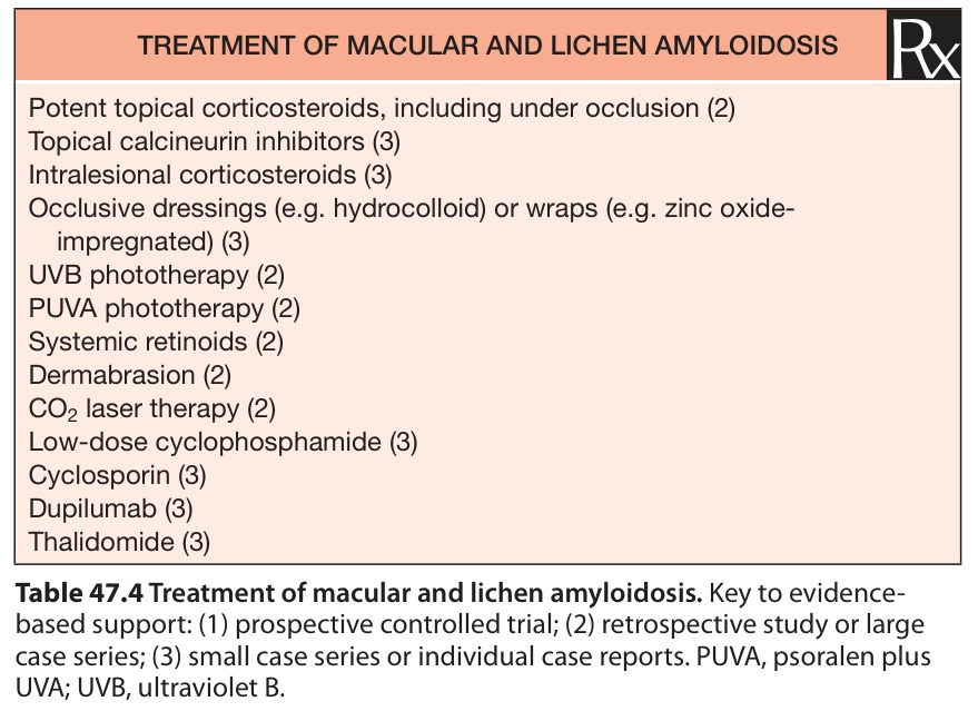
Table 47.4 Treatment of macular and lichen amyloidosis. Key to evidencebased support: (1) prospective controlled trial; (2) retrospective study or large case series; (3) small case series or individual case reports. PUVA, psoralen plus UVA; UVB, ultraviolet B.