SYSTEMIC AMYLOIDOSIS
Primary Systemic Amyloidosis
Multiple forms of systemic amyloidosis have been identified, but skin involvement is variable. The following discussion focuses on those in which the skin is a major target.
AL amyloidosis
AL amyloidosis is a life-threatening manifestation of an underlying plasma cell dyscrasia, although patients usually do not fulfill the criteria for multiple myeloma (e.g. hypercalcemia, osteolytic lesions; see Ch. 119). The fibrils are composed of AL protein, which consists of immunoglobulin light chains, usually λ-type (75%–80%).
AL amyloidosis is associated with a wide spectrum of organ involvement. The presenting symptoms are varied and nonspecific, such as fatigue, weight loss, paresthesias, dyspnea, and syncopal attacks due to orthostatic hypotension. As a result, there is often difficulty and delay in diagnosis.
In the oral cavity, amyloid deposits appear as rubbery swellings or an infiltration of the mucosa (Fig. 47.8A). An associated hemorrhagic component is frequently seen. The tongue can be uniformly enlarged and firm (Fig. 47.8B), or it may have hemorrhagic papules, plaques, and blisters on its surface; the latter may vary from translucent to yellow–brown in color. Xerostomia can result from infiltration of the salivary glands.
Following minor trauma, petechiae, purpura, and ecchymoses are often observed, especially on the eyelids and neck and in the axillae and anogenital region (due to amyloid infiltration of vessel walls; Fig. 47.9). Characteristically, periorbital purpura (the “raccoon eyes” sign) may be precipitated by coughing, the Valsalva maneuver, or proctoscopy for a rectal biopsy, as well as after pinching or rubbing the skin (pinch purpura). Even removal of adhesive tape can lead to purpura.
Clinically evident skin involvement occurs in ~25% of individuals with AL amyloidosis. Skin infiltration by amyloid presents as waxy, translucent, or purpuric papules (Fig. 47.8D,E & 47.10), nodules, and plaques that resemble nodular amyloidosis. A smooth, erythematous, waxy infiltration with scattered superimposed papules can appear on the palms and volar aspect of the fingertips (Fig. 47.8C). Smooth, skincolored papules, a few millimeters in diameter, may be seen on the face, neck, and scalp and in the anogenital region.
Less often, diffuse cutaneous infiltration can result in an infiltrated sclerodermoid appearance or cause the scalp skin to be thrown into folds resembling cutis verticis gyrata with associated alopecia. Bullous lesions, especially hemorrhagic blisters resembling those of porphyria cutanea tarda and epidermolysis bullosa acquisita, may be the initial sign of the disease. In addition, nail dystrophy can occur and may resemble lichen planus, with longitudinal ridging and thinning; histologically, amyloid deposits are found around blood vessels and in the dermis of the nail bed and matrix. Rarely, patients also have evidence of acquired acral cutis laxa. The presence of macroglossia together with carpal tunnel syndrome is a classic presentation and should trigger an investigation for amyloidosis.
Other signs and symptoms of AL amyloidosis depend upon the organs involved. Renal involvement manifests as proteinuria (albuminuria) which leads to hypoalbuminemia and edema, i.e. nephrotic syndrome. Cardiac involvement may eventually lead to a restrictive cardiomyopathy and congestive heart failure, but with a preserved ejection fraction. This results in dyspnea, hepatomegaly, and bilateral lower extremity as well as presacral edema. Autonomic and sensory neuropathies are common. Sensory involvement is usually bilateral and symmetrical, while autonomic involvement can result in postural hypotension, impotence, and disturbances in gastrointestinal motility (e.g. gastroparesis). Hepatomegaly due to amyloid infiltration or congestive heart failure may be present.
In up to 80%–90% of patients with AL amyloidosis, amyloid deposits can be demonstrated in random rectal mucosal biopsies or in abdominal subcutaneous fat aspirates; the latter is preferred because of the potential risk for bleeding with the former. Gingival or tongue biopsies may also be obtained to demonstrate the presence of amyloid, but in the absence of involvement clinically, they are less sensitive. In addition, bone marrow biopsies are examined for the presence of amyloid deposits. Based upon the finding that all of these amyloid deposits contain amyloid P (which is derived from serum amyloid protein [SAP]), SAP scintigraphy is also employed, primarily outside the US. It utilizes radioiodine-labeled SAP and is a sensitive, non-invasive method for locating amyloid deposits and monitoring disease. A more practical method for assessing response to therapy is measurement of serum free light chains.
The approach to a patient with suspected or confirmed AL amyloidosis is outlined in Fig. 47.11.
In AL amyloidosis, cutaneous lesions are characterized by amyloid deposits in the dermis and subcutis. Amyloid is also often found around sweat glands and within blood vessel walls (Fig. 47.12). Rarely, an infiltrate of amyloid-producing monoclonal plasma cells can be found adjacent to the amyloid deposits. An unusual pattern of amyloid deposition around elastic fibers, in which the fibers appear coated with a thick mantle of amyloid-staining material, is occasionally seen and has been termed amyloid elastosis. The latter is rarely observed in primary cutaneous amyloidosis.
The differential diagnosis of the waxy papules includes papular mucinosis, nodular amyloidosis, and lipoid proteinosis, and when the lesions are primarily on the face, they can be confused with adnexal tumors (see Ch. 111). Conditions in the sclerodermoid differential diagnosis (see Table 43.7) need to be considered when there is diffuse infiltration of the skin.
Without therapy, the prognosis for AL amyloidosis is poor: median survival is ~13 months, especially if there is cardiac involvement. Treatment strategies have paralleled those for multiple myeloma, including melphalan and systemic corticosteroids. The current FDA-approved treatment regimen for AL amyloidosis is the combination of cyclophosphamide, bortezomib, and dexamethasone (CyborD) plus daratumumab (anti-CD38 antibody). If the patient is younger and has minimal cardiac involvement, high-dose melphalan followed by autologous hematopoietic stem cell transplantation is a potentially curative therapeutic option. In one study with an 8-year follow-up period, a complete hematologic response was observed in 40% of transplanted patients and was associated with a prolonged survival. A survival benefit and clinical improvement in organ function was also seen in those who did not achieve a complete hematologic response.
Supportive measures are administered according to the organs affected and the consequent manifestations. Nephrotic syndrome and congestive heart failure require diuretic therapy and treatment of associated arrhythmias. Neuropathy and gastrointestinal involvement are treated symptomatically.
Secondary Systemic Amyloidosis (AA Amyloidosis)
Secondary systemic amyloidosis occurs as a complication of severe chronic inflammatory diseases of an infectious or non-infectious nature, such as tuberculosis, lepromatous leprosy, rheumatoid arthritis, and ankylosing spondylitis. It has also been observed in patients with hidradenitis suppurativa, dystrophic epidermolysis bullosa, generalized psoriasis, chronic pustular psoriasis, systemic sclerosis, dermatomyositis, autoinflammatory syndromes such as familial Mediterranean fever and Muckle–Wells syndrome, and systemic lupus erythematosus. Secondary systemic amyloidosis is characterized by the deposition of a distinctive non-immunoglobulin protein designated AA (amyloid A protein). The precursor is an acute phase protein which is synthesized by the liver and appears to have a regulatory function in lipoprotein metabolism during inflammation. AA amyloidosis usually affects the kidneys, liver, spleen, adrenals, and heart. Cutaneous lesions due to amyloid deposits are rarely seen in this type of amyloidosis, although amyloid can be detected within aspirates of subcutaneous fat.
Successful treatment of the underlying infectious or inflammatory disease may halt the progression of the reactive amyloidosis. For example, the use of systemic immunomodulators (“biologic agents”) such as TNF inhibitors in patients with rheumatoid arthritis or ankylosing spondylitis and secondary amyloidosis has been shown to be clinically beneficial, resulting in a significant reduction in acute phase reactants and proteinuria. A controlled clinical study found
Amyloid deposits are seen in a perivascular location. Courtesy Lorenzo Cerroni, MD.
that eprodisate may slow the loss of renal function in AA amyloidosis. Interestingly, this oral drug acts by changing the conformational configuration of the amyloid protein, rather than by affecting its production.
Hemodialysis-Associated Amyloidosis (Aβ2M Amyloidosis)
Hemodialysis-associated amyloidosis results from decreased excretion of β-microglobulin and is seen in patients receiving long-term hemodialysis for renal failure. Because β-microglobulin is not readily filtered through dialysis membranes, it is retained within the circulation and has a tendency to deposit in synovial membranes. Thus, the predominant manifestations are musculoskeletal, including carpal tunnel syndrome, bone cysts, and a destructive spondylo-arthropathy. Occasionally, skin lesions have been observed, usually as subcutaneous nodules. With improvement in dialysis membranes, the incidence has declined.
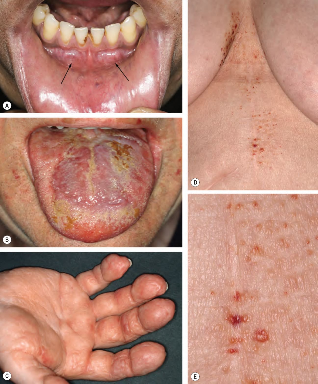
Fig. 47.8 Range of mucocutaneous findings in primary systemic AL amyloi- dosis.A Intraoral rubbery swellings (arrows). B Macroglossia with irregular thickening of the tongue. C Waxy infiltration of the palm and volar aspects of the fingers. D, E Multiple waxy, translucent papules on the breast, central chest, and upper abdomen; several of the lesions are purpuric. Courtesy Lorenzo Cerroni, MD.
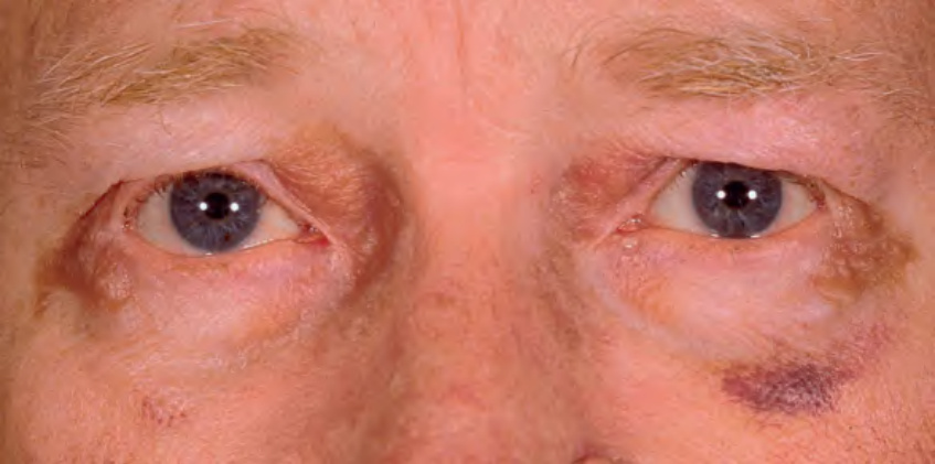
Fig. 47.9 Primary systemic AL amyloidosis. Purpura and yellow–brown plaques in a periorbital distribution. Courtesy Joyce Rico, MD.
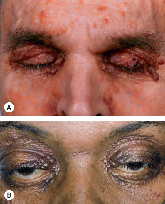
Fig. 47.10 Primary systemic AL amyloi- dosis.A Numerous waxy translucent facial papules. Some have a yellow to yellow–brown color. B Some of the waxy papules have become purpuric, but this can be more difficult to appreciate because of the pigmentation of the skin (compare to Fig. 47.9). The periorbital region is a characteristic site of involvement. A, Courtesy Jean L. Bolognia, MD; B, Courtesy Judit Stenn, MD.
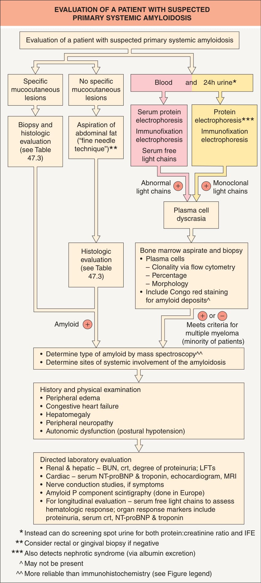
Fig. 47.11 Evaluation of a patient with suspected primary systemic AL amyloi- dosis. Because the amyloid is formed from immunoglobulin light chains, an abnormality of free light chains in the blood or urine (e.g. increased concentration or abnormal ratio in the blood, positive immunofixation electrophoresis [IFE] of the urine) is required for diagnosis. Mass spectroscopy has become more routine given the potential for misdiagnosis of older patients with wild-type ATTR deposits in the heart or ALect2 deposits in the kidney and liver in the setting of a monoclonal gammopathy of unknown significance. A positive cardiac pyrophosphate scan points to the diagnosis of ATTR amyloidosis. BUN, blood urea nitrogen; crt, creatinine; LFTs, liver function tests; NT-proBNP, N-terminal pro-brain natriuretic peptide.
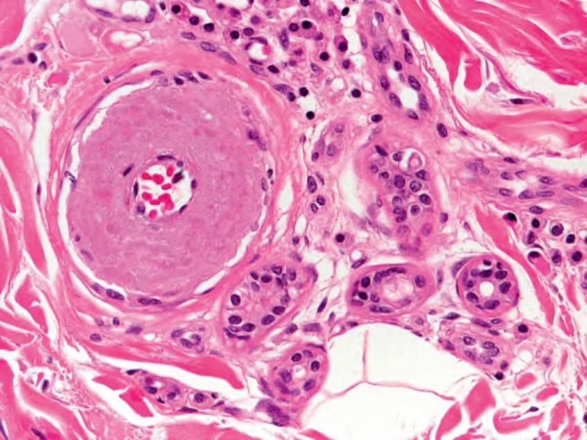
Fig. 47.12 Primary systemic AL amyloidosis – histopathologic features.