INTRODUCTION
The cutaneous deposition disorders are a heterogeneous group of conditions characterized by the presence of primarily endogenous substances within the dermis or the subcutis. Deposition disorders can be associated with localized or generalized cutaneous findings, and skin involvement is sometimes the earliest sign of a deposition disease. Histologic examination of cutaneous lesions, along with the use of special stains including immunohistochemistry, is a very helpful diagnostic tool (Table 48.1). In addition, specific enzymatic assays or genetic mutational analysis can then be performed to establish the precise diagnosis.
This chapter will focus on gout, pseudogout, lipoid proteinosis, colloid milium, and the mucopolysaccharidoses. Additional deposition disorders such as mucinoses, amyloidosis, porphyria, and calcinosis cutis are reviewed in Chapters 46–50.
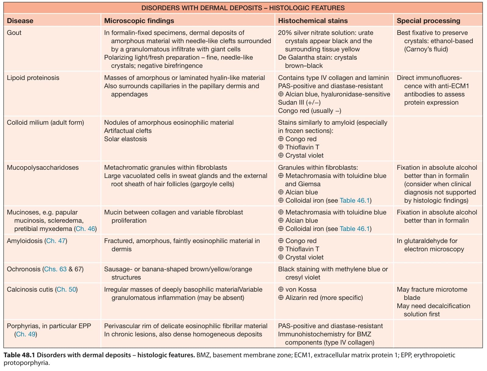
Table 48.1 Disorders with dermal deposits – histologic features. BMZ, basement membrane zone; ECM1, extracellular matrix protein 1; EPP, erythropoietic protoporphyria.