GOUT
Synonyms Urate crystal arthropathy Podagra (first MTP joint) Urate deposition disease
Key features
Gout is a metabolic disease in which the underlying abnormality is hyperuricemia
Deposits of needle-like crystals of monosodium urate are found in several sites, most commonly the skin (as tophi) and the joints
In fresh preparations of synovial fluid and expressed material from tophi, the crystals exhibit negative birefringence when examined by polarized light
In routinely fixed histologic sections, amorphous deposits that contain needle-like clefts are seen in the dermis and subcutis
Introduction
Gout is a metabolic disease in which needle-like crystals of monosodium urate (the ionized form of uric acid) from supersaturated fluids are deposited within tissue. Clinical manifestations include gouty arthritis, accumulation of crystals in connective tissue (tophi), uric acid nephrolithiasis, and renal impairment.
Epidemiology
Gout is the most common form of crystal-induced arthritis, and its prevalence seems to have increased over the past several decades. Men 40–50 years of age are most commonly affected. The male-to-female ratio is 9 : 1, and most women with gout are postmenopausal. Gout is found in up to 4% of adults and ~20% of patients have a family history of gout.
Pathogenesis
Gout is a consequence of deposits of urate crystals that have precipitated from supersaturated body fluids. Uric acid is the end product of the catabolism of purines. When the body increases its production of uric acid or if the kidneys do not excrete sufficient amounts, the result is hyperuricemia (Table 48.2). Migration and activation of neutrophils that are attempting to phagocytize urate crystals lead to the generation of neutrophil extracellular traps (NETs). The latter activate the NLRP3 inflammasome (see Fig. 4.2) via RIPK3-MLKL signaling, with subsequent production of proinflammatory cytokines. RIPK3 and MLKL represent two core proteins of the necroptosis pathway. Of note, NETs can also promote auto-resolution of the inflammatory phase of gout.
Clinical Features
The typical patient with gout is a middle-aged or older man, who may have a family history of the disease. Risk factors include obesity, excessive alcohol intake, renal insufficiency, and certain medications, most commonly diuretics. The natural history of gout involves four clinical stages: (1) asymptomatic hyperuricemia; (2) acute gouty arthritis; (3) intercritical gout (the intervals between attacks, which progressively become shorter in duration); and (4) chronic tophaceous gout.
Acute gouty arthritis
The most common symptom of gout is the development of severe joint pain over a period of 6–12 hours, accompanied by tenderness, erythema, warmth, and swelling. Initial attacks are often monoarticular, but in up to 40% of patients they may be polyarticular. In 75% of patients, the first metatarsophalangeal joint is involved (referred to as podagra); additional sites include the knee, ankle, and foot, and less commonly, the hand, wrist, and elbow. Fever and systemic symptoms may accompany an acute attack of gout. As the swelling subsides, the skin acquires a violaceous hue often followed by desquamation as edema subsides.
Chronic tophaceous gout
Cutaneous deposits of monosodium urate, known as “tophi”, usually appear an average of 10 years after the onset of gout. These tophi present as firm dermal or subcutaneous papules and nodules or as a fusiform swelling. Their contours may be smooth or multilobulated (Fig. 48.1) and they vary from skin-colored to white–yellow to red (Fig. 48.2). The surface may be ulcerated and there may be associated drainage of material, which varies from clear fluid with white flecks to a thick chalky material.
The most common locations for tophi are the skin overlying joints and the helix of the ear. Unusual sites include the eyes, nose, larynx, breast, and heart valves. However, tophi develop in fewer than 10% of patients with gout. Depending upon the size and location of the tophi, complete, partial, or minimal resolution can occur when the serum uric acid level is normalized.
Elevated levels of uric acid in the urine are associated with uric acid nephrolithiasis and may cause acute renal failure when uric acid precipitates in the renal tubules and collecting ducts. The most common setting for the latter is tumor lysis syndrome following chemotherapy for a rapidly dividing chemosensitive malignancy, e.g. leukemias, lymphomas.
The initial diagnosis of gout is usually made in the setting of acute arthritis, based upon the presence of urate crystals in an aspirate of joint fluid. Hyperuricemia is usually present, but alone it is insufficient for establishing a definitive diagnosis of gout. Additional laboratory abnormalities during an acute attack include elevated white blood cell count (in the joint fluid and blood) and ESR.
The erythema reflects surrounding inflammation.
When examined by polarized light microscopy, the needle-shaped urate crystals change color from yellow to blue based upon their alignment relative to the axis of the “red plate” compensator, i.e. they exhibit negative birefringence. In patients with recurrent attacks, radiographs of involved joints often show “punched-out” erosions with sclerotic “overhanging” margins, but no osteophytes. A 24-hour urine collection for uric acid allows the identification of patients at risk for nephrolithiasis (see Table 48.2).
Pathology
The key histologic feature of formalin-fixed gouty tophi is the presence of deposits of amorphous material within the dermis and subcutis, corresponding to sites of crystal deposition. These deposits contain needle-like clefts (which represent dissolved urate crystals) surrounded by a granulomatous infiltrate with multinucleated giant cells (Fig. 48.3). Secondary calcification or even ossification can occasionally be seen in older lesions.
In order to preserve the crystals, the biopsy specimen needs to be placed in an ethanol-based fixative, such as Carnoy’s fluid. Then, under regular or polarized light, brightly refractile brown sheaths of fine needle-like crystals can be seen. Upon staining with 20% silver nitrate solution, the crystals appear black and the surrounding tissue yellow, whereas with the De Galantha stain, the crystals appear brown to black.
Differential Diagnosis
The differential diagnosis of acute gouty arthritis includes pseudogout (see below), osteoarthritis, psoriatic arthritis, reactive arthritis (previously referred to as Reiter disease), and septic arthritis. Given its destructive nature, septic arthritis needs to be considered even in patients in whom the diagnosis of gout has been previously established. The presence of needle-shaped crystals with negative birefringence in an aspirate of joint fluid is diagnostic of gout, while Gram stain and culture exclude septic arthritis. Occasionally, patients are misdiagnosed as having cellulitis.
The differential diagnosis of cutaneous gouty tophi includes xanthomas, rheumatoid nodules, and calcinosis cutis. Examination of expressed fluid or chalky material by polarized light can provide a “bedside” diagnosis. By ultrasonography, central clear spaces are seen within tophi whereas rheumatoid nodules contain central echodense areas.
Treatment
For acute gout, short-acting oral nonsteroidal anti-inflammatory drugs (NSAIDs) are taken within 24 hours of onset at the highest safe dosage as long as symptoms persist, e.g. indomethacin 50 mg three times daily for several days. Contraindications include peptic ulceration, renal insufficiency, and anticoagulation.
Colchicine, a derivative of the autumn crocus, inhibits the transport of phagocytized urate crystals to lysosomes within neutrophils via its binding to microtubules. Colchicine also interferes with leukocyte migration, chemotaxis, and adhesion. For patients with normal renal function who are not receiving strong CYP3A4 inhibitors, the recommended dosage for acute gout flares is 1.2 mg orally followed by 0.6 mg 1 hour later. Low-dose NSAIDs and colchicine (0.6 mg twice daily) are both utilized for prophylaxis. Outside of the US, colchicine tablets often contain 0.5 mg. Side effects of colchicine are outlined in Table 130.9. Intra-articular or systemic corticosteroids may be used for a period of 1–3 weeks if NSAIDs and colchicine are ineffective or contraindicated.
Urate-lowering therapy (ULT) consists of the xanthine oxidase inhibitors allopurinol and febuxostat which block uric acid production. Allopurinol is the preferred first-line ULT, but low starting doses (≤100 mg/day or lower if chronic kidney disease) are recommended. ULT is indicated for patients with tophaceous gout, radiographic damage due to gout, or frequent gout flares. Concomitant anti-inflammatory prophylaxis therapy for 3–6 months is also recommended when initiating ULT.
Side effects of allopurinol include diarrhea, thrombocytopenia, hepatitis, and skin eruptions that can range from morbilliform to erythroderma to Stevens–Johnson syndrome/toxic epidermal necrolysis (SJS/TEN). HLA-B*58:01-positive individuals are at markedly increased risk of developing severe cutaneous adverse reactions, including both SJS/TEN and DRESS. Of note, this allele is most common in those of Han Chinese, Korean, or Thai descent. Major side effects of febuxostat are liver enzyme elevations and cardiovascular thromboembolic events. Drug–drug interactions, e.g. with azathioprine, can occur with both of these medications (see Fig. 130.5). Uricosuric agents such as oral probenecid can be added or used as second-line therapy for patients with normal renal function and no renal stones. Overall, maintenance of a serum uric acid level of ≤6 mg/ dl is recommended. A newer agent, lesinurad, is a selective inhibitor of reabsorption of uric acid.
Some patients may benefit from a reduction in purine-rich foods (e.g. organ meats [liver, heart], fish), alcohol consumption, and body weight. In the setting of potential tumor lysis syndrome, intravenous rasburicase can be administered, which rapidly converts uric acid to the more soluble allantoin. Bimonthly intravenous pegloticase, which has the same mechanism of action, can be used for patients with chronic gout refractory to conventional therapy. IL-1 blocking agents (e.g. anakinra, canakinumab) have been used for recurrent acute gout unresponsive to other therapies, but are not currently approved for this indication (see Fig. 45.13).
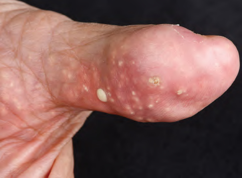
Fig. 48.1 Tophaceous gout of a digit. The deposits create a multilobulated appearance and there is evidence of transepidermal elimination. A small incision over the yellow–white papules followed by microscopic examination of the expressed material would allow for a bedside diagnosis. Courtesy Lorenzo Cerroni, MD.
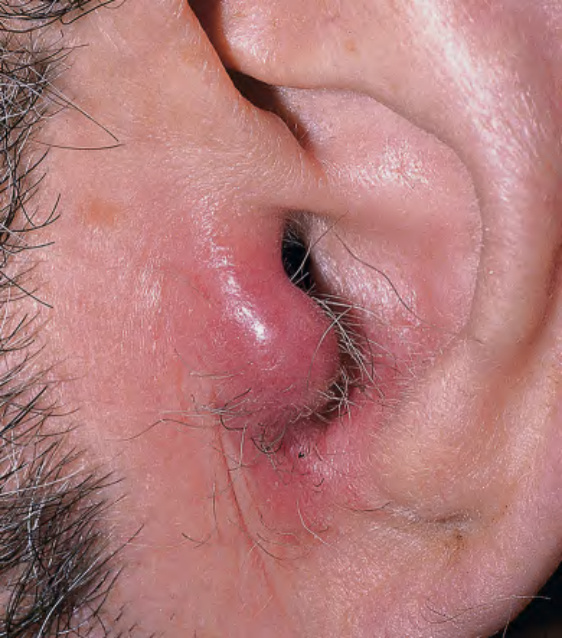
Fig. 48.2 Tophaceous gout of the tragus.
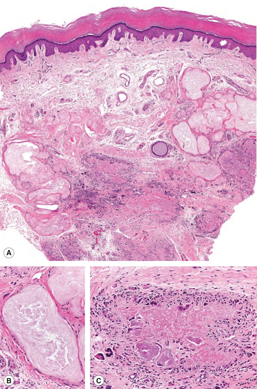
Fig. 48.3 Tophaceous gout – histopathologic features.A Within the dermis and subcutis, there are deposits of amorphous, acellular material. These deposits contain needle-like clefts (B). Histiocytes and multinucleated giant cells surround some of the deposits (C). Courtesy Lorenzo Cerroni, MD.
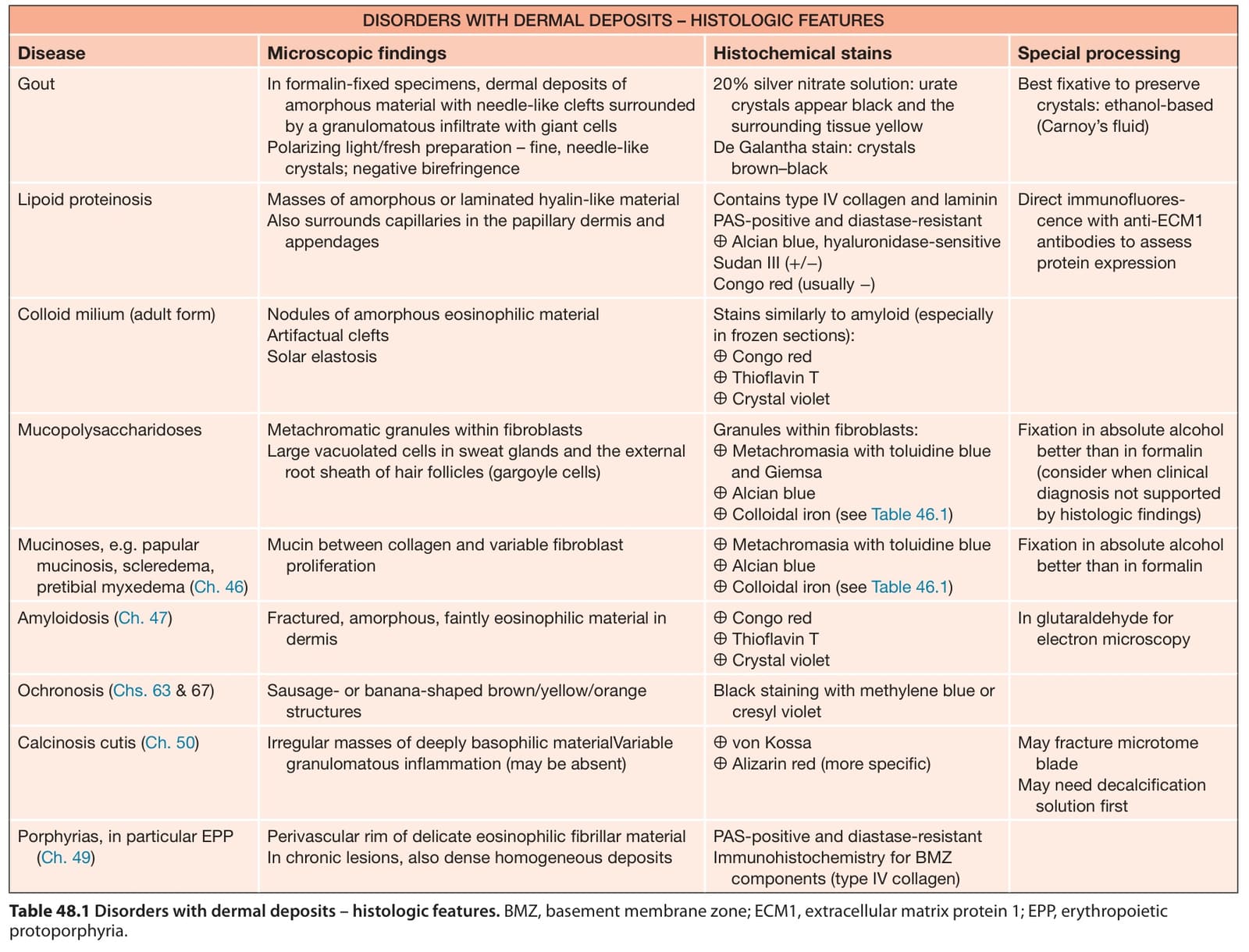
Table 48.1 Disorders with dermal deposits – histologic features. BMZ, basement membrane zone; ECM1, extracellular matrix protein 1; EPP, erythropoietic protoporphyria.
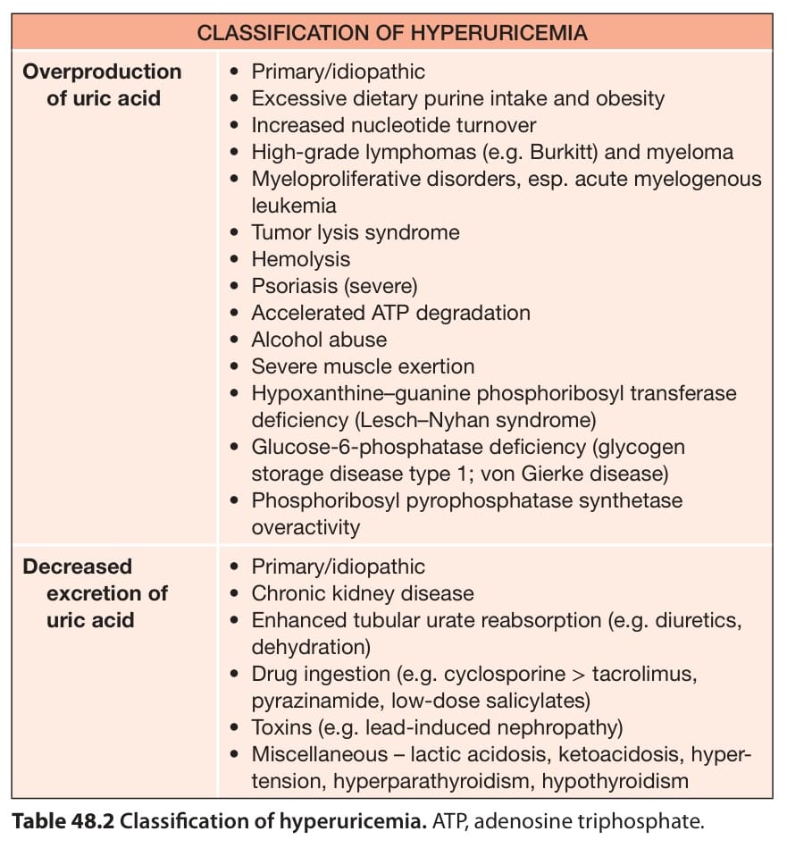
Table 48.2 Classification of hyperuricemia. ATP, adenosine triphosphate.