LIPOID PROTEINOSIS
Synonyms: Hyalinosis cutis et mucosae Urbach–Wiethe disease
Key features
A rare autosomal recessive deposition disorder due to mutations in
ECM1, which encodes extracellular matrix protein 1
Hyalin-like material is deposited within multiple organs, including the skin, oral mucosa, larynx, and brain
Papules and nodules favor the face, as do pitted scars; diffuse waxy thickening of the skin can be associated with verrucous changes, especially on the elbows, knees, and hands
History
Lipoid proteinosis was first described by Siebenmann in 1908. Twenty years later, it was established as a distinct entity by two Viennese physicians, Erich Urbach, a dermatologist, and Camillo Wiethe, an otorhinolaryngologist.
Epidemiology
Most patients are of European ancestry, including descendants of Dutch immigrants who settled in South Africa in the middle of the 17th century. Not only do Afrikaners have the highest incidence of this genetic disease, they also exhibit a founder effect, i.e. the population shares a common mutation.
Pathogenesis
In 2002, lipoid proteinosis was shown to be due to loss-of-function mutations in ECM1, which encodes extracellular matrix protein 1. ECM1 is a secretory glycoprotein that can act as a negative regulator of endochondral bone formation, a promoter of angiogenesis, and an inhibitor of matrix metalloproteinase 9. In addition, ECM1a (see below) is found within the basement membrane zone and can enhance the binding of collagen IV to laminin 332. It also interacts with components of the extracellular matrix such as fibronectin and glycosaminoglycans (e.g. hyaluronic acid, chondroitin sulfate).
ECM1 has three major splice variants that result in different isoforms: (1) ECM1a, a protein of 540 amino acids encoded by a 10-exon gene; (2) ECM1b, a protein of 415 amino acids reflecting a lack of exon 7; and (3) ECM1c, a protein of 559 amino acids due to an additional exon 5a within intron 5. Patients with mutations in exon 7 still express the ECM1b isoform and tend to have milder disease as compared to those with mutations in exon 6, who tend to have more severe disease and reduced expression of all three isoforms. A majority of pathogenic mutations occur within exon 6 or 7; occasionally, they occur in exon 2, 4, or 9. The degree of dermal immunofluorescent staining (utilizing an anti-ECM1 antibody) points to the location; reduced staining suggests mutations in exon 7 and an absence of staining mutations in exon 6.
Clinical Features
The first clinical sign of lipoid proteinosis is often a weak cry or hoarseness due to infiltration of the laryngeal mucosa. Hoarseness remains throughout life. Cutaneous lesions usually appear during the first 2 years of life, in two overlapping stages. In the first stage, vesicles and hemorrhagic crusts develop within the mouth and on the skin of the face and extremities, often in association with trauma. The cutaneous lesions may resolve with scarring, including “ice-pick”-like scars.
During the second stage, there is an increase in the hyalin deposits within the dermis (Fig. 48.4). The skin becomes diffusely thickened and waxy and develops a yellow color. Papules, plaques, and nodules appear on the face (including the eyelid margin) as well as in the axillae and on the scrotum. Verrucous lesions may develop on the extensor surfaces, especially the elbows (see Fig. 48.4C), knees, and hands. Generalized scaling has also been observed as has alopecia of the eyebrows and eyelashes. Corneal ulcers may occur due to infiltration of the eyelids.
In addition to involvement of the tongue (diffuse infiltration), the frenulum (restricted motion of the tongue), and the oropharynx, recurrent parotitis can occur due to occlusion of the salivary duct. Dental anomalies include hyperplasia or aplasia of the upper incisors, premolars or molars; patients usually lose their teeth at an early age. Neurologic manifestations are common and consist of behavioral and learning difficulties, seizures, and, less often, intracerebral hemorrhage. A pathognomonic radiographic finding is bilateral, intracranial, sickle-shaped calcification within the amygdalae. More recently, an increased prevalence of renal cysts has been described.
In general, the disease has a stable or slowly progressive course that is compatible with a normal lifespan, except for the risk of respiratory
obstruction in infancy. An elevated ESR may be seen due to increased α- or γ-globulins.
Pathology
The major clinical manifestations are related to the deposition of an amorphous or laminated material around blood vessels and within the connective tissues. The amorphous deposits consist primarily of non-collagen proteins, while the concentric layers of basement membranelike material contain collagen (types II and IV) and laminin. In addition, the deposits are PAS-positive and diastase-resistant, indicating the presence of neutral glycosaminoglycans (GAGs; mucopolysaccharides).
H&E-stained sections of early lesions reveal pink, hyalin-like deposits around the capillaries within the papillary dermis and at the periphery of eccrine sweat glands. Older lesions are characterized by hyperkeratosis, occasionally papillomatosis, and a thickened dermis in which bundles of pink hyalin deposits are found in a diffuse pattern (Fig. 48.5); the bundles are often oriented perpendicularly to the dermal–epidermal junction. There are smaller scattered deposits of hyalin in the lower dermis. Hyalin deposits also surround the hair follicles, sebaceous glands, and arrector pili muscles as well as the eccrine glands.
In addition to type IV collagen, laminin, and neutral GAGs, hyaluronic acid is present in the dermis, as demonstrated by Alcian blue and hyaluronidase staining (see Ch. 46). Fat stains (e.g. Sudan III) give inconsistent results when the tissue is formalin-fixed, and rarely the deposits stain with Congo red. Ultrastructurally, reduplication of the basement membrane of vessels and occasionally the dermal–epidermal junction is seen; the deposits appear to be composed of small granules and short filaments of low electron density that measure 3–5 nm in diameter.
There are few reports of the histopathologic features of the vesicular first phase and they vary from intraepidermal acantholysis to cell-poor subepidermal blisters.
Differential Diagnosis
The clinical differential diagnosis includes erythropoietic protoporphyria (EPP), amyloidosis, papular mucinosis (scleromyxedema), colloid milium, and non-Langerhans cell histiocytoses. During the initial phase, hydroa vacciniforme may be considered. In infants, the possibility of hyaline fibromatosis syndrome (juvenile hyaline fibromatosis and infantile systemic hyalinosis) needs to be considered, which is due to mutations in the gene that encodes capillary morphogenesis protein 2 (see Ch. 98). Histologically, the differential diagnosis includes EPP and colloid milium. In cutaneous lesions of EPP, colloid milium, and amyloidosis (see Table 48.1), the hyalinization is milder, more focal and superficial, and it rarely involves the eccrine glands. The deposits in colloid milium do not have the striking perivascular pattern seen in lipoid proteinosis.
Treatment
Currently, there is no known cure or effective treatment for lipoid proteinosis. Oral, intralesional (including submucosal), and topical corticosteroids have been used with some beneficial effect, as have humectants. Plastic surgery, dermabrasion, and laser resurfacing have been successfully performed, and there are limited reports of the use of systemic retinoids (e.g. acitretin 0.5 mg/kg/day for 6 months), D-penicillamine, and oral dimethyl sulfoxide (DMSO).
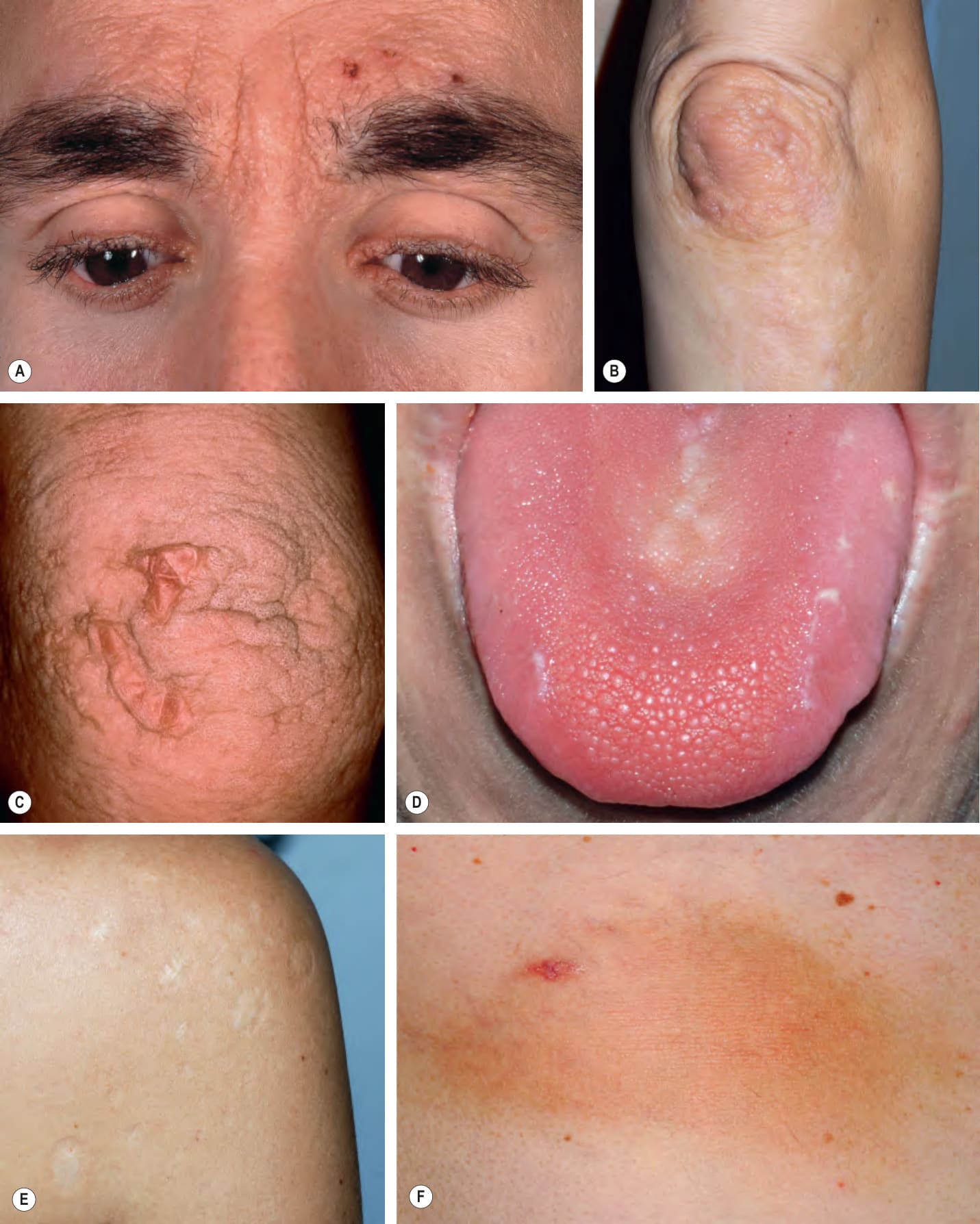
Fig. 48.4 Lipoid proteinosis – clinical features.A Beaded eyelid papules (moniliform blepharosis), hemorrhagic crusts, and confluent waxy papules of the glabella leading to an early leonine facies. B Skin-colored papulonodules of the elbow as well as irregular hypopigmented scars of the extensor forearm. C Characteristic verrucous changes of the elbow. D A firm tongue with numerous tiny papules on the dorsal surface. E Multiple, round hypopigmented and depressed scars on the shoulder. F Waxy, yellowish plaque on the back. B, D, E, Courtesy Julie V. Schaffer, MD; C, Courtesy Judit Stenn, MD.
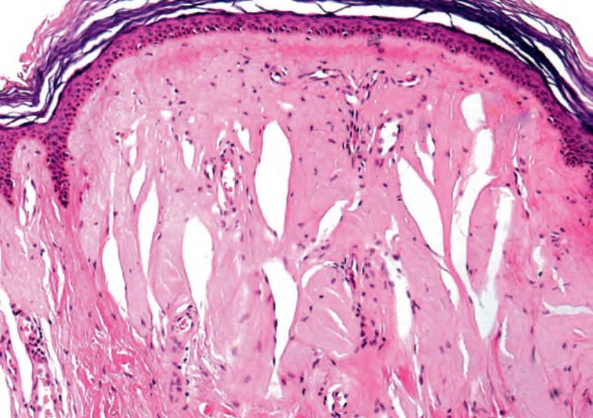
Fig. 48.5 Lipoid proteinosis – histopathologic features. Hyperkeratosis, a flattened epidermis, and vertically oriented eosinophilic deposits in the dermis. Note the cleft formation. Courtesy Lorenzo Cerroni, MD.
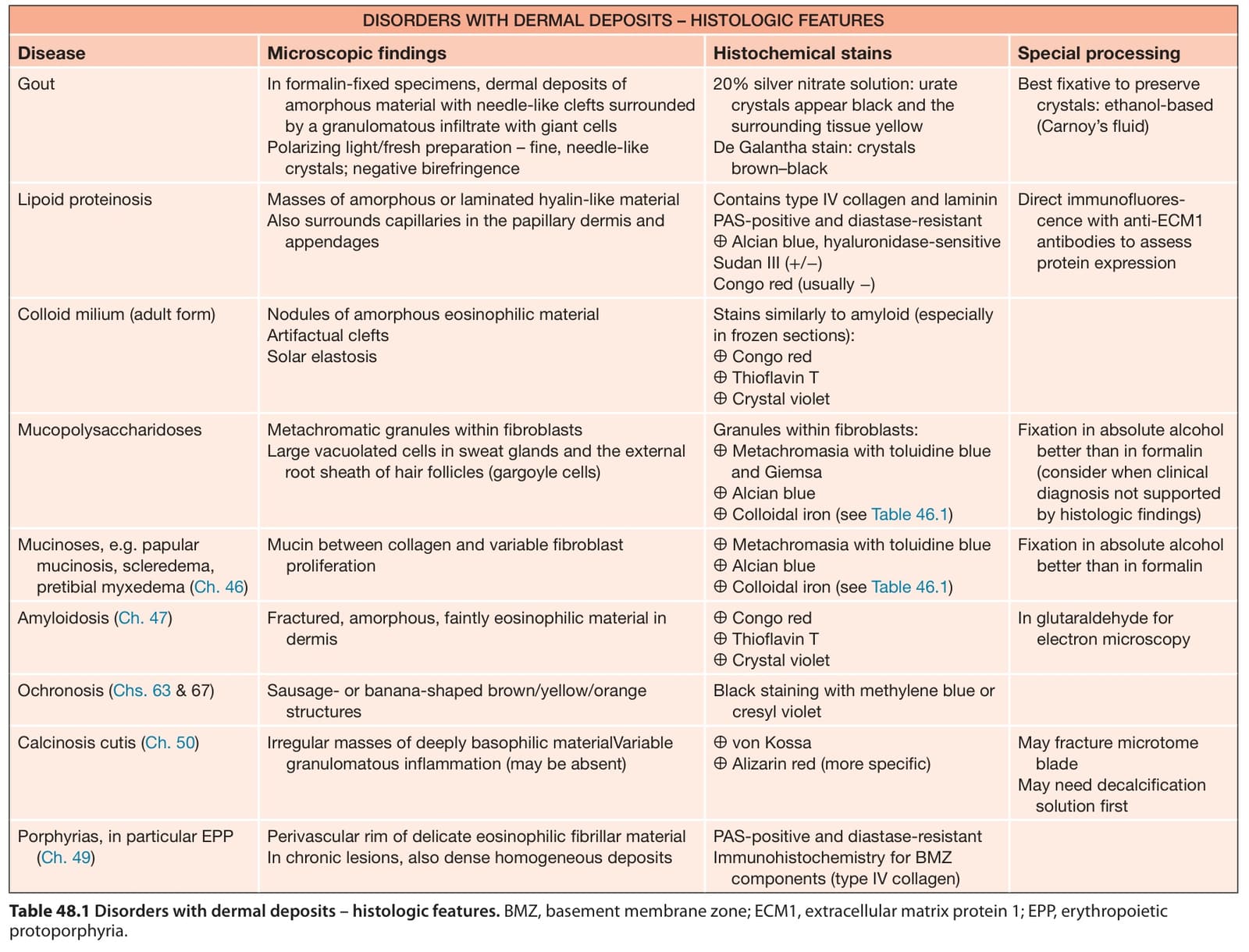
Table 48.1 Disorders with dermal deposits – histologic features. BMZ, basement membrane zone; ECM1, extracellular matrix protein 1; EPP, erythropoietic protoporphyria.