COLLOID MILIUM
Synonyms: Colloid degeneration of the skin Elastosis colloidalis conglomerata Miliary colloidoma
Key features
A rare disorder in which dome-shaped, translucent papules develop in sun-exposed skin
Four variants are recognized: adult, juvenile, and pigmented colloid milium and nodular colloid degeneration
Histologically, amorphous, eosinophilic, granular deposits are found within the superficial dermis
Introduction and Epidemiology
Adult colloid milium is the most common form of the disease. It usually occurs in middle-aged, fair-skinned individuals who have had significant cumulative sun exposure, with a male-to-female ratio of 4 : 1. The adult form has also been observed in outdoor oil refinery workers, suggesting an additional effect of petrochemicals. In general, juvenile colloid milium develops before puberty and both an autosomal recessive and an autosomal dominant inheritance pattern have been reported.
Pathogenesis
Although excessive sun exposure clearly plays a role in adult colloid milium, the precise pathogenesis is not known. Photoinduced damage to dermal elastic fibers, as in solar elastosis, has been suggested. In juvenile colloid milium, ultraviolet light-induced damage to epidermal keratinocytes has been observed and this may reflect an inherited susceptibility. Pigmented colloid milium is a variant of the adult form in which there is a combination of excessive sun exposure plus topical application of hydroquinone or phenols.
Clinical Features
In both the adult and juvenile forms, multiple, dome-shaped, translucent to yellow or yellow–brown papules are seen in areas of chronic sun exposure (Fig. 48.6), e.g. the face (especially periorbitally), ears, posterolateral neck, and the dorsal aspect of the hands and forearms. With minor trauma, lesions can become hemorrhagic. By dermoscopy, a diffuse orange background is seen along with linear-curved blurred telangiectasias and a few milia-like cysts. In pigmented colloid milium, grouped to confluent gray–brown papules are present on the face, whereas in nodular colloid degeneration, skin-colored to yellowish nodules or plaques develop, often on the face.
Pathology
In adult colloid milium, nodules composed of homogeneous eosinophilic colloid material are seen in the papillary dermis (Fig. 48.7). Within the nodules, there may be clefts and fissures. A grenz zone is often present, as is evidence of solar elastosis beneath the nodules. Although the colloid masses contain degenerated elastic fibers, they often exhibit a staining profile similar to amyloidosis, i.e. positive staining with crystal violet, Congo red, and thioflavin T stains; such staining is more consistent in frozen sections. In addition, PAS staining is positive. A negative pan-cytokeratin stain is helpful in distinguishing adult colloid milium from primary cutaneous amyloidosis (amyloid K).
In juvenile colloid milium, amorphous eosinophilic deposits are also found within the papillary dermis, but when compared to the adult form, there is less clefting and no grenz zone. As with colloid bodies (which are also keratinocyte-derived), nonspecific staining for immunoglobulins, complement, and fibrin is seen. Similar to primary cutaneous amyloidosis, staining for keratin is positive due to the presence of amyloid K.
In pigmented colloid milium, lightly pigmented islands of colloid material are found in the upper dermis. In nodular colloid degeneration,
homogenized amorphous dermal collagen with small fissures and clefts is seen. Whether this entity is a variant of nodular amyloidosis is still under debate. Ultrastructurally, colloid milium is characterized by amorphous and granular material which lacks the straight, non-branching filaments seen in amyloidosis.
Differential Diagnosis
Clinically, the differential diagnosis of juvenile and adult forms of colloid milium includes erythropoietic protoporphyria (EPP), mucinoses (e.g. papular, self-healing), lipoid proteinosis, adnexal tumors, and primary systemic amyloidosis (adults). Pigmented colloid milium needs to be distinguished from exogenous ochronosis. Histologically, colloid milium must be distinguished from EPP, lipoid proteinosis, and primary cutaneous amyloidosis. In cutaneous lesions of EPP, there are hyalin deposits around and within the superficial blood vessel walls, whereas the dermal deposits of lipoid proteinosis are accentuated around the blood vessels. Primary cutaneous amyloidosis may be indistinguishable from the juvenile type of colloid milium, as both stain positively for keratin; some authors consider juvenile colloid milium to be a variant of lichen amyloidosis. Unlike lipoid proteinosis and lichen amyloidosis, the dermal deposits of adult colloid milium do not stain positively for laminin and type IV collagen or cytokeratin, respectively.
Treatment
Treatment modalities are limited and include laser therapy (e.g. long-pulsed Er:YAG, fractional photothermolysis), dermabrasion, intense pulsed light, photodynamic therapy, cryotherapy, and chemical peels.
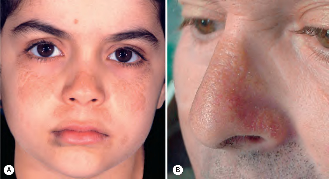
Fig. 48.6 Juvenile and adult colloid milium.A Coalescing brown papules in sun-exposed sites of the face. Note the sparing of the philtrum. B Yellowish, translucent papules and multi-lobulated plaques are present on the nose, admixed with telangiectasias. A, Courtesy Lorenzo Cerroni, MD, and Sergio Chimenti, MD; B, Courtesy Luis Requena, MD.
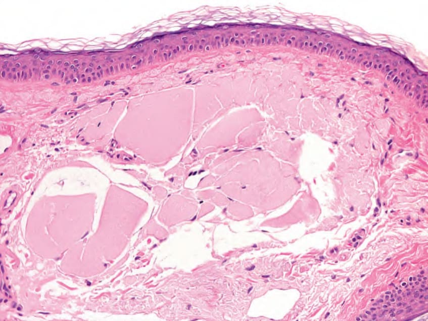
Fig. 48.7 Adult colloid milium – histopathologic features. Nodular mass of eosinophilic, homogeneous material within the upper dermis with a thin subepidermal grenz zone. Note fissures and clefts within the mass with a few fibroblasts along some of the clefts. Courtesy Lorenzo Cerroni, MD.